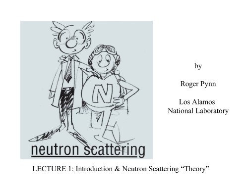
An Introduction to Neutron Scattering - Spallation Neutron Source
An Introduction to Neutron Scattering - Spallation Neutron Source
An Introduction to Neutron Scattering - Spallation Neutron Source

Create successful ePaper yourself
Turn your PDF publications into a flip-book with our unique Google optimized e-Paper software.
y<br />
Roger Pynn<br />
Los Alamos<br />
National Labora<strong>to</strong>ry<br />
LECTURE 1: <strong>Introduction</strong> & <strong>Neutron</strong> <strong>Scattering</strong> “Theory”
Overview<br />
1. <strong>Introduction</strong> and theory of neutron scattering<br />
1. Advantages/disadvantages of neutrons<br />
2. Comparison with other structural probes<br />
3. Elastic scattering and definition of the structure fac<strong>to</strong>r, S(Q)<br />
4. Coherent & incoherent scattering<br />
5. Inelastic scattering<br />
6. Magnetic scattering<br />
7. Overview of science studied by neutron scattering<br />
8. References<br />
2. <strong>Neutron</strong> scattering facilities and instrumentation<br />
3. Diffraction<br />
4. Reflec<strong>to</strong>metry<br />
5. Small angle neutron scattering<br />
6. Inelastic scattering
Why do <strong>Neutron</strong> <strong>Scattering</strong>?<br />
• To determine the positions and motions of a<strong>to</strong>ms in condensed matter<br />
– 1994 Nobel Prize <strong>to</strong> Shull and Brockhouse cited these areas<br />
(see http://www.nobel.se/physics/educational/poster/1994/neutrons.html)<br />
• <strong>Neutron</strong> advantages:<br />
– Wavelength comparable with intera<strong>to</strong>mic spacings<br />
– Kinetic energy comparable with that of a<strong>to</strong>ms in a solid<br />
– Penetrating => bulk properties are measured & sample can be contained<br />
– Weak interaction with matter aids interpretation of scattering data<br />
– Iso<strong>to</strong>pic sensitivity allows contrast variation<br />
– <strong>Neutron</strong> magnetic moment couples <strong>to</strong> B => neutron “sees” unpaired electron spins<br />
• <strong>Neutron</strong> Disadvantages<br />
– <strong>Neutron</strong> sources are weak => low signals, need for large samples etc<br />
– Some elements (e.g. Cd, B, Gd) absorb strongly<br />
– Kinematic restrictions (can’t access all energy & momentum transfers)
The 1994 Nobel Prize in Physics – Shull & Brockhouse<br />
<strong>Neutron</strong>s show where the a<strong>to</strong>ms are….<br />
…and what the a<strong>to</strong>ms do.
The <strong>Neutron</strong> has Both Particle-Like and Wave-Like Properties<br />
• Mass: m n = 1.675 x 10 -27 kg<br />
• Charge = 0; Spin = ½<br />
• Magnetic dipole moment: μ n = - 1.913 μ N<br />
• Nuclear magne<strong>to</strong>n: μ N = eh/4πm p = 5.051 x 10 -27 J T -1<br />
• Velocity (v), kinetic energy (E), wavevec<strong>to</strong>r (k), wavelength (λ),<br />
temperature (T).<br />
• E = m n v 2 /2 = k B T = (hk/2π) 2 /2m n ; k = 2 π/λ = m n v/(h/2π)<br />
Energy (meV) Temp (K) Wavelength (nm)<br />
Cold 0.1 – 10 1 – 120 0.4 – 3<br />
Thermal 5 – 100 60 – 1000 0.1 – 0.4<br />
Hot 100 – 500 1000 – 6000 0.04 – 0.1<br />
l (nm) = 395.6 / v (m/s)<br />
E (meV) = 0.02072 k 2 (k in nm -1 )
Comparison of Structural Probes<br />
Note that scattering methods<br />
provide statistically averaged<br />
information on structure<br />
rather than real-space pictures<br />
of particular instances<br />
Macromolecules, 34, 4669 (2001)
Thermal <strong>Neutron</strong>s, 8 keV X-Rays & Low Energy<br />
Electrons:- Absorption by Matter<br />
Note for neutrons:<br />
• H/D difference<br />
• Cd, B, Sm<br />
• no systematic A<br />
dependence
Interaction Mechanisms<br />
• <strong>Neutron</strong>s interact with a<strong>to</strong>mic nuclei via very short range (~fm) forces.<br />
• <strong>Neutron</strong>s also interact with unpaired electrons via a magnetic dipole<br />
interaction.
Brightness & Fluxes for <strong>Neutron</strong> &<br />
X-Ray <strong>Source</strong>s<br />
Brightness<br />
(s -1 m -2 ster -1 )<br />
<strong>Neutron</strong>s 10 15<br />
Rotating<br />
<strong>An</strong>ode<br />
Bending<br />
Magnet<br />
10 16<br />
10 24<br />
Wiggler 10 26<br />
Undula<strong>to</strong>r<br />
(APS)<br />
10 33<br />
dE/E<br />
(%)<br />
Divergence<br />
(mrad 2 )<br />
Flux<br />
(s -1 m -2 )<br />
2 10 x 10 10 11<br />
3 0.5 x 10 5 x 10 10<br />
0.01 0.1 x 5 5 x 10 17<br />
0.01 0.1 x 1 10 19<br />
0.01 0.01 x 0.1 10 24
Φ = number of<br />
σ<br />
=<br />
<strong>to</strong>talnumber<br />
dσ<br />
number of<br />
=<br />
dΩ<br />
2<br />
d σ<br />
=<br />
dΩdE<br />
of<br />
number of<br />
incident neutrons per<br />
Cross Sections<br />
cm<br />
neutrons scatteredper<br />
per second<br />
second / Φ<br />
neutronsscatteredper<br />
second in<strong>to</strong><br />
dΩ<br />
Φ dΩ<br />
neutronsscatteredper<br />
second in<strong>to</strong><br />
Φ dΩ<br />
dE<br />
2<br />
dΩ<br />
& dE<br />
σ measured in barns:<br />
1 barn = 10 -24 cm 2<br />
Attenuation = exp(-Nσt)<br />
N = # of a<strong>to</strong>ms/unit volume<br />
t = thickness
<strong>Scattering</strong> by a Single (fixed) Nucleus<br />
If v is the velocity of<br />
passing through an area dS per second after scattering is<br />
v dS ψ<br />
scat<br />
Since the number of<br />
2<br />
= v dS b<br />
2<br />
dσ<br />
v b dΩ<br />
= = b<br />
dΩ<br />
ΦdΩ<br />
2<br />
2<br />
the neutron (same before and after scattering),<br />
the<br />
/r<br />
2<br />
= v b<br />
2<br />
dΩ<br />
incident neutrons passing through unit areasis<br />
soσ<br />
<strong>to</strong>tal<br />
= 4πb<br />
2<br />
:<br />
• range of nuclear force (~ 1fm)<br />
is scattering is<br />
elastic<br />
• we consider only scattering far<br />
from nuclear resonances where<br />
neutron absorption is negligible<br />
:<br />
number of<br />
Φ = vψ<br />
incident<br />
neutrons<br />
2<br />
= v
Adding up <strong>Neutron</strong>s Scattered by Many Nuclei<br />
0<br />
)<br />
(<br />
,<br />
)<br />
(<br />
)<br />
'<br />
(<br />
,<br />
2<br />
i<br />
2<br />
)<br />
'<br />
(<br />
i<br />
'.<br />
2<br />
)<br />
'.(<br />
i<br />
i<br />
scat<br />
R<br />
.<br />
k<br />
i<br />
i<br />
'<br />
by<br />
defined<br />
is<br />
Q<br />
r transfer<br />
wavevec<strong>to</strong><br />
the<br />
where<br />
get<br />
<strong>to</strong><br />
dS/r<br />
d<br />
use<br />
can<br />
we<br />
R<br />
r<br />
that<br />
so<br />
away<br />
enough<br />
far<br />
measure<br />
we<br />
If<br />
R<br />
-<br />
r<br />
1<br />
R<br />
-<br />
r<br />
b<br />
-<br />
is<br />
wave<br />
scattered<br />
the<br />
so<br />
e<br />
is<br />
ave<br />
incident w<br />
the<br />
R<br />
at<br />
located<br />
nucleus<br />
a<br />
At<br />
0<br />
0<br />
0<br />
i<br />
0<br />
k<br />
k<br />
Q<br />
e<br />
b<br />
b<br />
e<br />
b<br />
b<br />
d<br />
d<br />
e<br />
e<br />
b<br />
d<br />
dS<br />
vd<br />
vdS<br />
d<br />
d<br />
e<br />
e<br />
j<br />
i<br />
j<br />
i<br />
i<br />
i<br />
i<br />
R<br />
R<br />
.<br />
Q<br />
i<br />
j<br />
j<br />
i<br />
i<br />
R<br />
R<br />
.<br />
k<br />
k<br />
i<br />
j<br />
j<br />
i<br />
i<br />
R<br />
.<br />
k<br />
k<br />
i<br />
r<br />
k<br />
i<br />
i<br />
scat<br />
R<br />
r<br />
k<br />
i<br />
R<br />
.<br />
k<br />
i<br />
r<br />
r<br />
r<br />
r<br />
r<br />
r<br />
r<br />
r<br />
r<br />
r<br />
r<br />
r<br />
r<br />
r<br />
r<br />
r<br />
r<br />
r<br />
r<br />
r<br />
r<br />
r<br />
r<br />
r<br />
r<br />
r<br />
r<br />
−<br />
=<br />
=<br />
=<br />
Ω<br />
=<br />
Ω<br />
>><br />
Ω<br />
=<br />
Ω<br />
=<br />
Ω<br />
∴<br />
⎥<br />
⎥<br />
⎦<br />
⎤<br />
⎢<br />
⎢<br />
⎣<br />
⎡<br />
=<br />
−<br />
−<br />
−<br />
−<br />
−<br />
−<br />
∑<br />
∑<br />
∑<br />
∑<br />
σ<br />
ψ<br />
σ<br />
ψ
Coherent and Incoherent <strong>Scattering</strong><br />
The scattering length, b i , depends on the nuclear iso<strong>to</strong>pe, spin relative <strong>to</strong> the<br />
neutron & nuclear eigenstate. For a single nucleus:<br />
b<br />
i<br />
b b<br />
i<br />
=<br />
but<br />
j<br />
δb<br />
2<br />
i<br />
=<br />
dσ<br />
∴<br />
dΩ<br />
b<br />
δb<br />
=<br />
=<br />
+ δb<br />
b<br />
2<br />
=<br />
b<br />
i<br />
b<br />
0<br />
i<br />
+<br />
−<br />
2<br />
b<br />
where δb<br />
averages <strong>to</strong> zero<br />
and<br />
b<br />
∑<br />
i,<br />
j<br />
( δb<br />
e<br />
2<br />
i<br />
+ δb<br />
δb<br />
δb<br />
=<br />
i<br />
b<br />
2<br />
r r r<br />
−iQ.(<br />
R −R<br />
Coherent <strong>Scattering</strong><br />
(scattering depends on the<br />
direction of Q)<br />
i<br />
j<br />
j<br />
j<br />
)<br />
) + δbδb<br />
vanishes<br />
−<br />
+ (<br />
b<br />
b<br />
2<br />
i<br />
2<br />
j<br />
−<br />
unless<br />
b<br />
2<br />
) N<br />
Note: N = number of a<strong>to</strong>ms in scattering system<br />
i<br />
=<br />
Incoherent <strong>Scattering</strong><br />
(scattering is uniform in all directions)<br />
j
Nuclide<br />
1 H<br />
2 H<br />
C<br />
O<br />
Al<br />
Values of σ coh and σ inc<br />
s coh<br />
1.8<br />
5.6<br />
5.6<br />
4.2<br />
1.5<br />
s inc<br />
80.2<br />
2.0<br />
0.0<br />
0.0<br />
0.0<br />
Nuclide<br />
V<br />
Fe<br />
Co<br />
Cu<br />
36 Ar<br />
s coh<br />
0.02<br />
11.5<br />
1.0<br />
7.5<br />
24.9<br />
• Difference between H and D used in experiments with soft matter (contrast variation)<br />
• Al used for windows<br />
• V used for sample containers in diffraction experiments and as calibration for energy<br />
resolution<br />
• Fe and Co have nuclear cross sections similar <strong>to</strong> the values of their magnetic cross sections<br />
• Find scattering cross sections at the NIST web site at:<br />
http://webster.ncnr.nist.gov/resources/n-lengths/<br />
s inc<br />
5.0<br />
0.4<br />
5.2<br />
0.5<br />
0.0
so<br />
or<br />
ie<br />
Coherent Elastic <strong>Scattering</strong> measures the Structure<br />
Fac<strong>to</strong>r S(Q) I.e. correlations of a<strong>to</strong>mic positions<br />
dσ<br />
=<br />
dΩ<br />
Now<br />
where<br />
b<br />
∑<br />
i<br />
2<br />
e<br />
r<br />
N.<br />
S(<br />
Q)<br />
r r<br />
−iQ.<br />
R<br />
r 1<br />
S( Q)<br />
=<br />
N<br />
∫<br />
r<br />
g( R)<br />
=<br />
i<br />
=<br />
∫<br />
r<br />
dr<br />
'<br />
∑<br />
i≠0<br />
r<br />
dr.<br />
e<br />
∫<br />
for an assembly of similar a<strong>to</strong>mswhere<br />
r r<br />
−iQ.<br />
r<br />
∑<br />
r r r<br />
δ(<br />
R−<br />
R + R<br />
i<br />
i<br />
r r<br />
δ(<br />
r −R<br />
) =<br />
0<br />
∫<br />
)<br />
i<br />
∫<br />
r<br />
dr.<br />
e<br />
r r<br />
−iQ.<br />
r<br />
r<br />
r 1 r r 2<br />
−iQ.<br />
r r<br />
S(<br />
Q)<br />
= ∫ dr.<br />
e ρN<br />
( r)<br />
N<br />
r r r r<br />
−iQ.(<br />
r −r<br />
')<br />
r r 1<br />
dr.<br />
e ρN<br />
( r)<br />
ρN<br />
( r')<br />
=<br />
N<br />
r r r r r<br />
−iQ.<br />
R<br />
S(<br />
Q)<br />
= 1+<br />
dR.<br />
g(<br />
R)<br />
. e<br />
ρ<br />
r<br />
( r)<br />
r<br />
dR<br />
∫ ∫<br />
whereρ<br />
r<br />
dre<br />
r<br />
is a function of R only.<br />
N<br />
r 1<br />
S(<br />
Q)<br />
=<br />
N<br />
r r<br />
−iQ.<br />
R<br />
N<br />
ρ<br />
is<br />
N<br />
thenuclear<br />
r<br />
( r)<br />
ρ<br />
∑<br />
N<br />
i,<br />
j<br />
e<br />
r r r<br />
−iQ.(<br />
R −R<br />
r r<br />
( r − R)<br />
i<br />
j<br />
)<br />
ensemble<br />
number density<br />
g(R) is known as the static pair correlation function. It gives the probability that there is an<br />
a<strong>to</strong>m, i, at distance R from the origin of a coordinate system at time t, given that there is also a<br />
(different) a<strong>to</strong>m at the origin of the coordinate system
S(Q) and g(r) for Simple Liquids<br />
• Note that S(Q) and g(r)/ρ both tend <strong>to</strong> unity at large values of their arguments<br />
• The peaks in g(r) represent a<strong>to</strong>ms in “coordination shells”<br />
• g(r) is expected <strong>to</strong> be zero for r < particle diameter – ripples are truncation<br />
errors from Fourier transform of S(Q)
<strong>Neutron</strong>s can also gain or lose energy in the scattering process: this is<br />
called inelastic scattering
Inelastic neutron scattering measures a<strong>to</strong>mic motions<br />
The concept of a pair correlation function can be generalized:<br />
G(r,t) = probability of finding a nucleus at (r,t) given that there is one at r=0 at t=0<br />
G s (r,t) = probability of finding a nucleus at (r,t) if the same nucleus was at r=0 at t=0<br />
Then one finds:<br />
2 ⎛ d σ ⎞<br />
⎜<br />
d dE ⎟<br />
⎝ Ω.<br />
⎠<br />
2 ⎛ d σ ⎞<br />
⎜<br />
d dE<br />
⎟<br />
⎝ Ω.<br />
⎠<br />
coh<br />
inc<br />
= b<br />
= b<br />
where<br />
r 1<br />
S(<br />
Q,<br />
ω)<br />
=<br />
2πh<br />
2<br />
coh<br />
2<br />
inc<br />
∫∫<br />
k'<br />
r<br />
NS(<br />
Q,<br />
ω)<br />
k<br />
k'<br />
r<br />
NSi<br />
( Q,<br />
ω)<br />
k<br />
r<br />
G(<br />
r,<br />
t)<br />
e<br />
r r<br />
i(<br />
Q.<br />
r −ωt)<br />
r<br />
drdt<br />
and<br />
(h/2π)Q & (h/2π)ω are the momentum &<br />
energy transferred <strong>to</strong> the neutron during the<br />
scattering process<br />
r 1<br />
Si<br />
( Q,<br />
ω)<br />
=<br />
2πh<br />
∫∫<br />
G<br />
s<br />
r<br />
( r,<br />
t)<br />
e<br />
r r<br />
i(<br />
Q.<br />
r −ωt<br />
)<br />
r<br />
drdt<br />
Inelastic coherent scattering measures correlated motions of a<strong>to</strong>ms<br />
Inelastic incoherent scattering measures self-correlations e.g. diffusion
Magnetic <strong>Scattering</strong><br />
• The magnetic moment of the neutron interacts with B fields<br />
caused, for example, by unpaired electron spins in a material<br />
– Both spin and orbital angular momentum of electrons contribute <strong>to</strong> B<br />
– Expressions for cross sections are more complex than for nuclear scattering<br />
• Magnetic interactions are long range and non-central<br />
– Nuclear and magnetic scattering have similar magnitudes<br />
– Magnetic scattering involves a form fac<strong>to</strong>r – FT of electron spatial distribution<br />
• Electrons are distributed in space over distances comparable <strong>to</strong> neutron wavelength<br />
• Elastic magnetic scattering of neutrons can be used <strong>to</strong> probe electron distributions<br />
– Magnetic scattering depends only on component of B perpendicular <strong>to</strong> Q<br />
– For neutrons spin polarized along a direction z (defined by applied H field):<br />
• Correlations involving B z do not cause neutron spin flip<br />
• Correlations involving B x or B y cause neutron spin flip<br />
– Coherent & incoherent nuclear scattering affects spin polarized neutrons<br />
• Coherent nuclear scattering is non-spin-flip<br />
• Nuclear spin-incoherent nuclear scattering is 2/3 spin-flip<br />
• Iso<strong>to</strong>pic incoherent scattering is non-spin-flip
Magnetic <strong>Neutron</strong> <strong>Scattering</strong> is a Powerful Tool<br />
• In early work Shull and his collabora<strong>to</strong>rs:<br />
– Provided the first direct evidence of antiferromagnetic ordering<br />
– Confirmed the Neel model of ferrimagnetism in magnetite (Fe 3 O 4 )<br />
– Obtained the first magnetic form fac<strong>to</strong>r (spatial distribution of magnetic<br />
electrons) by measuring paramagnetic scattering in Mn compounds<br />
– Produced polarized neutrons by Bragg reflection (where nuclear and magnetic<br />
scattering scattering cancelled for one neutronspin state)<br />
– Determined the distribution of magnetic moments in 3d alloys by measuring<br />
diffuse magnetic scattering<br />
– Measured the magnetic critical scattering at the Curie point in Fe<br />
• More recent work using polarized neutrons has:<br />
– Discriminated between longitudinal & transverse magnetic fluctuations<br />
– Provided evidence of magnetic soli<strong>to</strong>ns in 1-d magnets<br />
– Quantified electron spin fluctuations in correlated-electron materials<br />
– Provided the basis for measuring slow dynamics using the neutron spin-echo<br />
technique…..etc
Ene rgy Trans fe r (me V)<br />
10000<br />
100<br />
1<br />
0.01<br />
10-4<br />
10-6<br />
Ne utro ns in Co n de ns e d Matte r Re s e arc h<br />
SSPSS pa lla tio - Chopper n - Ch opp Spectrometer<br />
er<br />
ILL - w itho u t s pin -ec ho<br />
ILL - w ith s p in -e ch o<br />
Elastic <strong>Scattering</strong><br />
[Larger Objects Resolved]<br />
Micelles Polymers<br />
Proteins in Solution<br />
Viruses<br />
Metallu rgical<br />
Systems<br />
Colloids<br />
Aggregate<br />
Motion<br />
Polymers<br />
and<br />
Biological<br />
Systems<br />
Coherent<br />
M odes in<br />
Glasses<br />
and Liquids<br />
Molecular<br />
M otions<br />
Mem branes<br />
Proteins<br />
Critic al<br />
Sca ttering<br />
Diffusive<br />
Modes<br />
Spin<br />
Wa ves<br />
Elec tron-<br />
phonon<br />
Intera ctions<br />
C rystal and<br />
Magnetic<br />
Structures<br />
Crystal<br />
Fields<br />
Slower<br />
M otions<br />
Res olved<br />
Molecular<br />
Reorie nta tion<br />
Tunneling<br />
Spectroscopy<br />
Surface<br />
Effects ?<br />
Itine rant<br />
Magnets<br />
Hyd rog en<br />
Modes<br />
Molecular<br />
Vibrations<br />
La ttice<br />
Vibra tions<br />
and<br />
A nharmonicity<br />
Amorphous<br />
Systems<br />
N eutron<br />
Induced<br />
Excitations<br />
Momentum<br />
Distributions<br />
Accurate Liquid Structu res<br />
Precision Crystallography<br />
<strong>An</strong>harmonicity<br />
0 .0 01 0.01 0 .1 1 10 100<br />
Q (Å -1 )<br />
<strong>Neutron</strong> scattering experiments measure the number of neutrons scattered at different values of<br />
the wavevec<strong>to</strong>r and energy transfered <strong>to</strong> the neutron, denoted Q and E. The phenomena probed<br />
depend on the values of Q and E accessed.
Next Lecture<br />
2. <strong>Neutron</strong> <strong>Scattering</strong> Instrumentation and Facilities – how is<br />
neutron scattering measured?<br />
1. <strong>Source</strong>s of neutrons for scattering – reac<strong>to</strong>rs & spallation sources<br />
1. <strong>Neutron</strong> spectra<br />
2. Monochromatic-beam and time-of-flight methods<br />
2. Instrument components<br />
1. Crystal monochroma<strong>to</strong>rs and analysers<br />
2. <strong>Neutron</strong> guides<br />
3. <strong>Neutron</strong> detec<strong>to</strong>rs<br />
4. <strong>Neutron</strong> spin manipulation<br />
5. Choppers<br />
6. etc<br />
3. A zoo of specialized neutron spectrometers
References<br />
• <strong>Introduction</strong> <strong>to</strong> the Theory of Thermal <strong>Neutron</strong> <strong>Scattering</strong><br />
by G. L. Squires<br />
Reprint edition (February 1997)<br />
Dover Publications<br />
ISBN 048669447<br />
• <strong>Neutron</strong> <strong>Scattering</strong>: A Primer<br />
by Roger Pynn<br />
Los Alamos Science (1990)<br />
(see www.mrl.ucsb.edu/~pynn)
y<br />
Roger Pynn<br />
Los Alamos<br />
National Labora<strong>to</strong>ry<br />
LECTURE 2: <strong>Neutron</strong> <strong>Scattering</strong> Instrumentation & Facilities
Overview<br />
1. Essential messages from Lecture #1<br />
2. <strong>Neutron</strong> <strong>Scattering</strong> Instrumentation and Facilities – how is<br />
neutron scattering measured?<br />
1. <strong>Source</strong>s of neutrons for scattering – reac<strong>to</strong>rs & spallation sources<br />
1. <strong>Neutron</strong> spectra<br />
2. Monochromatic-beam and time-of-flight methods<br />
2. Instrument components<br />
3. A “zoo” of specialized neutron spectrometers
Recapitulation of Key Messages From Lecture #1<br />
• <strong>Neutron</strong> scattering experiments measure the number of neutrons scattered<br />
by a sample as a function of the wavevec<strong>to</strong>r change (Q) and the energy<br />
change (E) of the neutron<br />
• Expressions for the scattered neutron intensity involve the positions and<br />
motions of a<strong>to</strong>mic nuclei or unpaired electron spins in the scattering sample<br />
• The scattered neutron intensity as a function of Q and E is proportional <strong>to</strong><br />
the space and time Fourier Transform of the probability of finding two a<strong>to</strong>ms<br />
separated by a particular distance at a particular time<br />
• Sometimes the change in the spin state of the neutron during scattering is<br />
also measured <strong>to</strong> give information about the locations and orientations of<br />
unpaired electron spins in the sample
What Do We Need <strong>to</strong> Do a Basic <strong>Neutron</strong> <strong>Scattering</strong> Experiment?<br />
• A source of neutrons<br />
• A method <strong>to</strong> prescribe the wavevec<strong>to</strong>r of the neutrons incident on the sample<br />
• (<strong>An</strong> interesting sample)<br />
• A method <strong>to</strong> determine the wavevec<strong>to</strong>r of the scattered neutrons<br />
– Not needed for elastic scattering<br />
• A neutron detec<strong>to</strong>r<br />
Incident neutrons of wavevec<strong>to</strong>r k 0 Scattered neutrons of<br />
wavevec<strong>to</strong>r, k 1<br />
Q<br />
k 0<br />
k 1<br />
Sample<br />
Detec<strong>to</strong>r<br />
Remember: wavevec<strong>to</strong>r, k, &<br />
wavelength, λ, are related by:<br />
k = m n v/(h/2π) = 2π/λ
<strong>Neutron</strong> <strong>Scattering</strong> Requires Intense <strong>Source</strong>s of <strong>Neutron</strong>s<br />
• <strong>Neutron</strong>s for scattering experiments can be produced either by nuclear<br />
fission in a reac<strong>to</strong>r or by spallation when high-energy pro<strong>to</strong>ns strike a heavy<br />
metal target (W, Ta, or U).<br />
– In general, reac<strong>to</strong>rs produce continuous neutron beams and spallation sources produce<br />
beams that are pulsed between 20 Hz and 60 Hz<br />
– The energy spectra of neutrons produced by reac<strong>to</strong>rs and spallation sources are different,<br />
with spallation sources producing more high-energy neutrons<br />
– <strong>Neutron</strong> spectra for scattering experiments are tailored by modera<strong>to</strong>rs – solids or liquids<br />
maintained at a particular temperature – although neutrons are not in thermal equilibrium<br />
with modera<strong>to</strong>rs at a short-pulse spallation sources<br />
• Both reac<strong>to</strong>rs and spallation sources are expensive <strong>to</strong> build and require<br />
sophisticated operation.<br />
– SNS at ORNL will cost about $1.5B <strong>to</strong> construct & ~$140M per year <strong>to</strong> operate<br />
• Either type of source can provide neutrons for 30-50 neutron spectrometers<br />
– Small science at large facilities
About 1.5 Useful <strong>Neutron</strong>s Are Produced by Each Fission Event in<br />
a Nuclear Reac<strong>to</strong>r Whereas About 25 <strong>Neutron</strong>s Are Produced by<br />
spallation for Each 1-GeV Pro<strong>to</strong>n Incident on a Tungsten Target<br />
Artist’s view of spallation<br />
Nuclear Fission<br />
<strong>Spallation</strong>
<strong>Neutron</strong>s From Reac<strong>to</strong>rs and <strong>Spallation</strong> <strong>Source</strong>s Must Be<br />
Moderated Before Being Used for <strong>Scattering</strong> Experiments<br />
• Reac<strong>to</strong>r spectra are Maxwellian<br />
• Intensity and peak-width ~ 1/(E) 1/2 at<br />
high neutron energies at spallation<br />
sources<br />
• Cold sources are usually liquid<br />
hydrogen (though deuterium is also<br />
used at reac<strong>to</strong>rs & methane is sometimes<br />
used at spallation sources)<br />
• Hot source at ILL (only one in the<br />
world) is graphite, radiation heated.
The ESRF* & ILL* With Grenoble & the Beldonne Mountains<br />
*ESRF = European Synchrotron Radiation Facility; ILL = Institut Laue-Langevin
The National Institute of Standards and Technology (NIST) Reac<strong>to</strong>r Is a<br />
20 MW Research Reac<strong>to</strong>r With a Peak Thermal Flux of 4x10 14 N/sec. It Is<br />
Equipped With a Unique Liquid-hydrogen Modera<strong>to</strong>r That Provides <strong>Neutron</strong>s<br />
for Seven <strong>Neutron</strong> Guides
<strong>Neutron</strong> <strong>Source</strong>s Provide <strong>Neutron</strong>s for Many<br />
Spectrometers: Schematic Plan of the ILL Facility
A ~ 30 X 20 m 2 Hall at the ILL Houses About 30 Spectrometers.<br />
<strong>Neutron</strong>s Are Provided Through Guide Tubes
Pro<strong>to</strong>n<br />
Radiography<br />
800-MeV Linear<br />
Accelera<strong>to</strong>r<br />
LANSCE<br />
Visi<strong>to</strong>r’s Center<br />
Los Alamos <strong>Neutron</strong> Science Center<br />
Manuel Lujan Jr.<br />
<strong>Neutron</strong> <strong>Scattering</strong><br />
Center<br />
Pro<strong>to</strong>n S<strong>to</strong>rage<br />
Ring<br />
Weapons<br />
<strong>Neutron</strong> Research
<strong>Neutron</strong> Production at LANSCE<br />
• Linac produces 20 H - (a pro<strong>to</strong>n + 2 electrons) pulses per second<br />
– 800 MeV, ~800 μsec long pulses, average current ~100 μA<br />
• Each pulse consists of repetitions of 270 nsec on, 90 nsec off<br />
• Pulses are injected in<strong>to</strong> a Pro<strong>to</strong>n S<strong>to</strong>rage Ring with a period of 360 nsec<br />
– Thin carbon foil strips electrons <strong>to</strong> convert H - <strong>to</strong> H + (I.e. a pro<strong>to</strong>n)<br />
– ~3 x 10 13 pro<strong>to</strong>ns/pulse ejected on<strong>to</strong> neutron production target
Components of a <strong>Spallation</strong> <strong>Source</strong><br />
A half-mile long pro<strong>to</strong>n linac…<br />
…and a neutron production target<br />
….a pro<strong>to</strong>n accumulation ring….
A Comparison of <strong>Neutron</strong> Flux Calculations for the ESS SPSS (50 Hz, 5 MW)<br />
& LPSS (16 Hz, 5W) With Measured <strong>Neutron</strong> Fluxes at the ILL<br />
Instantaneous flux [n/cm 2 /s/str/Å]<br />
Flux [n/cm 2 /s/str/Å]<br />
10 17<br />
10 16<br />
10 15<br />
10 14<br />
10 13<br />
10 12<br />
10 11<br />
10 10<br />
2.0x10 14<br />
1.5x10 14<br />
1.0x10 14<br />
5.0x10 13<br />
ILL hot source<br />
ILL thermal source<br />
ILL cold source<br />
0 1 2 3 4<br />
Wavelength [Å]<br />
0.0<br />
0 500 1000 1500 2000 2500 3000<br />
Time [μs]<br />
l = 4 Å<br />
poisoned modera<strong>to</strong>r<br />
decoupled modera<strong>to</strong>r<br />
coupled modera<strong>to</strong>r<br />
long pulse<br />
peak flux<br />
poisoned m.<br />
decoupled m.<br />
coupled m.<br />
long pulse<br />
Flux [n/cm 2 /s/str/Å]<br />
10 17<br />
10 16<br />
10 15<br />
10 14<br />
10 13<br />
10 12<br />
10 11<br />
10 10<br />
ILL hot source<br />
ILL thermal source<br />
ILL cold source<br />
average flux<br />
poisoned m.<br />
decoupled m.<br />
coupled m.<br />
and long pulse<br />
0 1 2 3 4<br />
Wavelength [Å]<br />
Pulsed sources only make sense if<br />
one can make effective use of the<br />
flux in each pulse, rather than the<br />
average neutron flux
Simultaneously Using <strong>Neutron</strong>s With Many Different Wavelengths<br />
Enhances the Efficiency of <strong>Neutron</strong> <strong>Scattering</strong> Experiments<br />
k i<br />
k i<br />
k f<br />
k f<br />
I<br />
I<br />
Dl res = Dl bw<br />
Dl bw a T<br />
Dl res a DT<br />
Potential Performance Gain relative <strong>to</strong> use of a Single Wavelength<br />
is the Number of Different Wavelength Slices used<br />
l<br />
l<br />
I<br />
<strong>Neutron</strong><br />
<strong>Source</strong> Spectrum<br />
l
The Time-of-flight Method Uses Multiple Wavelength Slices<br />
at a Reac<strong>to</strong>r or a Pulsed <strong>Spallation</strong> <strong>Source</strong><br />
Detec<strong>to</strong>r<br />
Distance<br />
Modera<strong>to</strong>r<br />
Dl res = 3956 dT p / L<br />
Dl bw = 3956 DT / L<br />
DT<br />
When the neutron wavelength is determined by time-of-flight, ΔT/δT p different<br />
wavelength slices can be used simultaneously.<br />
dT p<br />
DT<br />
Time<br />
L
The Actual ToF Gain From <strong>Source</strong> Pulsing<br />
Often Does Not Scale Linearly With Peak <strong>Neutron</strong> Flux<br />
low rep. rate => large dynamic range<br />
short pulse => good resolution —<br />
neither may be necessary or useful<br />
large dynamic range may result in intensity<br />
changes across the spectrum<br />
at a traditional short-pulse source the wavelength<br />
resolution changes with wavelength<br />
k i<br />
k i<br />
Q<br />
k i k f<br />
k i<br />
Q<br />
k f<br />
k f<br />
I<br />
I<br />
k f<br />
l<br />
l<br />
l
A Comparison of Reac<strong>to</strong>rs & <strong>Spallation</strong> <strong>Source</strong>s<br />
Short Pulse <strong>Spallation</strong> <strong>Source</strong><br />
Energy deposited per useful neutron is<br />
~20 MeV<br />
<strong>Neutron</strong> spectrum is “slowing down”<br />
spectrum – preserves short pulses<br />
Constant, small δλ/λ at large neutron<br />
energy => excellent resolution<br />
especially at large Q and E<br />
Copious “hot” neutrons=> very good for<br />
measurements at large Q and E<br />
Low background between pulses =><br />
good signal <strong>to</strong> noise<br />
Single pulse experiments possible<br />
Reac<strong>to</strong>r<br />
Energy deposited per useful neutron is<br />
~ 180 MeV<br />
<strong>Neutron</strong> spectrum is Maxwellian<br />
Resolution can be more easily tailored<br />
<strong>to</strong> experimental requirements, except<br />
for hot neutrons where monochroma<strong>to</strong>r<br />
crystals and choppers are less effective<br />
Large flux of cold neutrons => very<br />
good for measuring large objects and<br />
slow dynamics<br />
Pulse rate for TOF can be optimized<br />
independently for different<br />
spectrometers<br />
<strong>Neutron</strong> polarization easier
Why Isn’t There a Universal <strong>Neutron</strong> <strong>Scattering</strong> Spectrometer?<br />
Mononchromatic<br />
incident beam<br />
Detec<strong>to</strong>r<br />
• Conservation of momentum => Q = k f – k i<br />
• Conservation of energy => E = ( h 2 m/ 8 p 2 ) (k f 2 - ki 2 )<br />
Elastic <strong>Scattering</strong> — detect all<br />
scattered neutrons<br />
Inelastic <strong>Scattering</strong> — detect<br />
neutrons as a function of<br />
scattered energy (color)<br />
• <strong>Scattering</strong> properties of sample depend only on Q and E, not on neutron l<br />
Many types of neutron scattering spectrometer are required because<br />
the accessible Q and E depend on neutron energy and because resolution and<br />
detec<strong>to</strong>r coverage have <strong>to</strong> be tailored <strong>to</strong> the science for such a signal-limited technique.
Brightness & Fluxes for <strong>Neutron</strong> &<br />
X-ray <strong>Source</strong>s<br />
Brightness<br />
(s -1 m -2 ster -1 )<br />
<strong>Neutron</strong>s 10 15<br />
Rotating<br />
<strong>An</strong>ode<br />
Bending<br />
Magnet<br />
10 16<br />
10 24<br />
Wiggler 10 26<br />
Undula<strong>to</strong>r<br />
(APS)<br />
10 33<br />
dE/E<br />
(%)<br />
Divergence<br />
(mrad 2 )<br />
Flux<br />
(s -1 m -2 )<br />
2 10 x 10 10 11<br />
3 0.5 x 10 5 x 10 10<br />
0.01 0.1 x 5 5 x 10 17<br />
0.01 0.1 x 1 10 19<br />
0.01 0.01 x 0.1 10 24
• Uncertainties in the neutron<br />
wavelength & direction of travel<br />
imply that Q and E can only be<br />
defined with a certain precision<br />
Instrumental Resolution<br />
• When the box-like resolution<br />
volumes in the figure are convolved,<br />
the overall resolution is Gaussian<br />
(central limit theorem) and has an<br />
elliptical shape in (Q,E) space<br />
• The <strong>to</strong>tal signal in a scattering<br />
experiment is proportional <strong>to</strong> the<br />
phase space volume within the<br />
elliptical resolution volume – the better the resolution, the lower the count<br />
rate
Examples of Specialization of Spectrometers:<br />
Optimizing the Signal for the Science<br />
• Small angle scattering [Q = 4π sinθ/λ; (δQ/Q) 2 = (δλ/λ) 2 + (cotθ δθ) 2 ]<br />
– Small diffraction angles <strong>to</strong> observe large objects => long (20 m) instrument<br />
– poor monochromatization (δλ/λ ~ 10%) sufficient <strong>to</strong> match obtainable angular<br />
resolution (1 cm 2 pixels on 1 m 2 detec<strong>to</strong>r at 10 m => δθ ~ 10 -3 at θ ~ 10 -2 ))<br />
• Back scattering [θ = π/2; λ = 2 d sin θ; δλ/λ = cot θ +…]<br />
– very good energy resolution (~neV) => perfect crystal analyzer at θ ~ π/2<br />
– poor Q resolution => analyzer crystal is very large (several m 2 )
Typical <strong>Neutron</strong> <strong>Scattering</strong> Instruments<br />
Note: relatively massive shielding; long<br />
flight paths for time-of-flight spectrometers;<br />
many or multi-detec<strong>to</strong>rs<br />
but… small science at a large facility
Components of <strong>Neutron</strong> <strong>Scattering</strong> Instruments<br />
• Monochroma<strong>to</strong>rs<br />
– Monochromate or analyze the energy of a neutron beam using Bragg’s law<br />
• Collima<strong>to</strong>rs<br />
– Define the direction of travel of the neutron<br />
• Guides<br />
– Allow neutrons <strong>to</strong> travel large distances without suffering intensity loss<br />
• Detec<strong>to</strong>rs<br />
– <strong>Neutron</strong> is absorbed by 3 He and gas ionization caused by recoiling particles<br />
is detected<br />
• Choppers<br />
– Define a short pulse or pick out a small band of neutron energies<br />
• Spin turn coils<br />
– Manipulate the neutron spin using Lamor precession<br />
• Shielding<br />
– Minimize background and radiation exposure <strong>to</strong> users
Most <strong>Neutron</strong> Detec<strong>to</strong>rs Use 3 He<br />
• 3 He + n -> 3 H + p + 0.764 MeV<br />
• Ionization caused by tri<strong>to</strong>n and pro<strong>to</strong>n<br />
is collected on an electrode<br />
• 70% of neutrons are absorbed<br />
when the product of gas<br />
pressure x thickness x neutron<br />
wavelength is 16 atm. cm. Å<br />
• Modern detec<strong>to</strong>rs are often “position<br />
sensitive” – charge division is used<br />
<strong>to</strong> determine where the ionization<br />
cloud reached the cathode.<br />
A selection of neutron detec<strong>to</strong>rs –<br />
thin-walled stainless steel tubes<br />
filled with high-pressure 3 He.
Essential Components of Modern <strong>Neutron</strong><br />
<strong>Scattering</strong> Spectrometers<br />
Horizontally & vertically focusing<br />
monochroma<strong>to</strong>r (about 15 x 15 cm 2 )<br />
Pixelated detec<strong>to</strong>r covering a wide range<br />
of scattering angles (vertical & horizontal)<br />
<strong>Neutron</strong> guide<br />
(glass coated<br />
either with Ni<br />
or “supermirror”)
Rotating Choppers Are Used <strong>to</strong> Tailor <strong>Neutron</strong><br />
Pulses at Reac<strong>to</strong>rs and <strong>Spallation</strong> <strong>Source</strong>s<br />
• T-zero choppers made of Fe-Co are used at spallation sources<br />
<strong>to</strong> absorb the prompt high-energy pulse of neutrons<br />
• Cd is used in frame overlap choppers <strong>to</strong> absorb slower<br />
neutrons<br />
Fast neutrons from one pulse can catch-up with<br />
slower neutrons from a succeeding pulse and spoil<br />
the measurement if they are not removed.This is<br />
called “frame-overlap”
Larmor Precession & Manipulation of the <strong>Neutron</strong> Spin<br />
• In a magnetic field, H, the neutron<br />
spin precesses at a rate<br />
υ<br />
L<br />
= −γH<br />
/ 2π<br />
= −2916.<br />
4H<br />
Hz<br />
where γ is the neutron’s gyromagnetic<br />
ratio & H is in Oesteds<br />
• This effect can be used <strong>to</strong> manipulate<br />
the neutron spin<br />
• A “spin flipper” – which turns the spin<br />
through 180 degrees is illustrated<br />
• The spin is usually referred <strong>to</strong> as “up”<br />
when the spin (not the magnetic<br />
moment) is parallel <strong>to</strong> a (weak ~ 10 –<br />
50 Oe) magnetic guide field
3. Diffraction<br />
Next Lecture<br />
1. Diffraction by a lattice<br />
2. Single-crystal diffraction and powder diffraction<br />
3. Use of monochromatic beams and time-of-flight <strong>to</strong> measure powder<br />
diffraction<br />
4. Rietveld refinement of powder patterns<br />
5. Examples of science with powder diffraction<br />
• Refinement of structures of new materials<br />
• Materials texture<br />
• Strain measurements<br />
• Structures at high pressure<br />
• Microstrain peak broadening<br />
• Pair distribution functions (PDF)
LECTURE 3: Diffraction<br />
by<br />
Roger Pynn<br />
Los Alamos<br />
National Labora<strong>to</strong>ry
2. Diffraction<br />
This Lecture<br />
1. Diffraction by a lattice<br />
2. Single-crystal compared with powder diffraction<br />
3. Use of monochromatic beams and time-of-flight <strong>to</strong> measure powder<br />
diffraction<br />
4. Rietveld refinement of powder patterns<br />
5. Examples of science with powder diffraction<br />
• Refinement of structures of new materials<br />
• Materials texture<br />
• Strain measurements<br />
• Pair distribution functions (PDF)
dσ<br />
dΩ<br />
⎛<br />
⎜<br />
⎝<br />
Q<br />
=<br />
dσ<br />
⎞<br />
⎟<br />
dΩ<br />
⎠<br />
where<br />
the<br />
i,<br />
j<br />
From Lecture 1:<br />
number of neutrons scattered through angle 2θ<br />
per second in<strong>to</strong> dΩ<br />
number of incident neutrons per square cm per second<br />
coh<br />
k 0<br />
=<br />
k’<br />
∑<br />
b<br />
coh<br />
i<br />
b<br />
coh<br />
j<br />
e<br />
r r r r<br />
i(<br />
k −k<br />
')<br />
. ( R −R<br />
wavevec<strong>to</strong>r<br />
transfer<br />
2θ<br />
Incident neutrons of wavevec<strong>to</strong>r k 0<br />
0<br />
i<br />
j<br />
)<br />
=<br />
∑<br />
i,<br />
j<br />
b<br />
coh<br />
i<br />
b<br />
coh<br />
j<br />
r<br />
Q is defined by<br />
e<br />
r r r<br />
−iQ.<br />
( R −R<br />
i<br />
r r r<br />
Q = k '−k<br />
For elastic scattering k 0 =k’=k:<br />
Q = 2k sin θ<br />
Q = 4π sin θ/λ<br />
Sample<br />
2θ<br />
j<br />
)<br />
0<br />
Detec<strong>to</strong>r<br />
Scattered neutrons of<br />
wavevec<strong>to</strong>r, k’
<strong>Neutron</strong> Diffraction<br />
• <strong>Neutron</strong> diffraction is used <strong>to</strong> measure the differential cross section, dσ/dΩ<br />
and hence the static structure of materials<br />
– Crystalline solids<br />
– Liquids and amorphous materials<br />
– Large scale structures<br />
• Depending on the scattering angle,<br />
structure on different length scales, d,<br />
is measured:<br />
2π / Q<br />
= d = λ / 2sin(<br />
θ )<br />
• For crystalline solids & liquids, use<br />
wide angle diffraction. For large structures,<br />
e.g. polymers, colloids, micelles, etc.<br />
use small-angle neutron scattering
Diffraction by a Lattice of A<strong>to</strong>ms<br />
.<br />
G<br />
vec<strong>to</strong>r,<br />
lattice<br />
reciprocal<br />
a<br />
<strong>to</strong><br />
equal<br />
is<br />
Q,<br />
r,<br />
wavevec<strong>to</strong><br />
scattering<br />
when the<br />
occurs<br />
only<br />
lattice<br />
(frozen)<br />
a<br />
from<br />
scattering<br />
So<br />
.<br />
2<br />
)<br />
.(<br />
then<br />
If<br />
.<br />
2<br />
.<br />
Then<br />
ns.<br />
permutatio<br />
cyclic<br />
and<br />
2<br />
Define<br />
cell.<br />
unit<br />
the<br />
of<br />
on vec<strong>to</strong>rs<br />
translati<br />
primitive<br />
the<br />
are<br />
,<br />
,<br />
where<br />
can write<br />
we<br />
lattice,<br />
Bravais<br />
a<br />
In<br />
.<br />
2<br />
)<br />
.(<br />
such that<br />
s<br />
Q'<br />
for<br />
zero<br />
-<br />
non<br />
only<br />
is<br />
S(Q)<br />
s,<br />
vibration<br />
thermal<br />
Ignoring<br />
position.<br />
m<br />
equilibriu<br />
the<br />
from<br />
thermal)<br />
(e.g.<br />
nt<br />
displaceme<br />
any<br />
is<br />
and<br />
i<br />
a<strong>to</strong>m<br />
of<br />
position<br />
m<br />
equilibriu<br />
the<br />
is<br />
where<br />
with<br />
1<br />
)<br />
(<br />
hkl<br />
*<br />
3<br />
*<br />
2<br />
*<br />
1<br />
*<br />
3<br />
2<br />
0<br />
*<br />
1<br />
3<br />
2<br />
1<br />
3<br />
3<br />
2<br />
2<br />
1<br />
1<br />
,<br />
)<br />
.(<br />
π<br />
πδ<br />
π<br />
π<br />
M<br />
j<br />
i<br />
Q<br />
a<br />
l<br />
a<br />
k<br />
a<br />
h<br />
G<br />
Q<br />
a<br />
a<br />
a<br />
a<br />
V<br />
a<br />
a<br />
a<br />
a<br />
a<br />
m<br />
a<br />
m<br />
a<br />
m<br />
i<br />
M<br />
j<br />
i<br />
Q<br />
u<br />
i<br />
u<br />
i<br />
R<br />
e<br />
N<br />
Q<br />
S<br />
hkl<br />
ij<br />
j<br />
i<br />
i<br />
i<br />
i<br />
i<br />
i<br />
i<br />
j<br />
i<br />
R<br />
R<br />
Q<br />
i j<br />
i<br />
=<br />
−<br />
+<br />
+<br />
=<br />
=<br />
=<br />
∧<br />
=<br />
+<br />
+<br />
=<br />
=<br />
−<br />
+<br />
=<br />
= ∑<br />
−<br />
−<br />
r<br />
r<br />
r<br />
r<br />
r<br />
r<br />
v<br />
r<br />
r<br />
r<br />
r<br />
r<br />
r<br />
r<br />
r<br />
r<br />
r<br />
r<br />
r<br />
v<br />
r<br />
r<br />
r<br />
r<br />
r<br />
r<br />
r<br />
r<br />
r<br />
v<br />
r<br />
r<br />
a 1<br />
a 2<br />
a 3
For Periodic Arrays of Nuclei, Coherent <strong>Scattering</strong> Is Reinforced Only in<br />
Specific Directions Corresponding <strong>to</strong> the Bragg Condition:<br />
λ = 2 d hkl sin(θ) or 2 k sin(θ) = G hkl
Working<br />
⎛ dσ<br />
⎞<br />
⎜ ⎟<br />
⎝ dΩ<br />
⎠<br />
and W<br />
is<br />
Bragg <strong>Scattering</strong> from Crystals<br />
through the<br />
( 2π<br />
)<br />
V<br />
where the unit - cell structure fac<strong>to</strong>r<br />
r<br />
r r<br />
iQ.<br />
d −Wd<br />
F ( Q)<br />
= b e e<br />
hkl<br />
Bragg<br />
d<br />
=<br />
∑<br />
d<br />
N<br />
d<br />
0<br />
the Debye - Waller<br />
3<br />
math (see, for example, Squires' book), we find :<br />
r r<br />
δ ( Q − G ) F<br />
r 2<br />
( Q)<br />
∑<br />
hkl<br />
hkl<br />
is<br />
hkl<br />
fac<strong>to</strong>r that<br />
given<br />
accounts<br />
for thermal<br />
motions<br />
a<strong>to</strong>ms<br />
• Using either single crystals or powders, neutron diffraction can be used <strong>to</strong><br />
measure F 2 (which is proportional <strong>to</strong> the intensity of a Bragg peak) for<br />
various values of (hkl).<br />
• Direct Fourier inversion of diffraction data <strong>to</strong> yield crystal structures is not<br />
possible because we only measure the magnitude of F, and not its phase =><br />
models must be fit <strong>to</strong> the data<br />
• <strong>Neutron</strong> powder diffraction has been particularly successful at determining<br />
structures of new materials, e.g. high T c materials<br />
by<br />
of
We would be better off<br />
if diffraction measured<br />
phase of scattering<br />
rather than amplitude!<br />
Unfortunately, nature<br />
did not oblige us.<br />
Picture by courtesy of D. Sivia
Reciprocal Space – <strong>An</strong> Array of Points (hkl)<br />
that is Precisely Related <strong>to</strong> the Crystal Lattice<br />
a*<br />
• • • • • • • • • • •<br />
• • • •<br />
k<br />
• • • • • • •<br />
k 0<br />
• • • • • • • • • • •<br />
G hkl<br />
• • • • • • • • • • •<br />
O<br />
• • • • • • • • • • •<br />
b* Ghkl = 2p/dhkl (hkl)=(260)<br />
a* = 2p(b x c)/V 0 , etc.<br />
A single crystal has <strong>to</strong> be aligned precisely <strong>to</strong> record Bragg scattering
Powder – A Polycrystalline Mass<br />
All orientations of<br />
crystallites possible<br />
Single crystal reciprocal lattice<br />
- smeared in<strong>to</strong> spherical shells<br />
Typical Sample: 1cc powder of<br />
10mm crystallites - 10 9 particles<br />
if 1mm crystallites - 10 12 particles
Powder Diffraction gives <strong>Scattering</strong> on<br />
Debye-Scherrer Cones<br />
Incident beam<br />
x-rays or neutrons<br />
Bragg’s Law l = 2dsinQ<br />
Powder pattern – scan 2Q or l<br />
Sample<br />
(220)<br />
(200)<br />
(111)
Measuring <strong>Neutron</strong> Diffraction Patterns with a<br />
Monochromatic <strong>Neutron</strong> Beam<br />
Since we know the neutron wavevec<strong>to</strong>r, k,<br />
the scattering angle gives G hkl directly:<br />
G hkl = 2 k sin θ<br />
Use a continuous beam<br />
of mono-energetic<br />
neutrons.
<strong>Neutron</strong> Powder Diffraction using Time-of-Flight<br />
Pulsed<br />
source<br />
L o = 9-100m<br />
F<br />
Detec<strong>to</strong>r<br />
bank<br />
Sample<br />
L 1 ~ 1-2m<br />
l = 2dsinQ<br />
2Q - fixed<br />
Measure scattering as<br />
a function of time-offlight<br />
t = const*l
Time-of-Flight Powder Diffraction<br />
Use a pulsed beam with a<br />
broad spectrum of neutron<br />
energies and separate<br />
different energies (velocities)<br />
by time of flight.
Counts<br />
10.0 0.05 155.9 CPD RRRR PbSO4 1.909A neutron data 8.8<br />
Scan no. = 1 Lambda = 1.9090 Observed Profile<br />
X10E 3<br />
.5 1.0 1.5 2.0 2.5<br />
Compare X-ray & <strong>Neutron</strong> Powder Patterns<br />
X-ray Diffraction - CuKa<br />
Phillips PW1710<br />
• Higher resolution<br />
• Intensity fall-off at small d<br />
spacings<br />
• Better at resolving small<br />
lattice dis<strong>to</strong>rtions<br />
1.0<br />
D-spacing, A<br />
2.0 3.0 4.0<br />
10.000 0.025 159.00 CPD RRRR PbSO4 Cu Ka X-ray data 22.9.<br />
Scan no. = 1 Lambda1,lambda2 = 1.540 Observed Profile<br />
Counts<br />
X10E 4<br />
.0 .5 1.0 1.5<br />
1.0<br />
D-spacing, A<br />
2.0 3.0 4.0<br />
<strong>Neutron</strong> Diffraction - D1a, ILL<br />
l=1.909 Å<br />
• Lower resolution<br />
• Much higher intensity at small<br />
d-spacings<br />
• Better a<strong>to</strong>mic positions/thermal<br />
parameters
There’s more than meets the eye in a powder pattern*<br />
Intensity<br />
Rietveld Model<br />
I c = I b + SY p<br />
I o<br />
I c<br />
Y p<br />
TOF<br />
*Discussion of Rietveld method adapted from viewgraphs by R. Vondreele (LANSCE)<br />
I b
The Rietveld Model for Refining Powder Patterns<br />
I c = I o {Sk h F 2 h m h L h P(D h ) + I b }<br />
I o - incident intensity - variable for fixed 2Q<br />
k h - scale fac<strong>to</strong>r for particular phase<br />
F 2 h<br />
- structure fac<strong>to</strong>r for particular reflection<br />
m h - reflection multiplicity<br />
L h - correction fac<strong>to</strong>rs on intensity - texture, etc.<br />
P(D h ) - peak shape function – includes instrumental resolution,<br />
crystallite size, microstrain, etc.
How good is this function?<br />
Protein Rietveld refinement - Very low angle fit<br />
1.0-4.0° peaks - strong asymmetry<br />
“perfect” fit <strong>to</strong> shape
Examples of Science using Powder Diffraction<br />
• Refinement of structures of new materials<br />
• Materials texture<br />
• Strain measurements<br />
• Pair distribution functions (PDF)
High-resolution <strong>Neutron</strong> Powder Diffraction in CMR manganates*<br />
• In Sr 2LaMn 2O 7 – synchrotron data indicated<br />
two phases at low temperature. Simultaneous<br />
refinement of neutron powder data at 2 λ’s<br />
allowed two almost-isostructural phases (one<br />
FM the other AFM) <strong>to</strong> be refined. Only<br />
neutrons see the magnetic reflections<br />
• High resolution powder diffraction<br />
with Nd 0.7Ca 0.3MnO 3 showed<br />
splitting of (202) peak due <strong>to</strong> a<br />
transition <strong>to</strong> a previously unknown<br />
monoclinic phase. The data showed<br />
the existence of 2 Mn sites with<br />
different Mn-O distances. The<br />
different sites are likely occupied<br />
by Mn3+ and Mn4+ respectively<br />
* E. Suard & P. G. Radaelli (ILL)
Texture: “Interesting Preferred Orientation”<br />
Loose powder<br />
Metal wire<br />
Random powder - all crystallite orientations<br />
equally probable - flat pole figure<br />
Pole figure - stereographic projection of a<br />
crystal axis down some sample direction<br />
(100) random texture<br />
(100) wire texture<br />
Crystallites oriented along wire axis - pole figure<br />
peaked in center and at the rim (100’s are 90º<br />
apart)<br />
Orientation Distribution Function - probability<br />
function for texture
Texture Measurement by Diffraction<br />
Non-random crystallite<br />
orientations in sample<br />
Incident beam<br />
x-rays or neutrons<br />
Sample<br />
(220)<br />
Debye-Scherrer cones<br />
• uneven intensity due <strong>to</strong> texture<br />
• different pattern of unevenness for different hkl’s<br />
• intensity pattern changes as sample is turned<br />
(200)<br />
(111)
Texture Determination of Titanium Wire Plate<br />
(Wright-Patterson AFB/LANSCE Collaboration)<br />
Possible “pseudo-single crystal” turbine blade material<br />
Ti Wire -<br />
(100) texture<br />
• Bulk measurement<br />
• <strong>Neutron</strong> time-of-flight data<br />
• Rietveld refinement of texture<br />
TD<br />
• Spherical harmonics <strong>to</strong> L max = 16<br />
• Very strong wire texture in plate<br />
ND<br />
Ti Wire Plate - does it<br />
still have wire texture?<br />
RD<br />
ND<br />
TD<br />
reconstructed (100) pole figure
Definitions of Stress and Strain<br />
• Macroscopic strain – <strong>to</strong>tal strain measured by an extensometer<br />
• Elastic lattice strain – response of lattice planes <strong>to</strong> applied<br />
stress, measured by diffraction<br />
d - d<br />
e hkl =<br />
d<br />
hkl 0<br />
• Intergranular strain – deviation of elastic lattice strain from<br />
linear behavior<br />
• Residual strains – internal strains present with no applied force<br />
• Thermal residual strains – strains that develop on cooling from<br />
processing temperature due <strong>to</strong> anisotropic coefficients of<br />
thermal expansion<br />
0<br />
s<br />
e I =ehkl -<br />
E<br />
applied<br />
hkl
<strong>Neutron</strong> Diffraction Measurements of Lattice Strain*<br />
Text box<br />
The <strong>Neutron</strong> Powder<br />
Diffrac<strong>to</strong>meter at LANSCE<br />
<strong>Neutron</strong> measurements :-<br />
• Non destructive, bulk, phase sensitive<br />
• Time consuming, Limited spatial resolution<br />
s<br />
-90° Detec<strong>to</strong>r<br />
measures e parallel<br />
<strong>to</strong> loading direction<br />
Incident neutrons<br />
* Discussion of residual strain adapted from viewgraphs by M. Bourke (LANSCE)<br />
+90° Detec<strong>to</strong>r<br />
measures e ^<br />
<strong>to</strong> loading direction<br />
s
Why use Metal Matrix Composites ?<br />
F117 (JSF)<br />
Landing Gear<br />
Higher Pay Loads<br />
High temperatures<br />
High pressures<br />
Reduced Engine Weights<br />
Reduced Fuel Consumption<br />
Better Engine Performance<br />
MMC Applications<br />
Ro<strong>to</strong>rs<br />
Fan Blades<br />
Structural Rods<br />
Impellers<br />
Landing Gears<br />
National AeroSpace Plane<br />
Engine
W-Fe :- Fabrication, Microstructure & Composition<br />
• 200 mm diameter continuous Tungsten fibers,<br />
• Hot pressed at 1338 K for 1 hour in<strong>to</strong> Kanthal<br />
( 73 Fe, 21 Cr, 6 Al wt%) – leads <strong>to</strong> residual strains after cooling<br />
• Specimens :- 200 * 25 * 2.5 mm 3<br />
10 vol.%<br />
20 vol.%<br />
30 vol.%<br />
70 vol.%<br />
Normalized Intensity<br />
Powder diffraction data includes Bragg peaks<br />
from both Kanthal and Tungsten.<br />
K<br />
W<br />
d spacing (A)<br />
K<br />
W
<strong>Neutron</strong> Diffraction Measures Mean Residual Phase<br />
Strains when Results for a MMC are compared <strong>to</strong> an<br />
Undeformed Standard<br />
Residual Strain (me)<br />
Residual strain (me)<br />
4000<br />
3000<br />
2000<br />
1000<br />
0<br />
-1000<br />
-2000<br />
-3000<br />
-4000<br />
0 20 40 60 80 100<br />
Symbols show data points<br />
<strong>An</strong>gle <strong>to</strong> the fiber (deg)<br />
Kanthal<br />
10 vol.% W<br />
20 vol.% W<br />
30 vol.% W<br />
70 vol.% W<br />
Tungsten<br />
10 vol.% W<br />
20 vol.% W<br />
30 vol.% W<br />
70 vol.% W<br />
Curves are fits of the form =< e 11 >cos 2 a + < e 22 >sin 2 a<br />
Pressure Vessels and Piping, Vol. 373, “Fatigue, Fracture & Residual Stresses”, Book No H01154
Load Sharing in MMCs can also be<br />
Measured by <strong>Neutron</strong> Diffraction<br />
• Initial co-deformation results<br />
from confinement of W fibers<br />
by the Kanthal matrix<br />
• Change of slope (125 MPa) is<br />
the typical load sharing<br />
behavior of MMCs:<br />
– Kanthal yields and ceases <strong>to</strong> bear<br />
further load<br />
Applied stress [MPa]<br />
– Tungsten fibers reinforce and<br />
strengthen the composite<br />
50<br />
0<br />
-1000 -500 0 500 1000 1500 2000 2500 3000<br />
400<br />
350<br />
300<br />
250<br />
200<br />
150<br />
100<br />
Kanthal<br />
Tungsten<br />
Elastic lattice strain [με]
Deformation Mechanism in U/Nb Alloys<br />
Atypical “Double Plateau Stress Strain Curve.<br />
Applied Stress (MPa)<br />
500<br />
400<br />
300<br />
200<br />
100<br />
Tensile Axis<br />
0<br />
0 0.02 0.04 0.06<br />
Q ||<br />
-90° Detec<strong>to</strong>r<br />
U-6 wt% Nb<br />
Typical Metal<br />
Macroscopic Strain<br />
NPD Diffraction Geometry<br />
Q ⊥<br />
+ 90 ° Detec<strong>to</strong>r<br />
Diffraction Results<br />
• Important Programmatic <strong>An</strong>swers.<br />
1. Material is Single Phase Monoclinic (a’’).<br />
2. Lack of New Peaks Rules out Stress Induced Phase Transition.<br />
3. Changes in Peak Intensity Indicate That Twinning is the Deformation Mechanism.
Pair Distribution Functions<br />
• Modern materials are often disordered.<br />
• Standard crystallographic methods lose the aperiodic<br />
(disorder) information.<br />
• We would like <strong>to</strong> be able <strong>to</strong> sit on an a<strong>to</strong>m and look at<br />
our neighborhood.<br />
• The PDF method allows us <strong>to</strong> do that (see next slide):<br />
– First we do a neutron or x-ray diffraction experiment<br />
– Then we correct the data for experimental effects<br />
– Then we Fourier transform the data <strong>to</strong> real-space
Obtaining the Pair Distribution Function*<br />
Raw data<br />
* See http://www.pa.msu.edu/cmp/billinge-group/<br />
Structure function<br />
2<br />
G( r)<br />
∫ Q[<br />
S(<br />
Q)<br />
1]<br />
sinQrdQ<br />
0<br />
∞<br />
= −<br />
π<br />
Structure and PDF of a High Temperature<br />
Superconduc<strong>to</strong>r<br />
The structure of La 2-x Sr x CuO 4<br />
looks like this: (copper [orange]<br />
sits in the middle of octahedra<br />
of oxygen ions [shown shaded<br />
with pale blue].)<br />
The resulting PDFs look like this.<br />
The peak at 1.9A is the Cu-O<br />
bond.<br />
So what can we learn about<br />
charge-stripes from the PDF?
Effect of Doping on the Octahedra<br />
• Doping holes (positive<br />
charges) by adding Sr<br />
shortens Cu-O bonds<br />
• Localized holes in stripes<br />
implies a coexistence of<br />
short and long Cu-O inplane<br />
bonds => increase in<br />
Cu-O bond distribution<br />
width with doping.<br />
• We see this in the PDF: σ 2<br />
is the width of the CuO<br />
bond distribution which<br />
increases with doping then<br />
decreases beyond optimal<br />
doping<br />
Polaronic<br />
Fermiliquid<br />
like<br />
Bozin et al. Phys. Rev. Lett. Submitted;<br />
cond-mat/9907017
LECTURE 3: Surface Reflection<br />
by<br />
Roger Pynn<br />
Los Alamos<br />
National Labora<strong>to</strong>ry
Surface Reflection Is Very Different From<br />
Most <strong>Neutron</strong> <strong>Scattering</strong><br />
• We worked out the neutron cross section by adding scattering<br />
from different nuclei<br />
– We ignored double scattering processes because these are usually very weak<br />
• This approximation is called the Born Approximation<br />
• Below an angle of incidence called the critical angle, neutrons<br />
are perfectly reflected from a smooth surface<br />
– This is NOT weak scattering and the Born Approximation is not applicable <strong>to</strong><br />
this case<br />
• Specular reflection is used:<br />
– In neutron guides<br />
– In multilayer monochroma<strong>to</strong>rs and polarizers<br />
– To probe surface and interface structure in layered systems
This Lecture<br />
• Reflectivity measurements<br />
– <strong>Neutron</strong> wavevec<strong>to</strong>r inside a medium<br />
– Reflection by a smooth surface<br />
– Reflection by a film<br />
– The kinematic approximation<br />
– Graded interface<br />
– Science examples<br />
• Polymers & vesicles on a surface<br />
• Lipids at the liquid air interface<br />
• Boron self-diffusion<br />
• Iron on MgO<br />
– Rough surfaces<br />
• Shear aligned worm-like micelles
What Is the <strong>Neutron</strong> Wavevec<strong>to</strong>r Inside a Medium?<br />
[ ]<br />
materials.<br />
most<br />
from<br />
reflected<br />
externally<br />
are<br />
neutrons<br />
1,<br />
n<br />
generally<br />
Since<br />
2<br />
/<br />
1<br />
:<br />
get<br />
we<br />
)<br />
A<br />
10<br />
(~<br />
small<br />
very<br />
is<br />
and<br />
),<br />
definition<br />
(by<br />
index<br />
refractive<br />
Since<br />
material<br />
a<br />
in<br />
r<br />
wavevec<strong>to</strong><br />
the<br />
is<br />
and<br />
vevec<strong>to</strong>r<br />
neutron wa<br />
is<br />
where<br />
4<br />
/<br />
)<br />
(<br />
2<br />
Simlarly<br />
.<br />
/<br />
2<br />
so<br />
)<br />
(<br />
0<br />
)<br />
(<br />
/<br />
)<br />
(<br />
2<br />
:<br />
equation<br />
s<br />
r'<br />
Schrodinge<br />
obeys<br />
neutron<br />
The<br />
(SLD)<br />
Density<br />
Length<br />
<strong>Scattering</strong><br />
nuclear<br />
the<br />
called<br />
is<br />
1<br />
where<br />
2<br />
:<br />
is<br />
medium<br />
the<br />
inside<br />
potential<br />
average<br />
the<br />
So<br />
nucleus.<br />
single<br />
a<br />
for<br />
)<br />
(<br />
2<br />
)<br />
(<br />
:<br />
by<br />
given<br />
is<br />
potential<br />
neutron<br />
-<br />
nucleus<br />
the<br />
Rule,<br />
Golden<br />
s<br />
Fermi'<br />
by<br />
given<br />
with that<br />
S(Q)<br />
for<br />
expression<br />
our<br />
Comparing<br />
2<br />
2<br />
-<br />
6<br />
-<br />
0<br />
0<br />
2<br />
0<br />
2<br />
2<br />
2<br />
2<br />
0<br />
.<br />
2<br />
2<br />
2<br />
2<br />
<<br />
=<br />
=<br />
=<br />
−<br />
=<br />
−<br />
=<br />
=<br />
=<br />
=<br />
−<br />
+<br />
∇<br />
=<br />
=<br />
=<br />
∑<br />
π<br />
ρ<br />
λ<br />
ρ<br />
πρ<br />
ψ<br />
ψ<br />
ρ<br />
ρ<br />
ρ<br />
π<br />
δ<br />
π<br />
-<br />
n<br />
n<br />
k/k<br />
k<br />
in vacuo<br />
k<br />
k<br />
V<br />
E<br />
m<br />
k<br />
mE<br />
k<br />
e<br />
r<br />
in vacuo<br />
r<br />
V<br />
E<br />
m<br />
b<br />
volume<br />
m<br />
V<br />
r<br />
b<br />
m<br />
r<br />
V<br />
r<br />
k<br />
i<br />
i<br />
i<br />
o h<br />
h<br />
h<br />
h<br />
r<br />
h<br />
r<br />
r<br />
r
Typical Values<br />
• Let us calculate the scattering length density for quartz – SiO 2<br />
• Density is 2.66 gm.cm -3 ; Molecular weight is 60.08 gm. mole -1<br />
• Number of molecules per Å 3 = N = 10 -24 (2.66/60.08)*N avagadro<br />
= 0.0267 molecules per Å 3<br />
• ρ=Σb/volume = N(b Si + 2b O ) = 0.0267(4.15 + 11.6) 10 -5 Å -2 =<br />
4.21 x10 -6 Å -2<br />
• This means that the refractive index n = 1 – λ 2 2.13 x 10 -7 for<br />
quartz<br />
• To make a neutron “bottle” out of quartz we require k= 0 I.e.<br />
k 0 2 = 4πρ or λ=(π/ρ) 1/2 .<br />
• Plugging in the numbers -- λ = 864 Å or a neutron velocity of<br />
4.6 m/s (you could out-run it!)
Only Those Thermal or Cold <strong>Neutron</strong>s With Very Low<br />
Velocities Perpendicular <strong>to</strong> a Surface Are Reflected<br />
α<br />
α’<br />
nickel<br />
for<br />
)<br />
(<br />
1<br />
.<br />
0<br />
)<br />
(<br />
:<br />
Note<br />
quartz<br />
for<br />
)<br />
(<br />
02<br />
.<br />
0<br />
)<br />
(<br />
sin<br />
)<br />
/<br />
2<br />
(<br />
A<br />
10<br />
05<br />
.<br />
2<br />
quartz<br />
For<br />
4<br />
is<br />
reflection<br />
external<br />
for <strong>to</strong>tal<br />
of<br />
value<br />
critical<br />
The<br />
4<br />
or<br />
4<br />
sin<br />
'<br />
sin<br />
i.e.<br />
4<br />
)<br />
sin<br />
(cos<br />
)<br />
'<br />
sin<br />
'<br />
(cos<br />
4<br />
Since<br />
Law<br />
s<br />
Snell'<br />
obey<br />
<strong>Neutron</strong>s<br />
'<br />
/cos<br />
cos<br />
n<br />
i.e<br />
'<br />
cos<br />
'<br />
cos<br />
cos<br />
:<br />
so<br />
surface<br />
the<br />
<strong>to</strong><br />
parallel<br />
locity<br />
neutron ve<br />
the<br />
change<br />
cannot<br />
surface<br />
The<br />
/<br />
critical<br />
critical<br />
critical<br />
1<br />
-<br />
3<br />
0<br />
0<br />
2<br />
0<br />
2<br />
2<br />
2<br />
0<br />
2<br />
2<br />
2<br />
2<br />
2<br />
0<br />
2<br />
2<br />
2<br />
2<br />
0<br />
2<br />
0<br />
0<br />
0<br />
0<br />
0<br />
A<br />
A<br />
k<br />
x<br />
k<br />
k<br />
k<br />
k<br />
k<br />
k<br />
k<br />
k<br />
k<br />
k<br />
k<br />
n<br />
k<br />
k<br />
k<br />
n<br />
k<br />
k<br />
o<br />
o<br />
critical<br />
critical<br />
z<br />
z<br />
z<br />
z<br />
zl<br />
zl<br />
λ<br />
α<br />
λ<br />
α<br />
α<br />
λ<br />
π<br />
πρ<br />
πρ<br />
πρ<br />
α<br />
α<br />
πρ<br />
α<br />
α<br />
α<br />
α<br />
πρ<br />
α<br />
α<br />
α<br />
α<br />
α<br />
≈<br />
≈<br />
⇒<br />
=<br />
=<br />
=<br />
−<br />
=<br />
−<br />
=<br />
−<br />
+<br />
=<br />
+<br />
−<br />
=<br />
=<br />
=<br />
=<br />
=<br />
−
Reflection of <strong>Neutron</strong>s by a Smooth Surface: Fresnel’s Law<br />
n = 1-λ 2 ρ/2π<br />
T<br />
T<br />
R<br />
R<br />
I<br />
I<br />
T<br />
R<br />
I<br />
k<br />
a<br />
k<br />
a<br />
k<br />
a<br />
a<br />
a<br />
a<br />
r<br />
r<br />
r<br />
&<br />
=<br />
+<br />
=<br />
+<br />
⇒<br />
=<br />
(1)<br />
0<br />
z<br />
at<br />
&<br />
of<br />
continuity<br />
ψ<br />
ψ<br />
)<br />
/(<br />
)<br />
(<br />
/<br />
by<br />
given<br />
is<br />
e<br />
reflectanc<br />
so<br />
sin<br />
sin<br />
sin<br />
sin<br />
)<br />
(<br />
)<br />
(<br />
(3)<br />
&<br />
(1)<br />
cos<br />
cos<br />
:<br />
Law<br />
s<br />
Snell'<br />
(2)<br />
&<br />
(1)<br />
(3)<br />
sin<br />
sin<br />
)<br />
(<br />
(2)<br />
cos<br />
cos<br />
cos<br />
:<br />
surface<br />
the<br />
<strong>to</strong><br />
parallel<br />
and<br />
lar<br />
perpendicu<br />
components<br />
Tz<br />
Iz<br />
Tz<br />
Iz<br />
I<br />
R<br />
Iz<br />
Tz<br />
R<br />
I<br />
R<br />
I<br />
T<br />
R<br />
I<br />
T<br />
R<br />
I<br />
k<br />
k<br />
k<br />
k<br />
a<br />
a<br />
r<br />
k<br />
k<br />
n<br />
a<br />
a<br />
a<br />
a<br />
n<br />
nk<br />
a<br />
k<br />
a<br />
a<br />
nk<br />
a<br />
k<br />
a<br />
k<br />
a<br />
+<br />
−<br />
=<br />
=<br />
=<br />
′<br />
≈<br />
′<br />
=<br />
+<br />
−<br />
=><br />
′<br />
=<br />
=><br />
′<br />
−<br />
=<br />
−<br />
−<br />
′<br />
=<br />
+<br />
α<br />
α<br />
α<br />
α<br />
α<br />
α<br />
α<br />
α<br />
α<br />
α<br />
α
What do the Amplitudes a R and a T Look Like?<br />
• For reflection from a flat substrate, both a R and a T are complex when k 0 < 4πρ<br />
I.e. below the critical edge. For a I = 1, we find:<br />
1<br />
0.5<br />
-0.5<br />
-1<br />
0.005 0.01 0.015 0.02 0.025<br />
Real (red) & imaginary (green) parts of a R<br />
plotted against k 0 . The modulus of a R is<br />
plotted in blue. The critical edge is at<br />
k 0 ~ 0.009 A -1 . Note that the reflected wave is<br />
completely out of phase with the incident wave<br />
at k 0 = 0<br />
2<br />
1.5<br />
1<br />
0.5<br />
-0.5<br />
-1<br />
0.005 0.01 0.015 0.02 0.025<br />
Real (red) and imaginary (green) parts<br />
of a T . The modulus of a T is plotted in<br />
blue. Note that a T tends <strong>to</strong> unity at<br />
large values of k 0 as one would expect
V(z)<br />
One can also think about <strong>Neutron</strong> Reflection from a Surface as a<br />
substrate<br />
z<br />
1-d Problem<br />
Film Vacuum<br />
V(z)= 2 π ρ(z) h 2 /m n<br />
k 2 =k 0 2 - 4π ρ(z)<br />
Where V(z) is the potential seen by<br />
the neutron & ρ(z) is the scattering<br />
length density
Fresnel’s Law for a Thin Film<br />
• r=(k 1z-k 0z)/(k 1z+k 0z) is Fresnel’s law<br />
• Evaluate with ρ=4.10 -6 A -2 gives the<br />
red curve with critical wavevec<strong>to</strong>r<br />
given by k 0z = (4πρ) 1/2<br />
• If we add a thin layer on <strong>to</strong>p of the<br />
substrate we get interference fringes &<br />
the reflectance is given by:<br />
r<br />
=<br />
r<br />
01<br />
1+<br />
+ r<br />
r<br />
01<br />
12<br />
r<br />
12<br />
i2k<br />
1z<br />
i2k<br />
and we measure the reflectivity R = r.r*<br />
e<br />
e<br />
t<br />
1z<br />
t<br />
0.01 0.02 0.03 0.04 0.05<br />
• If the film has a higher scattering length density than the substrate we get the<br />
green curve (if the film scattering is weaker than the substance, the green curve is<br />
below the red one)<br />
• The fringe spacing at large k 0z is ~ π/t (a 250 A film was used for the figure)<br />
-1<br />
-2<br />
-3<br />
-4<br />
-5<br />
Log(r.r*)<br />
0<br />
1<br />
2<br />
substrate<br />
k 0z<br />
Film thickness = t
Kinematic (Born) Approximation<br />
• We defined the scattering cross section in terms of an incident plane wave & a<br />
weakly scattered spherical wave (called the Born Approximation)<br />
• This picture is not correct for surface reflection, except at large values of Q z<br />
• For large Q z , one may use the definition of the scattering cross section <strong>to</strong><br />
calculate R for a flat surface (in the Born Approximation) as follows:<br />
R<br />
=<br />
=<br />
L<br />
because<br />
number of<br />
number of<br />
sin α<br />
From the definition<br />
dσ<br />
dΩ<br />
x<br />
L<br />
=<br />
σ<br />
y<br />
ρ<br />
2<br />
k<br />
x<br />
=<br />
=<br />
k<br />
0<br />
neutrons reflected by a sample of<br />
neutrons incident<br />
L<br />
x<br />
L<br />
y<br />
1<br />
cosα<br />
r<br />
r<br />
∫ dr∫<br />
dr<br />
'e<br />
sin α<br />
of<br />
so<br />
iQ.(<br />
r −r ')<br />
r v r<br />
∫<br />
dk<br />
2<br />
x<br />
4π<br />
Q<br />
on sample ( = ΦL<br />
L<br />
2<br />
2<br />
z<br />
It is easy <strong>to</strong> show that this is the same as the Fresnel form at large Q<br />
0<br />
1<br />
sin α<br />
sin α<br />
dα.<br />
a cross section we get for a smooth substrate :<br />
=<br />
dσ<br />
dΩ<br />
=<br />
dΩ<br />
ρ<br />
= −k<br />
L<br />
L<br />
x<br />
x<br />
L<br />
y<br />
y<br />
L δ ( Q<br />
x<br />
∫<br />
) δ ( Q<br />
size<br />
x<br />
dσ<br />
dΩ<br />
y<br />
)<br />
y<br />
L<br />
2<br />
0<br />
so<br />
x<br />
L<br />
y<br />
sinα<br />
)<br />
dk<br />
k<br />
x<br />
dk<br />
y<br />
sin α<br />
R = 16π<br />
2<br />
ρ<br />
z<br />
2<br />
/ Q<br />
4<br />
z
Reflection by a Graded Interface<br />
ion.<br />
justificat<br />
physical<br />
good<br />
have<br />
must<br />
data<br />
ty<br />
refelctivi<br />
fit<br />
<strong>to</strong><br />
refined<br />
models<br />
that<br />
means<br />
This<br />
n.<br />
informatio<br />
phase<br />
important<br />
lack<br />
generally<br />
we<br />
because<br />
(z),<br />
obtain<br />
o<br />
uniquely t<br />
inverted<br />
be<br />
cannot<br />
data<br />
ty<br />
reflectivi<br />
:<br />
point<br />
important<br />
an<br />
s<br />
illustrate<br />
equation<br />
e<br />
approximat<br />
This<br />
(z).<br />
1/cosh<br />
as<br />
such<br />
/dz<br />
d<br />
of<br />
forms<br />
convenient<br />
several<br />
for<br />
ly<br />
analytical<br />
solved<br />
be<br />
can<br />
This<br />
)<br />
(<br />
Q<br />
large<br />
at<br />
form<br />
correct<br />
the<br />
as<br />
well<br />
as<br />
interface,<br />
smooth<br />
a<br />
for<br />
answer<br />
right<br />
get the<br />
we<br />
,<br />
R<br />
ty<br />
reflectivi<br />
Fresnel<br />
by the<br />
prefac<strong>to</strong>r<br />
the<br />
replace<br />
we<br />
If<br />
parts.<br />
by<br />
ng<br />
intergrati<br />
after<br />
follows<br />
equality<br />
second<br />
the<br />
where<br />
)<br />
(<br />
16<br />
)<br />
(<br />
16<br />
:<br />
gives<br />
of<br />
dependence<br />
-<br />
z<br />
the<br />
keeping<br />
but<br />
viewgraph<br />
previous<br />
the<br />
of<br />
line<br />
bot<strong>to</strong>m<br />
the<br />
Repeating<br />
2<br />
2<br />
z<br />
F<br />
2<br />
4<br />
2<br />
2<br />
2<br />
2<br />
ρ<br />
ρ<br />
ρ<br />
ρ<br />
π<br />
ρ<br />
π<br />
ρ<br />
∫<br />
∫<br />
∫<br />
=<br />
=<br />
=<br />
dz<br />
e<br />
dz<br />
z<br />
d<br />
R<br />
R<br />
dz<br />
e<br />
dz<br />
z<br />
d<br />
Q<br />
dz<br />
e<br />
z<br />
Q<br />
R<br />
z<br />
iQ<br />
F<br />
z<br />
iQ<br />
z<br />
z<br />
iQ<br />
z<br />
z<br />
z<br />
z
The Goal of Reflectivity Measurements Is <strong>to</strong> Infer a<br />
Density Profile Perpendicular <strong>to</strong> a Flat Interface<br />
• In general the results are not unique, but independent<br />
knowledge of the system often makes them very reliable<br />
• Frequently, layer models are used <strong>to</strong> fit the data<br />
• Advantages of neutrons include:<br />
– Contrast variation (using H and D, for example)<br />
– Low absorption – probe buried interfaces, solid/liquid interfaces etc<br />
– Non-destructive<br />
– Sensitive <strong>to</strong> magnetism<br />
– Thickness length scale 10 – 5000 Å
Direct Inversion of Reflectivity Data is Possible*<br />
• Use different “fronting” or “backing” materials for two<br />
measurement of the same unknown film<br />
– E.g. D 2 O and H 2 O “backings” for an unknown film deposited on a quartz<br />
substrate or Si & Al 2 O 3 as substrates for the same unknown sample<br />
– Allows Re(R) <strong>to</strong> be obtained from two simultaneous equations for<br />
R<br />
1<br />
2<br />
and<br />
R<br />
2<br />
2<br />
– Re(R) can be Fourier inverted <strong>to</strong> yield a unique SLD profile<br />
• <strong>An</strong>other possibility is <strong>to</strong> use a magnetic “backing” and<br />
polarized neutrons<br />
SiO 2<br />
Unknown film<br />
H2O or D2O<br />
Si or Al 2 O 3 substrate<br />
* Majkrzak et al Biophys Journal, 79,3330 (2000)
Vesicles composed of DMPC molecules fuse creating almost a perfect<br />
lipid bilayer when deposited on the pure, uncoated quartz block*<br />
(blue curves)<br />
When PEI polymer was added only after quartz was covered by the lipid<br />
bilayer, the PEI appeared <strong>to</strong> diffuse under the bilayer (red curves)<br />
4<br />
Reflectivity, R*Q z<br />
10 -8<br />
10 -9<br />
<strong>Neutron</strong> Reflectivities<br />
Lipid Bilayer on Quartz<br />
Lipid Bilayer on Polymer<br />
on Quartz<br />
0.00 0.02 0.04 0.06 0.08 0.10 0.12<br />
Q z [Å -1 ]<br />
* Data courtesy of G. Smith (LANSCE)<br />
<strong>Scattering</strong> Length Density [Å -2 ]<br />
2 10 -6<br />
0<br />
-2 10 -6<br />
-4 10 -6<br />
-6 10 -6<br />
-8 10 -6<br />
2 O<br />
D 2 O<br />
<strong>Scattering</strong> Length Density<br />
Profiles<br />
Head<br />
σ = 5.9 Å<br />
Hydrogenated<br />
Tails<br />
Head<br />
Length, z [Å]<br />
Polymer<br />
σ = 4.3 Å<br />
Quartz<br />
Quartz<br />
0.0 20.0 40.0 60.0 80.0 100.0
4<br />
R*Q z<br />
10 -7<br />
10 -8<br />
10 -9<br />
1.3% PEG lipid<br />
in lipids<br />
<strong>Neutron</strong> Reflectivities<br />
Pure lipid<br />
Lipid + 9% PEG<br />
4.5% PEG lipid<br />
in lipids<br />
0 0.05 0.1 0.15 0.2 0.25<br />
Q z [Å -1 ]<br />
Polymer-Decorated Lipids at a Liquid-Air Interface*<br />
mushroom-<strong>to</strong>brush<br />
transition<br />
*Data courtesy of G. Smith (LANSCE)<br />
9.0% PEG lipid<br />
in lipids<br />
neutrons see contrast between<br />
heads (2.6), tails (-0.4),<br />
D 2 O (6.4) & PEG (0.24)<br />
x-rays see heads (0.65), but all<br />
else has same electron density<br />
within 10% (0.33)<br />
SLD [Å -2 ]<br />
Interface broadens as PEG<br />
concentration increases - this is<br />
main effect seen with x-rays<br />
R/R F<br />
8 10 -6<br />
6 10 -6<br />
4 10 -6<br />
2 10 -6<br />
0<br />
10 1<br />
10 0<br />
10 -1<br />
10 -2<br />
10 -3<br />
SLD Profiles of PEG-Lipids<br />
Black - pure lipid<br />
Blue - 1.3% PEG<br />
Green - 4.5% PEG<br />
Red - 9.0% PEG<br />
40 60 80 100 120 140 160<br />
Length [Å]<br />
X-Ray Reflectivities<br />
Lipid + 9% PEG<br />
Pure lipid<br />
0 0.1 0.2 0.3<br />
Q<br />
z<br />
0.4 0.5 0.6
~650Å<br />
~1350Å<br />
Reflectivity<br />
Non-Fickian Boron Self-Diffusion at an Interface*<br />
10<br />
0.1<br />
0.001<br />
10 -5<br />
10 -7<br />
10 -9<br />
10 -11<br />
0 0.02 0.04 0.06 0.08 0.1 0.12<br />
11 B<br />
10 B<br />
Si<br />
Boron Self Diffusion<br />
Q (Å -1<br />
)<br />
Unnanealed sample<br />
2.4 Hours<br />
5.4 Hours<br />
9.7 Hours<br />
22.3 Hours<br />
35.4 Hours<br />
*Data courtesy of G. Smith (LANSCE)<br />
-2 )<br />
<strong>Scattering</strong> Length Density (Å<br />
8 10 -6<br />
7 10 -6<br />
6 10 -6<br />
5 10 -6<br />
4 10 -6<br />
3 10 -6<br />
2 10 -6<br />
1 10 -6<br />
Non-Standard Fickian Model<br />
10 -4<br />
10 -5<br />
10 -6<br />
10 -7<br />
10 -8<br />
10 -9<br />
10 -10<br />
10 -11<br />
Data requires density<br />
step at interface<br />
0<br />
-800 -600 -400 -200 0 200 400 600 800<br />
Distance from<br />
11 B- 10 B Interface (Å)<br />
0.045 0.05 0.055 0.06 0.065 0.07 0.075 0.08<br />
-2 )<br />
<strong>Scattering</strong> Length Density (Å<br />
8 10 -6<br />
7 10 -6<br />
6 10 -6<br />
5 10 -6<br />
4 10 -6<br />
3 10 -6<br />
2 10 -6<br />
1 10 -6<br />
Fickian diffusion<br />
doesn’t fit the data<br />
Standard Fickian Model<br />
0<br />
-600 -400 -200 0 200 400 600 800<br />
Distance from<br />
11 B- 10 B Interface (Å)
Polarized <strong>Neutron</strong> Reflec<strong>to</strong>metry (PNR)<br />
Non-Spin-Flip Spin-Flip<br />
++ measures b + Mz - - measures b - Mz +- measures Mx + i My -+ measures Mx – i My
X-Ray Reflectivity<br />
Polarized <strong>Neutron</strong> Reflectivity<br />
10 0<br />
10 -1<br />
10 -2<br />
10 -3<br />
10 -4<br />
10 -5<br />
10 -6<br />
10 0<br />
10 -1<br />
10 -2<br />
10 -3<br />
10 -4<br />
Structure, Chemistry & Magnetism of Fe(001) on MgO(001)*<br />
0 0.1 0.2 0.3 0.4 0.5<br />
Q [Å -1 ]<br />
R ++ (obs)<br />
R ++ (fit)<br />
R -- (obs)<br />
R -- (fit)<br />
R SF (obs)<br />
R SF (fit)<br />
10 -5<br />
0.00 0.02 0.04 0.06<br />
Q [Å<br />
0.08 0.10 0.12<br />
-1 ]<br />
X-Ray<br />
<strong>Neutron</strong><br />
*Data courtesy of M. Fitzsimmons (LANSCE)<br />
Co-Refinement<br />
α-FeOOH (1.8µ B )<br />
Fe(001) (2.2µ<br />
B<br />
)<br />
fcc Fe (4µ B )<br />
H<br />
X-Ray<br />
β = 3.9(3)<br />
β = 5.2(3)<br />
β = 4.4(2)<br />
β = 3.1(3)<br />
1.9(2)nm<br />
10.7(2)nm Fe<br />
2.1(2)nm<br />
0.20(1)nm<br />
a-FeOOH<br />
0.44(2)nm<br />
0.07(1)nm<br />
interface<br />
0.03(1)nm<br />
MgO<br />
TEM<br />
<strong>Neutron</strong><br />
β = 4.4(8)<br />
β = 8.6(4)<br />
β = 6.9(4)<br />
β = 6.2(5)<br />
H
Reflection from Rough Surfaces<br />
z<br />
z = 0<br />
k 1<br />
θ 1<br />
• diffuse scattering is caused by surface roughness or inhomogeneities in<br />
the reflecting medium<br />
• a smooth surface reflects radiation in a single (specular) direction<br />
• a rough surface scatters in various directions<br />
• specular scattering is damped by surface roughness – treat as graded<br />
interface. For a single surface with r.m.s roughness σ:<br />
R<br />
=<br />
Fe R<br />
1<br />
t Izk z<br />
−2k<br />
σ<br />
2<br />
x<br />
θ 2<br />
k 2<br />
k t 1
When Does a “Rough” Surface Scatter Diffusely?<br />
• Rayleigh criterion<br />
path difference: Dr = 2 h sing<br />
phase difference: Df = (4ph/l) sing<br />
boundary between rough and smooth: Df = p/2<br />
that is h < l/(8sing) for a smooth surface<br />
γ<br />
γ<br />
γ<br />
γ<br />
where g = 4 p h sin g / l = Q z h<br />
h
fi fi fi<br />
Q =kf -ki<br />
Qx-Qz<br />
Transformation<br />
qi<br />
Time-of-Flight, Energy-Dispersive <strong>Neutron</strong> Reflec<strong>to</strong>metry<br />
ki fi<br />
z<br />
where<br />
fi<br />
Q<br />
-ki fi<br />
kf fi<br />
qf<br />
2 p<br />
Q x = l *(cos qf -cos qi )<br />
2 p<br />
Q z =<br />
l *(sin qf +sin qi )<br />
For specular reflectivity qf =qi. Then,<br />
Qz=<br />
4 psin( q)<br />
=2k z<br />
l<br />
Vandium-Carbon Multilayer — specular & diffuse scattering<br />
in θ f -TOF space and transformed <strong>to</strong> Q x -Q z<br />
x<br />
Raw data in θf -TOF space for a single layer.<br />
Note that large divergence does not imply poor Qz resolution<br />
TOF l . L / 4<br />
Raw Data from SPEAR
The Study of Diffuse <strong>Scattering</strong> From Rough Surfaces Has<br />
Not Made Much Headway Because Interpretation Is Difficult<br />
The theory (Dis<strong>to</strong>rted Wave<br />
2 2<br />
from a rough surface, works in some cases but breaks down when ξ / k >> 1,<br />
where ξ is the range of<br />
In some<br />
cases (e.g. faceted surfaces) one would also expect the approximation<br />
using an " average<br />
Born Approximation)<br />
correlations<br />
in thesurface<br />
surface" wavefunction<br />
used <strong>to</strong> describe scattering<br />
for perturbation<br />
theory<br />
R<br />
R<br />
micro−rough<br />
facet<br />
=<br />
R<br />
k z<br />
of<br />
<strong>to</strong> break down.<br />
=<br />
R<br />
smooth<br />
smooth<br />
e<br />
−2<br />
k<br />
2<br />
0<br />
s<br />
e<br />
2<br />
−2k<br />
t<br />
0k1<br />
s<br />
a<br />
2
Observation of Hexagonal Packing of Thread-like Micelles<br />
Under Shear: <strong>Scattering</strong> From Lateral Inhomogeneities<br />
NEUTRON<br />
BEAM<br />
INLET<br />
HOLES<br />
46Å<br />
QUARTZ or SILICON<br />
TEST SECTION<br />
TEFLON LIP OUTLET<br />
TRENCH<br />
RESERVOIRS<br />
TEFLON<br />
Up <strong>to</strong><br />
Microns<br />
Thread-like micelle<br />
H2O<br />
Specularly reflected beam<br />
Flow<br />
direction<br />
Quartz Single<br />
Crystal<br />
z<br />
x<br />
<strong>Scattering</strong> pattern<br />
implies hexagonal<br />
symmetry
LECTURE 5: Small <strong>An</strong>gle <strong>Scattering</strong><br />
by<br />
Roger Pynn<br />
Los Alamos<br />
National Labora<strong>to</strong>ry
This Lecture<br />
5. Small <strong>An</strong>gle <strong>Neutron</strong> <strong>Scattering</strong> (SANS)<br />
1. What is SANS and what does it measure?<br />
2. Form fac<strong>to</strong>rs and particle correlations<br />
3. Guinier approximation and Porod’s law<br />
4. Contrast and contrast variation<br />
5. Deuterium labelling<br />
6. Examples of science with SANS<br />
1. Particle correlations in colloidal suspensions<br />
2. Helium bubble size distribution in steel<br />
3. Verification of Gaussian statistics for a polymer chain in a melt<br />
4. Structure of 30S ribosome<br />
5. The fractal structure of sedimentary rocks<br />
Note: The NIST web site at www.ncnr.nist.gov has several good resources<br />
for SANS – calculations of scattering length densities & form fac<strong>to</strong>rs as well<br />
as tu<strong>to</strong>rials
Small <strong>An</strong>gle <strong>Neutron</strong> <strong>Scattering</strong> (SANS) Is Used <strong>to</strong><br />
Measure Large Objects (~1 nm <strong>to</strong> ~1 μm)<br />
• Complex fluids, alloys, precipitates,<br />
biological assemblies, glasses,<br />
ceramics, flux lattices, long-wave-<br />
length CDWs and SDWs, critical<br />
scattering, porous media, fractal<br />
structures, etc<br />
<strong>Scattering</strong> at small angles probes<br />
large length scales
Two Views of the Components of a Typical<br />
Reac<strong>to</strong>r-based SANS Diffrac<strong>to</strong>meter
The NIST 30m SANS Instrument Under Construction
Where Does SANS Fit As a Structural Probe?<br />
• SANS resolves structures<br />
on length scales of 1 – 1000<br />
nm<br />
• <strong>Neutron</strong>s can be used with<br />
bulk samples (1-2 mm thick)<br />
• SANS is sensitive <strong>to</strong> light<br />
elements such as H, C & N<br />
• SANS is sensitive <strong>to</strong><br />
iso<strong>to</strong>pes such as H and D
What Is the Largest Object That Can Be Measured by SANS?<br />
• <strong>An</strong>gular divergence of the neutron beam and its lack of monochromaticity contribute <strong>to</strong> finite<br />
transverse & longitudinal coherence lengths that limit the size of an object that can be seen<br />
by SANS<br />
• For waves emerging from slit center,<br />
path difference is hd/L if d
Instrumental Resolution for SANS<br />
Traditionally, neutron scatterers tend <strong>to</strong> think in terms of Q and E resolution<br />
4π<br />
Q =<br />
sin θ<br />
λ<br />
For SANS, ( δλ/<br />
λ)<br />
For equal source - sample & sample - detec<strong>to</strong>r distances<br />
apertures at source and sample of h, δθ<br />
The smallest v alue of θ<br />
At this<br />
δQ<br />
rms<br />
~ ( δθ<br />
The largest<br />
value of θ , angular<br />
rms<br />
⇒<br />
/ θ<br />
min<br />
δQ<br />
rms<br />
Q<br />
)Q<br />
observable<br />
is<br />
~ δθ<br />
This is equal <strong>to</strong> the transvers e coherence length for the neutron and achieves<br />
a maximum of about 5 μm<br />
at the ILL 40 m SANS instrument using 15 Å neutrons.<br />
Note that at the largest va lues of θ , set by the detec<strong>to</strong>r size and distance from the<br />
sample, wavelengt h resolution<br />
2<br />
2<br />
~ 5% andθ<br />
is small,<br />
min<br />
=<br />
δλ<br />
λ<br />
2<br />
determined<br />
resolution<br />
rms<br />
2<br />
+<br />
cos<br />
rms<br />
4π<br />
/ λ ~ ( 2π<br />
/ λ)<br />
h / L<br />
object is ~ 2π/<br />
δQ<br />
sin<br />
dominates.<br />
2<br />
θ.<br />
δθ<br />
so<br />
by the direct<br />
dominates<br />
rms<br />
=<br />
2<br />
θ<br />
2<br />
δQ<br />
2<br />
Q<br />
2<br />
5/<br />
12h/L.<br />
and<br />
~ λh<br />
/ L .<br />
=<br />
0.<br />
0025<br />
of<br />
L<br />
+<br />
and<br />
beam size : θ<br />
δθ<br />
θ<br />
equal<br />
min<br />
2<br />
2<br />
~ 1.<br />
5h<br />
/ L
ecall that<br />
where<br />
The <strong>Scattering</strong> Cross Section for SANS<br />
b<br />
nuclear density<br />
2<br />
ds<br />
dO<br />
=<br />
b<br />
2<br />
NS(<br />
Q)<br />
r r 1<br />
r<br />
−iQ.<br />
r<br />
where<br />
r<br />
S(<br />
Q)<br />
=<br />
r<br />
dr.<br />
e<br />
is the coherent nuclear scattering length and n<br />
r<br />
( r)<br />
is<br />
• Since the length scale probed at small Q is >> inter-a<strong>to</strong>mic spacing we may use the<br />
scattering length density (SLD), ρ, introduced for surface reflection and note that<br />
n nuc (r)b is the local SLD at position r.<br />
• A uniform scattering length density only gives forward scattering (Q=0), thus SANS<br />
measures deviations from average scattering length density.<br />
• If ρ is the SLD of particles dispersed in a medium of SLD ρ 0 , and n p (r) is the<br />
particle number density, we can separate the integral in the definition of S(Q) in<strong>to</strong><br />
an integral over the positions of the particles and an integral over a single particle.<br />
We also measure the particle SLD relative <strong>to</strong> that of the surrounding medium I.e:<br />
N<br />
∫<br />
nuc<br />
n<br />
nuc<br />
r<br />
( r )<br />
2<br />
the
SANS Measures Particle Shapes and Inter-particle Correlations<br />
dσ<br />
dΩ<br />
=<br />
=<br />
b<br />
3<br />
∫ d R ∫<br />
space space<br />
2 3<br />
∫ d r ∫<br />
space space<br />
d<br />
3<br />
R'<br />
n<br />
P<br />
d<br />
3<br />
r'<br />
n<br />
r<br />
( R)<br />
n<br />
N<br />
P<br />
r<br />
( r ) n<br />
r<br />
( R')<br />
dσ<br />
= ( ρ − ρ<br />
dΩ<br />
0<br />
)<br />
2<br />
N<br />
e<br />
v<br />
F(<br />
Q)<br />
r<br />
( r ').<br />
e<br />
r r r<br />
iQ.(<br />
R−<br />
R')<br />
r<br />
F(<br />
Q)<br />
2<br />
r r r<br />
iQ.(<br />
r −r<br />
')<br />
2<br />
where G P is the particle - particle correlation<br />
function (the probability<br />
that the re<br />
r<br />
v 2<br />
is a particle at R if there's<br />
one at the origin) and F( Q)<br />
is the particle form fac<strong>to</strong>r<br />
=<br />
∫<br />
( ρ − ρ<br />
N<br />
d<br />
particle<br />
3<br />
∫<br />
P<br />
space<br />
x.<br />
e<br />
d<br />
0<br />
3<br />
)<br />
r v<br />
iQ.<br />
x<br />
R.<br />
G<br />
2<br />
∫<br />
d<br />
particle<br />
P<br />
3<br />
x.<br />
e<br />
r v<br />
iQ.<br />
x<br />
r<br />
( R).<br />
e<br />
These expressions are the same as those for nuclear scattering except for the addition<br />
of a form fac<strong>to</strong>r that arises because the scattering is no longer from point-like particles<br />
2<br />
r r<br />
iQ.<br />
R<br />
:
<strong>Scattering</strong> for Spherical Particles<br />
particles.<br />
of<br />
number<br />
the<br />
times<br />
volume<br />
particle<br />
the<br />
of<br />
square<br />
the<br />
<strong>to</strong><br />
al<br />
proportion<br />
is<br />
)]<br />
r<br />
(<br />
)<br />
r<br />
G(<br />
when<br />
[i.e.<br />
particles<br />
spherical<br />
ed<br />
uncorrelat<br />
of<br />
assembly<br />
an<br />
from<br />
scattering<br />
<strong>to</strong>tal<br />
the<br />
0,<br />
Q<br />
as<br />
Thus,<br />
0<br />
Q<br />
at<br />
)<br />
(<br />
3<br />
)<br />
(<br />
cos<br />
sin<br />
3<br />
)<br />
(<br />
:<br />
Q<br />
of<br />
magnitude<br />
on the<br />
depends<br />
only<br />
F(Q)<br />
R,<br />
radius<br />
of<br />
sphere<br />
a<br />
For<br />
shape.<br />
particle<br />
by the<br />
determined<br />
is<br />
)<br />
(<br />
fac<strong>to</strong>r<br />
form<br />
particle<br />
The<br />
0<br />
1<br />
0<br />
3<br />
0<br />
2<br />
.<br />
2<br />
v<br />
r<br />
r<br />
r r<br />
r<br />
δ<br />
→<br />
→<br />
=<br />
→<br />
≡<br />
⎥<br />
⎦<br />
⎤<br />
⎢<br />
⎣<br />
⎡ −<br />
=<br />
= ∫<br />
V<br />
QR<br />
j<br />
QR<br />
V<br />
QR<br />
QR<br />
QR<br />
QR<br />
V<br />
Q<br />
F<br />
e<br />
r<br />
d<br />
Q<br />
F<br />
sphere<br />
V<br />
r<br />
Q<br />
i<br />
2 4 6 8 10<br />
0.2<br />
0.4<br />
0.6<br />
0.8<br />
1<br />
3j 1 (x)/x<br />
x<br />
Q<br />
and<br />
(a)<br />
axis<br />
major<br />
the<br />
between<br />
angle<br />
the<br />
is<br />
where<br />
)<br />
cos<br />
sin<br />
(<br />
:<br />
by<br />
R<br />
replace<br />
particles<br />
elliptical<br />
For<br />
2<br />
/<br />
1<br />
2<br />
2<br />
2<br />
2<br />
r<br />
ϑ<br />
ϑ<br />
ϑ b<br />
a<br />
R +<br />
→
Examples of Spherically-Averaged Form Fac<strong>to</strong>rs<br />
R<br />
H<br />
Fo rm Fact or fo r a Ve s icle<br />
2<br />
2<br />
R j1(<br />
QR)<br />
− r j1(<br />
Qr)<br />
F(<br />
Q,<br />
R,<br />
r)<br />
= 3V0<br />
2 3 3<br />
Q ( R − r )<br />
Form Fac<strong>to</strong>r for Cylinder with Q at angle q <strong>to</strong> cylinder axis<br />
4V<br />
F ( Q,<br />
θ,<br />
H , R)<br />
=<br />
r<br />
0<br />
t = R - r<br />
sin([ QH / 2]<br />
cosθ<br />
) J1(<br />
QR sinθ<br />
)<br />
2<br />
QH cosθ<br />
( QR sinθ<br />
)<br />
R
Determining Particle Size From Dilute Suspensions<br />
• Particle size is usually deduced from dilute suspensions in which inter-particle<br />
correlations are absent<br />
• In practice, instrumental resolution (finite beam coherence) will smear out<br />
minima in the form fac<strong>to</strong>r<br />
• This effect can be accounted for if the spheres are mono-disperse<br />
• For poly-disperse particles, maximum entropy techniques have been used<br />
successfully <strong>to</strong> obtain the distribution of particles sizes
Correlations Can Be Measured in Concentrated Systems<br />
• A series of experiments in the late 1980’s by Hayter et al and Chen et al<br />
produced accurate measurements of S(Q) for colloidal and micellar systems<br />
• To a large extent these data could be fit by S(Q) calculated from the mean<br />
spherical model using a Yukawa potential <strong>to</strong> yield surface charge and screening<br />
length
Size Distributions Have Been Measured<br />
for Helium Bubbles in Steel<br />
• The growth of He bubbles under neutron irradiation is a key fac<strong>to</strong>r limiting<br />
the lifetime of steel for fusion reac<strong>to</strong>r walls<br />
– Simulate by bombarding steel with alpha particles<br />
• TEM is difficult <strong>to</strong> use because bubble are small<br />
• SANS shows that larger bubbles grow as the steel is<br />
annealed, as a result of coalescence of small bubbles<br />
and incorporation of individual He a<strong>to</strong>ms<br />
SANS gives bubble volume (arbitrary units on the plots) as a function of bubble size<br />
at different temperatures. Red shading is 80% confidence interval.
Radius of Gyration Is the Particle “Size” Usually<br />
Deduced From SANS Measurements<br />
result.<br />
general<br />
this<br />
of<br />
case<br />
special<br />
a<br />
is<br />
sphere<br />
a<br />
of<br />
fac<strong>to</strong>r<br />
form<br />
for the<br />
expression<br />
that the<br />
ified<br />
easily ver<br />
is<br />
It<br />
/3.<br />
r<br />
is<br />
Q<br />
of<br />
ues<br />
lowest val<br />
at the<br />
data<br />
the<br />
of<br />
slope<br />
The<br />
.<br />
Q<br />
v<br />
ty)<br />
ln(Intensi<br />
plotting<br />
by<br />
or<br />
region)<br />
Guinier<br />
called<br />
-<br />
so<br />
(in the<br />
Q<br />
low<br />
at<br />
data<br />
SANS<br />
<strong>to</strong><br />
fit<br />
a<br />
from<br />
obtained<br />
usually<br />
is<br />
It<br />
.<br />
/<br />
is<br />
gyration<br />
of<br />
radius<br />
the<br />
is<br />
r<br />
where<br />
...<br />
6<br />
1<br />
...<br />
.<br />
sin<br />
.<br />
sin<br />
cos<br />
2<br />
1<br />
....<br />
)<br />
.<br />
(<br />
2<br />
1<br />
.<br />
)<br />
(<br />
:<br />
Q<br />
small<br />
at<br />
fac<strong>to</strong>r<br />
form<br />
the<br />
of<br />
definition<br />
in the<br />
l<br />
exponentia<br />
the<br />
expand<br />
and<br />
particle<br />
the<br />
of<br />
centroid<br />
the<br />
from<br />
measure<br />
we<br />
If<br />
2<br />
g<br />
2<br />
3<br />
3<br />
2<br />
g<br />
6<br />
0<br />
2<br />
2<br />
0<br />
3<br />
3<br />
2<br />
0<br />
0<br />
2<br />
2<br />
0<br />
3<br />
2<br />
3<br />
0<br />
.<br />
2<br />
2<br />
∫<br />
∫<br />
∫<br />
∫<br />
∫<br />
∫<br />
∫<br />
∫<br />
∫<br />
=<br />
≈<br />
⎥<br />
⎥<br />
⎦<br />
⎤<br />
⎢<br />
⎢<br />
⎣<br />
⎡<br />
+<br />
−<br />
=<br />
⎥<br />
⎥<br />
⎥<br />
⎥<br />
⎦<br />
⎤<br />
⎢<br />
⎢<br />
⎢<br />
⎢<br />
⎣<br />
⎡<br />
+<br />
−<br />
=<br />
+<br />
−<br />
+<br />
≈<br />
=<br />
−<br />
V<br />
V<br />
g<br />
r<br />
Q<br />
g<br />
V<br />
V<br />
V<br />
V<br />
r<br />
Q<br />
i<br />
V<br />
r<br />
d<br />
r<br />
d<br />
R<br />
r<br />
e<br />
V<br />
r<br />
Q<br />
V<br />
r<br />
d<br />
r<br />
d<br />
r<br />
d<br />
d<br />
Q<br />
V<br />
r<br />
d<br />
r<br />
Q<br />
r<br />
d<br />
r<br />
Q<br />
i<br />
V<br />
e<br />
r<br />
d<br />
Q<br />
F<br />
r<br />
g<br />
π<br />
π<br />
θ<br />
θ<br />
θ<br />
θ<br />
θ<br />
r<br />
r<br />
r<br />
r<br />
r<br />
r<br />
r<br />
r
Guinier Approximations: <strong>An</strong>alysis Road Map<br />
* Viewgraph courtesy of Rex Hjelm<br />
2<br />
P(Q) =<br />
d ln P(Q)<br />
d ln(Q)<br />
Generalized Guinier approximation<br />
1; α = 0<br />
απ Q –α ; α = 1,2 ΔM<br />
2 2<br />
α0 exp RαQ –<br />
3 – α<br />
Derivative-log analysis<br />
= Q<br />
P(Q)<br />
d P(Q)<br />
dQ<br />
= –α –<br />
2<br />
3 – α R α 2 Q 2<br />
• Guinier approximations provide a<br />
roadmap for analysis.<br />
• Information on particle<br />
composition, shape and size.<br />
• Generalization allows for<br />
analysis of complex mixtures,<br />
allowing identification of domains<br />
where each approximation<br />
applies.
Contrast & Contrast Matching<br />
Water<br />
O D2O HO 2O * Chart courtesy of Rex Hjelm<br />
Both tubes contain borosilicate beads +<br />
pyrex fibers + solvent. (A) solvent<br />
refractive index matched <strong>to</strong> pyrex;. (B)<br />
solvent index different from both beads<br />
and fibers – scattering from fibers<br />
dominates
Hydrogen<br />
Iso<strong>to</strong>pe<br />
Iso<strong>to</strong>pic Contrast for <strong>Neutron</strong>s<br />
<strong>Scattering</strong> Length<br />
b (fm)<br />
1 H -3.7409 (11)<br />
2 D 6.674 (6)<br />
3 T 4.792 (27)<br />
Nickel<br />
Iso<strong>to</strong>pe<br />
<strong>Scattering</strong> Lengths<br />
b (fm)<br />
58 Ni 15.0 (5)<br />
60 Ni 2.8 (1)<br />
61 Ni 7.60 (6)<br />
62 Ni -8.7 (2)<br />
64 Ni -0.38 (7)
Verification of of the Gaussian Coil Model for a Polymer Melt<br />
• One of the earliest important<br />
results obtained by SANS was<br />
the verification of that r g ~N -1/2<br />
for polymer chains in a melt<br />
• A better experiment was done<br />
3 years later using a small<br />
amount of H-PMMA in D-PMMA<br />
(<strong>to</strong> avoid the large incoherent<br />
background) covering a MW<br />
range of 4 decades
SANS Has Been Used <strong>to</strong> Study Bio-machines<br />
• Capel and Moore (1988) used the fact that<br />
prokaryotes can grow when H is replaced<br />
by D <strong>to</strong> produce reconstituted ribosomes<br />
with various pairs of proteins (but not<br />
rRNA) deuterated<br />
• They made 105 measurements of interprotein<br />
distances involving 93 30S protein<br />
pairs over a 12 year period. They also<br />
measured radii of gyration<br />
• Measurement of inter-protein distances<br />
is done by Fourier transforming the form<br />
fac<strong>to</strong>r <strong>to</strong> obtain G(R)<br />
• They used these data <strong>to</strong> solve the<br />
ribosomal structure, resolving ambiguities<br />
by comparison with electron microscopy
Porod <strong>Scattering</strong><br />
function.<br />
n<br />
correlatio<br />
for the<br />
form<br />
(Debye)<br />
h this<br />
fac<strong>to</strong>r wit<br />
form<br />
the<br />
evaluate<br />
<strong>to</strong><br />
and<br />
r<br />
small<br />
at<br />
V)]<br />
A/(2<br />
a<br />
[with<br />
..<br />
br<br />
ar<br />
-<br />
1<br />
G(r)<br />
expand<br />
<strong>to</strong><br />
is<br />
it<br />
obtain<br />
y <strong>to</strong><br />
<strong>An</strong>other wa<br />
smooth.<br />
is<br />
surface<br />
particle<br />
the<br />
provided<br />
shape<br />
particle<br />
any<br />
for<br />
Q<br />
as<br />
holds<br />
and<br />
law<br />
s<br />
Porod'<br />
is<br />
This<br />
surface.<br />
s<br />
sphere'<br />
the<br />
of<br />
area<br />
the<br />
is<br />
A<br />
where<br />
2<br />
)<br />
(<br />
2<br />
9<br />
Thus<br />
)<br />
resolution<br />
by<br />
out<br />
smeared<br />
be<br />
will<br />
ns<br />
oscillatio<br />
(the<br />
average<br />
on<br />
2<br />
/<br />
9<br />
Q<br />
as<br />
cos<br />
9<br />
cos<br />
sin<br />
9<br />
)<br />
(<br />
)<br />
(<br />
cos<br />
.<br />
sin<br />
9<br />
radius)<br />
sphere<br />
the<br />
is<br />
R<br />
where<br />
1/R<br />
Q<br />
(i.e.<br />
particle<br />
spherical<br />
a<br />
for<br />
Q<br />
of<br />
values<br />
large<br />
at<br />
)<br />
(<br />
F(Q)<br />
of<br />
behavior<br />
the<br />
examine<br />
us<br />
Let<br />
2<br />
4<br />
4<br />
2<br />
2<br />
2<br />
2<br />
2<br />
2<br />
2<br />
4<br />
2<br />
3<br />
2<br />
4<br />
2<br />
4<br />
2<br />
π<br />
π<br />
=<br />
+<br />
+<br />
=<br />
∞<br />
→<br />
=<br />
→<br />
=<br />
∞<br />
→<br />
→<br />
⎥<br />
⎦<br />
⎤<br />
⎢<br />
⎣<br />
⎡<br />
−<br />
=<br />
⎥<br />
⎦<br />
⎤<br />
⎢<br />
⎣<br />
⎡ −<br />
=<br />
>><br />
Q<br />
A<br />
QR<br />
V<br />
F(Q)<br />
V<br />
QR<br />
V<br />
QR<br />
QR<br />
QR<br />
V<br />
QR<br />
QR<br />
QR<br />
QR<br />
QR<br />
V<br />
(QR)<br />
F(Q)<br />
QR
<strong>Scattering</strong> From Fractal Systems<br />
• Fractals are systems that are “self-similar” under a change of scale I.e. R -> CR<br />
• For a mass fractal the number of particles within a sphere of radius R is<br />
proportional <strong>to</strong> R D where D is the fractal dimension<br />
Thus<br />
4pR<br />
2<br />
dR.<br />
G(<br />
R)<br />
= number of particles between distance R and R + dR = cR<br />
D−3<br />
∴ G(<br />
R)<br />
= ( c / 4π<br />
) R<br />
r<br />
r r r<br />
iQ.<br />
R 2π<br />
and S(<br />
Q)<br />
= ∫ dR.<br />
e G(<br />
R)<br />
= dR.<br />
R.<br />
sin QR.(<br />
c / 4 ) R<br />
Q ∫ π<br />
c 1<br />
= D<br />
2 Q<br />
dx.<br />
x<br />
const<br />
For a surface fractal, onecan<br />
prove that S(<br />
Q)<br />
∝ which reduces <strong>to</strong> the Porod<br />
6−<br />
Ds<br />
Q<br />
form for smooth surfaces<br />
∫<br />
D−2<br />
const<br />
. sin x = D<br />
Q<br />
of dimension 2.<br />
D−3<br />
D−1<br />
dR
ln(I)<br />
Typical Intensity Plot for SANS From Disordered<br />
Systems<br />
Zero Q intercept - gives particle volume if<br />
concentration is known<br />
Guinier region (slope = -r g 2 /3 gives particle “size”)<br />
ln(Q)<br />
Mass fractal dimension (slope = -D)<br />
Porod region - gives surface area and<br />
surface fractal dimension<br />
{slope = -(6-D s )}
Sedimentary Rocks Are One of the Most<br />
Extensive Fractal Systems*<br />
Variation of the average number of SEM<br />
features per unit length with feature size.<br />
Note the breakdown of fractality (D s =2.8<br />
<strong>to</strong> 2.9) for lengths larger than 4 microns<br />
*A. P. Radlinski (Austr. Geo. Survey)<br />
SANS & USANS data from sedimentary rock<br />
showing that the pore-rock interface is a surface<br />
fractal (D s =2.82) over 3 orders of magnitude in<br />
length scale
References<br />
• Viewgraphs describing the NIST 30-m SANS instrument<br />
– http://www.ncnr.nist.gov/programs/sans/tu<strong>to</strong>rials/30mSANS_desc.pdf
LECTURE 6: Inelastic <strong>Scattering</strong><br />
by<br />
Roger Pynn<br />
Los Alamos<br />
National Labora<strong>to</strong>ry
We Have Seen How <strong>Neutron</strong> <strong>Scattering</strong><br />
Can Determine a Variety of Structures<br />
crystals<br />
X-Ray<br />
β = 3.9(3)<br />
β = 5.2(3)<br />
β = 4.4(2)<br />
β = 3.1(3)<br />
0.20(1)nm<br />
<strong>Neutron</strong><br />
1.9(2)nm a-FeOOH β = 4.4(8)<br />
10.7(2)nm Fe<br />
2.1(2)nm<br />
0.44(2)nm<br />
0.07(1)nm<br />
interface<br />
0.03(1)nm<br />
MgO<br />
but what happens when the a<strong>to</strong>ms are moving?<br />
Can we determine the directions and<br />
time-dependence of a<strong>to</strong>mic motions?<br />
Can well tell whether motions are periodic?<br />
Etc.<br />
β = 8.6(4)<br />
β = 6.9(4)<br />
β = 6.2(5)<br />
surfaces & interfaces disordered/fractals biomachines<br />
These are the types of questions answered<br />
by inelastic neutron scattering
The <strong>Neutron</strong> Changes Both Energy & Momentum<br />
When Inelastically Scattered by Moving Nuclei
The Elastic & Inelastic <strong>Scattering</strong> Cross<br />
Sections Have an Intuitive Similarity<br />
• The intensity of elastic, coherent neutron scattering is proportional <strong>to</strong> the<br />
spatial Fourier Transform of the Pair Correlation Function, G(r) I.e. the<br />
probability of finding a particle at position r if there is simultaneously a<br />
particle at r=0<br />
• The intensity of inelastic coherent neutron scattering is proportional <strong>to</strong><br />
the space and time Fourier Transforms of the time-dependent pair<br />
correlation function function, G(r,t) = probability of finding a particle at<br />
position r at time t when there is a particle at r=0 and t=0.<br />
• For inelastic incoherent scattering, the intensity is proportional <strong>to</strong> the<br />
space and time Fourier Transforms of the self-correlation function, G s (r,t)<br />
I.e. the probability of finding a particle at position r at time t when the<br />
same particle was at r=0 at t=0
The Inelastic <strong>Scattering</strong> Cross Section<br />
Lovesey.<br />
and<br />
Marshal<br />
or<br />
Squires<br />
by<br />
books<br />
in the<br />
example,<br />
for<br />
found,<br />
be<br />
can<br />
Details<br />
us.<br />
ally tedio<br />
mathematic<br />
is<br />
opera<strong>to</strong>rs)<br />
mechanical<br />
quantum<br />
commuting<br />
-<br />
non<br />
as<br />
treated<br />
be<br />
<strong>to</strong><br />
have<br />
functions<br />
-<br />
and<br />
s<br />
'<br />
the<br />
(in which<br />
functions<br />
n<br />
correlatio<br />
the<br />
of<br />
evaluation<br />
The<br />
))<br />
(<br />
(<br />
))<br />
0<br />
(<br />
(<br />
1<br />
)<br />
,<br />
(<br />
and<br />
)<br />
,<br />
(<br />
)<br />
0<br />
,<br />
(<br />
1<br />
)<br />
,<br />
(<br />
:<br />
case<br />
scattering<br />
elastic<br />
for the<br />
those<br />
similar <strong>to</strong><br />
y<br />
intuitivel<br />
are<br />
that<br />
functions<br />
n<br />
correlatio<br />
the<br />
and<br />
)<br />
,<br />
(<br />
2<br />
1<br />
)<br />
,<br />
(<br />
and<br />
)<br />
,<br />
(<br />
2<br />
1<br />
)<br />
,<br />
(<br />
where<br />
)<br />
,<br />
(<br />
'<br />
.<br />
and<br />
)<br />
,<br />
(<br />
'<br />
.<br />
that<br />
Recall<br />
)<br />
.<br />
(<br />
)<br />
.<br />
(<br />
2<br />
2<br />
2<br />
2<br />
δ<br />
ρ<br />
δ<br />
δ<br />
ρ<br />
ρ<br />
π<br />
ω<br />
π<br />
ω<br />
ω<br />
σ<br />
ω<br />
σ<br />
ω<br />
ω<br />
r<br />
d<br />
t<br />
R<br />
R<br />
r<br />
R<br />
r<br />
N<br />
t<br />
r<br />
G<br />
r<br />
d<br />
t<br />
R<br />
r<br />
r<br />
N<br />
t<br />
r<br />
G<br />
dt<br />
r<br />
d<br />
e<br />
t<br />
r<br />
G<br />
Q<br />
S<br />
dt<br />
r<br />
d<br />
e<br />
t<br />
r<br />
G<br />
Q<br />
S<br />
Q<br />
NS<br />
k<br />
k<br />
b<br />
dE<br />
d<br />
d<br />
Q<br />
NS<br />
k<br />
k<br />
b<br />
dE<br />
d<br />
d<br />
j<br />
j<br />
j<br />
s<br />
N<br />
N<br />
t<br />
r<br />
Q<br />
i<br />
s<br />
i<br />
t<br />
r<br />
Q<br />
i<br />
i<br />
inc<br />
inc<br />
coh<br />
coh<br />
r<br />
r<br />
r<br />
r<br />
r<br />
r<br />
r<br />
r<br />
r<br />
r<br />
r<br />
r<br />
r<br />
r<br />
h<br />
r<br />
r<br />
r<br />
h<br />
r<br />
r<br />
r<br />
r<br />
r<br />
r<br />
r<br />
∑∫<br />
∫<br />
∫∫<br />
∫∫<br />
−<br />
+<br />
−<br />
=<br />
+<br />
=<br />
=<br />
=<br />
=<br />
⎟<br />
⎟<br />
⎠<br />
⎞<br />
⎜<br />
⎜<br />
⎝<br />
⎛<br />
Ω<br />
=<br />
⎟<br />
⎟<br />
⎠<br />
⎞<br />
⎜<br />
⎜<br />
⎝<br />
⎛<br />
Ω<br />
−<br />
−
Examples of S(Q,ω) and S s(Q,ω)<br />
• Expressions for S(Q,ω) and S s (Q,ω) can be worked out for a<br />
number of cases e.g:<br />
– Excitation or absorption of one quantum of lattice vibrational<br />
energy (phonon)<br />
– Various models for a<strong>to</strong>mic motions in liquids and glasses<br />
– Various models of a<strong>to</strong>mic & molecular translational & rotational<br />
diffusion<br />
– Rotational tunneling of molecules<br />
– Single particle motions at high momentum transfers<br />
– Transitions between crystal field levels<br />
– Magnons and other magnetic excitations such as spinons<br />
• Inelastic neutron scattering reveals details of the shapes of<br />
interaction potentials in materials
A Phonon is a Quantized Lattice Vibration<br />
• Consider linear chain of particles of mass M coupled by<br />
springs. Force on n’th particle is<br />
Fn = α 0 un<br />
+ α1(<br />
un−1<br />
+ un+<br />
1)<br />
+ α 2(<br />
un−2<br />
+ un+<br />
2)<br />
+ ...<br />
• Equation of motion is<br />
• Solution is:<br />
a<br />
First neighbor force constant displacements<br />
2π<br />
4π<br />
N 2π<br />
q =<br />
0 , ± , ± ,...... ±<br />
L L 2 L<br />
F = Mu&<br />
&<br />
i ( qna−ωt<br />
)<br />
2<br />
un ( t)<br />
= Aqe<br />
with ωq<br />
=<br />
Phonon Dispersion Relation:<br />
Measurable by inelastic neutron scattering<br />
n<br />
u<br />
n<br />
4<br />
M<br />
∑<br />
ν<br />
α<br />
ν<br />
sin<br />
2<br />
1<br />
( νqa)<br />
2<br />
1.4<br />
1.2<br />
1<br />
0.8<br />
0.6<br />
0.4<br />
0.2<br />
w<br />
-1 -0.5 0.5 1<br />
qa/2π
Inelastic Magnetic <strong>Scattering</strong> of <strong>Neutron</strong>s<br />
• In the simplest case, a<strong>to</strong>mic spins in a ferromagnet precess<br />
about the direction of mean magnetization<br />
r r r r<br />
+<br />
H = J ( l − l ')<br />
S . S = H + hω<br />
b b<br />
with<br />
hω<br />
q<br />
∑<br />
l,<br />
l'<br />
=<br />
2<br />
q Dq = ω h<br />
exchange coupling<br />
2S(<br />
J<br />
0<br />
−<br />
J<br />
q<br />
l<br />
)<br />
l'<br />
is the dispersion<br />
0<br />
where<br />
∑<br />
q<br />
ground state energy<br />
J<br />
q<br />
=<br />
q<br />
∑<br />
l<br />
q<br />
q<br />
r<br />
J ( l ) e<br />
relation for a ferromagnet<br />
Spin wave animation courtesy of A. Zheludev (ORNL)<br />
r r<br />
iq.<br />
l<br />
spin waves (magnons)<br />
Fluctuating spin is<br />
perpendicular <strong>to</strong> mean spin<br />
direction => spin-flip<br />
neutron scattering
Measured Inelastic <strong>Neutron</strong> <strong>Scattering</strong> Signals in Crystalline<br />
Solids Show Both Collective & Local Fluctuations*<br />
Spin waves – collective<br />
excitations<br />
* Courtesy of Dan Neumann, NIST<br />
Local spin resonances (e.g. ZnCr 2 O 4 )<br />
Crystal Field splittings<br />
(HoPd 2 Sn) – local excitations
Measured Inelastic <strong>Neutron</strong> <strong>Scattering</strong> Signals in Liquids<br />
Generally Show Diffusive Behavior<br />
“Simple” liquids (e.g. water)<br />
Quantum Fluids (e.g. He in porous silica)<br />
Complex Fluids (e.g. SDS)
Measured Inelastic <strong>Neutron</strong> <strong>Scattering</strong> in Molecular<br />
Systems Span Large Ranges of Energy<br />
Vibrational spectroscopy<br />
(e.g. C 60 )<br />
Molecular reorientation<br />
(e.g. pyrazine)<br />
Polymers Proteins<br />
Rotational tunneling<br />
(e.g. CH 3 I)
A<strong>to</strong>mic Motions for Longitudinal & Transverse Phonons<br />
Q Q<br />
Transverse phonon Longitudinal phonon<br />
r<br />
r<br />
= ( 0,<br />
0.<br />
1,<br />
0)<br />
a<br />
= ( 0.<br />
1,<br />
0,<br />
0)<br />
a<br />
e T<br />
r<br />
R<br />
l<br />
r<br />
Q =<br />
=<br />
r<br />
R<br />
l 0<br />
2π<br />
a<br />
a<br />
r<br />
+ e<br />
( 0.<br />
1,<br />
0,<br />
0)<br />
s<br />
e<br />
r r<br />
i(<br />
Q.<br />
R −ωt<br />
)<br />
l<br />
e L
Transverse Optic and Acoustic Phonons<br />
)<br />
.<br />
(<br />
0 t<br />
R<br />
Q<br />
i<br />
s<br />
lk<br />
lk<br />
l<br />
e<br />
e<br />
R<br />
R<br />
ω<br />
−<br />
+<br />
=<br />
r<br />
r<br />
r<br />
r<br />
r<br />
a<br />
e<br />
a<br />
e<br />
blue<br />
red<br />
)<br />
0<br />
,<br />
14<br />
.<br />
0<br />
,<br />
0<br />
(<br />
)<br />
0<br />
,<br />
1<br />
.<br />
0<br />
,<br />
0<br />
(<br />
=<br />
=<br />
r<br />
r<br />
a<br />
e<br />
a<br />
e<br />
blue<br />
red<br />
)<br />
0<br />
,<br />
14<br />
.<br />
0<br />
,<br />
0<br />
(<br />
)<br />
0<br />
,<br />
1<br />
.<br />
0<br />
,<br />
0<br />
(<br />
−<br />
=<br />
=<br />
r<br />
r<br />
Acoustic Optic<br />
Q
Phonons – the Classical Use for Inelastic <strong>Neutron</strong> <strong>Scattering</strong><br />
• Coherent scattering measures scattering from single phonons<br />
2 ⎛ d σ<br />
⎞<br />
⎜<br />
d dE ⎟<br />
⎝ Ω ⎠<br />
coh±<br />
1<br />
= σ<br />
coh<br />
• Note the following features:<br />
k<br />
k<br />
– Energy & momentum delta functions => see single phonons (labeled s)<br />
– Different thermal fac<strong>to</strong>rs for phonon creation (n s +1) & annihilation (n s )<br />
– Can see phonons in different Brillouin zones (different recip. lattice vec<strong>to</strong>rs, G)<br />
– Cross section depends on relative orientation of Q & a<strong>to</strong>mic motions (e s )<br />
– Cross section depends on phonon frequency (ω s ) and a<strong>to</strong>mic mass (M)<br />
– In general, scattering by multiple<br />
excitations is either insignificant<br />
or a small correction (the presence of<br />
other phonons appears in the Debye-<br />
Waller fac<strong>to</strong>r, W)<br />
'<br />
2<br />
π<br />
MV<br />
0<br />
e<br />
− 2W<br />
∑ ∑<br />
r r<br />
( Q.<br />
e<br />
ω<br />
s G s<br />
s<br />
)<br />
2<br />
( n<br />
s<br />
1 ± 1<br />
+ ) δ ( ω m ω<br />
2<br />
s<br />
r r r<br />
) δ ( Q − q − G )
The Workhorse of Inelastic <strong>Scattering</strong> Instrumentation at<br />
Reac<strong>to</strong>rs Is the Three-axis Spectrometer<br />
k F<br />
Q<br />
k I<br />
“scattering triangle”
The Accessible Energy and Wavevec<strong>to</strong>r<br />
Transfers Are Limited by Conservation Laws<br />
• <strong>Neutron</strong> cannot lose more than its initial kinetic energy &<br />
momentum must be conserved
Triple Axis Spectrometers Have Mapped Phonons<br />
Dispersion Relations in Many Materials<br />
• Point by point measurement in (Q,E)<br />
space<br />
• Usually keep either k I or k F fixed<br />
• Choose Brillouin zone (I.e. G) <strong>to</strong> maximize<br />
scattering cross section for phonons<br />
• Scan usually either at constant-Q<br />
(Brockhouse invention) or constant-E<br />
Phonon dispersion of 36 Ar
What Use Have Phonon Measurements Been?<br />
• Quantifying intera<strong>to</strong>mic potentials in metals, rare gas solids, ionic<br />
crystals, covalently bonded materials etc<br />
• Quantifying anharmonicity (I.e. phonon-phonon interactions)<br />
• Measuring soft modes at 2 nd order structural phase transitions<br />
• Electron-phonon interactions including Kohn anomalies<br />
• Ro<strong>to</strong>n dispersion in liquid He<br />
• Relating phonons <strong>to</strong> other properties such as superconductivity,<br />
anomalous lattice expansion etc
Examples of Phonon Measurements<br />
Phonons in 36 Ar – validation<br />
of LJ potential<br />
Phonons in 110 Cd<br />
Ro<strong>to</strong>n dispersion in 4 He<br />
Kohn anomalies<br />
in 110 Cd<br />
Soft mode
Time-of-flight Methods Can Give Complete Dispersion Curves at a<br />
Single Instrument Setting in Favorable Circumstances<br />
CuGeO 3 is a 1-d magnet. With the unique axis parallel <strong>to</strong> the incident<br />
neutron beam, the complete magnon dispersion can be obtained
Much of the Scientific Impact of <strong>Neutron</strong> <strong>Scattering</strong> Has Involved<br />
the Measurement of Inelastic <strong>Scattering</strong><br />
Ene rgy Trans fe r (me V)<br />
10000<br />
100<br />
1<br />
0.01<br />
10-4<br />
10-6<br />
<strong>Neutron</strong>s in Condens ed Matter Res earch<br />
<strong>Spallation</strong> SPSS - Chopper - Chopper Spectrometer<br />
ILL - without s pin-echo<br />
ILL - with s pin-e cho<br />
Elastic <strong>Scattering</strong><br />
[Larger Objects Resolved]<br />
Micelles Polymers<br />
Proteins in Solution<br />
Viruses<br />
Metallurgical<br />
Systems<br />
Colloids<br />
Aggregate<br />
Motion<br />
Polymers<br />
and<br />
Biological<br />
Systems<br />
Coherent<br />
Modes in<br />
Glasses<br />
and Liquids<br />
Molecular<br />
Motions<br />
Membranes<br />
Proteins<br />
Critical<br />
<strong>Scattering</strong><br />
Diffusive<br />
Modes<br />
Spin<br />
Waves<br />
Electron-<br />
phonon<br />
Interactions<br />
Crystal and<br />
Magnetic<br />
Structures<br />
Crystal<br />
Fields<br />
Slower<br />
Motions<br />
Resolved<br />
Molecular<br />
Reorientation<br />
Tunneling<br />
Spectroscopy<br />
Surface<br />
Effects?<br />
Itinerant<br />
Magnets<br />
Hydrogen<br />
Modes<br />
Molecular<br />
Vibrations<br />
Lattice<br />
Vibrations<br />
and<br />
<strong>An</strong>harmonicity<br />
Amorphous<br />
Systems<br />
<strong>Neutron</strong><br />
Induced<br />
Excitations<br />
Momentum<br />
Distributions<br />
Accurate Liquid Structures<br />
Precision Crystallography<br />
<strong>An</strong>harmonicity<br />
0.001 0.01 0.1 1 10 100<br />
Q (Å -1 )<br />
Energy & Wavevec<strong>to</strong>r Transfers accessible <strong>to</strong> <strong>Neutron</strong> <strong>Scattering</strong>
<strong>Neutron</strong> Spin Echo<br />
By<br />
Roger Pynn*<br />
Los Alamos National Labora<strong>to</strong>ry<br />
*With contributions from B. Farago (ILL), S. Longeville (Saclay),<br />
and T. Keller (Stuttgart)
What Do We Need for a Basic <strong>Neutron</strong> <strong>Scattering</strong> Experiment?<br />
• A source of neutrons<br />
• A method <strong>to</strong> prescribe the wavevec<strong>to</strong>r of the neutrons incident on the sample<br />
• (<strong>An</strong> interesting sample)<br />
• A method <strong>to</strong> determine the wavevec<strong>to</strong>r of the scattered neutrons<br />
• A neutron detec<strong>to</strong>r<br />
Detec<strong>to</strong>r<br />
Incident neutrons of wavevec<strong>to</strong>r k i Scattered neutrons of<br />
wavevec<strong>to</strong>r, k f<br />
Q<br />
k i<br />
k f<br />
Sample<br />
2<br />
h 2<br />
E = hω<br />
= ( ki<br />
− k<br />
2m<br />
We usually measure scattering<br />
as a function of energy (E) and<br />
wavevec<strong>to</strong>r (Q) transfer<br />
2<br />
f<br />
)
• Uncertainties in the neutron<br />
wavelength & direction of travel<br />
imply that Q and E can only be<br />
defined with a certain precision<br />
Instrumental Resolution<br />
• When the box-like resolution<br />
volumes in the figure are convolved,<br />
the overall resolution is Gaussian<br />
(central limit theorem) and has an<br />
elliptical shape in (Q,E) space<br />
• The <strong>to</strong>tal signal in a scattering<br />
experiment is proportional <strong>to</strong> the<br />
phase space volume within the<br />
elliptical resolution volume – the better the resolution, the smaller the<br />
resolution volume and the lower the count rate
The Goal of <strong>Neutron</strong> Spin Echo is <strong>to</strong> Break the Inverse<br />
Relationship between Intensity & Resolution<br />
• Traditional – define both incident & scattered wavevec<strong>to</strong>rs in order <strong>to</strong><br />
define E and Q accurately<br />
• Traditional – use collima<strong>to</strong>rs, monochroma<strong>to</strong>rs, choppers etc <strong>to</strong> define<br />
both k i and k f<br />
• NSE – measure as a function of the difference between appropriate<br />
components of k i and k f (original use: measure k i –k f i.e. energy change)<br />
• NSE – use the neutron’s spin polarization <strong>to</strong> encode the difference<br />
between components of k i and k f<br />
• NSE – can use large beam divergence &/or poor monochromatization <strong>to</strong><br />
increase signal intensity, while maintaining very good resolution
The Underlying Physics of <strong>Neutron</strong> Spin Echo (NSE)<br />
Technology is Larmor Precession of the <strong>Neutron</strong>’s Spin<br />
• The time evolution of the expectation value of the spin of a<br />
spin-1/2 particle in a magnetic field can be B<br />
determined classically as:<br />
r<br />
ds<br />
r r<br />
= γs<br />
∧ B ⇒ ω L<br />
dt<br />
= γ B<br />
s<br />
−1<br />
−1<br />
γ = −2913<br />
* 2π<br />
Gauss . s<br />
• The <strong>to</strong>tal precession angle of the spin, φ, depends on the time<br />
the neutron spends in the field: φ = ω t<br />
B(Gauss)<br />
10<br />
ω L (10 3 rad.s -1 )<br />
183<br />
L<br />
N (msec -1 )<br />
29<br />
Turns/m for<br />
4 Å neutrons<br />
~29
Larmor Precession allows the <strong>Neutron</strong> Spin <strong>to</strong> be Manipulated<br />
using π or π/2 Spin-Turn Coils: Both are Needed for NSE<br />
• The <strong>to</strong>tal precession angle of the spin, φ, depends on the time<br />
the neutron spends in the B field<br />
φ = ω t = γBd<br />
/ v<br />
L<br />
d<br />
B<br />
<strong>Neutron</strong> velocity, v<br />
1<br />
Number of turns =<br />
. B[ Gauss].<br />
d[<br />
cm].<br />
λ[<br />
<strong>An</strong>gstroms]<br />
135.65
How does a <strong>Neutron</strong> Spin Behave when the<br />
Magnetic Field Changes Direction?<br />
When H rotates with<br />
frequency ω, H 0 ->H 1<br />
->H 2 …, and the spin<br />
trajec<strong>to</strong>ry is described<br />
by a cone rolling on the<br />
plane in which H moves<br />
• Distinguish two cases: adiabatic and sudden<br />
• Adiabatic –tan(δ) > 1 – large ω – spin precesses around new<br />
field direction – this limit is used <strong>to</strong> design spin-turn devices
<strong>Neutron</strong> Spin Echo (NSE) uses Larmor Precession <strong>to</strong><br />
“Code” <strong>Neutron</strong> Velocities<br />
• A neutron spin precesses at the Larmor frequency in a<br />
magnetic field, B. ω L = γB<br />
• The <strong>to</strong>tal precession angle of the spin, φ, depends on the time<br />
the neutron spends in the field<br />
σ d r r<br />
H,<br />
φ = ω t = γBd<br />
L<br />
/<br />
v<br />
B<br />
<strong>Neutron</strong> velocity, v<br />
1<br />
Number of turns = . B[ Gauss].<br />
d[<br />
cm].<br />
λ[<br />
<strong>An</strong>gstroms]<br />
135.65<br />
The precession angle φ is a measure of the neutron’s speed v
The Principles of NSE are Very Simple<br />
• If a spin rotates anticlockwise & then clockwise by the same<br />
amount it comes back <strong>to</strong> the same orientation<br />
– Need <strong>to</strong> reverse the direction of the applied field<br />
– Independent of neutron speed provided the speed is constant<br />
• The same effect can be obtained by reversing the precession<br />
angle at the mid-point and continuing the precession in the<br />
same sense<br />
– Use a π rotation<br />
• If the neutron’s velocity, v, is changed by the sample, its spin<br />
will not come back <strong>to</strong> the same orientation<br />
– The difference will be a measure of the change in the neutron’s speed or<br />
energy
In NSE*, <strong>Neutron</strong> Spins Precess Before and After <strong>Scattering</strong> & a<br />
y Polarization Echo is Obtained if <strong>Scattering</strong> is Elastic<br />
z x π/2<br />
F S<br />
π/2<br />
π<br />
Initially,<br />
neutrons<br />
are polarized<br />
along z<br />
F<br />
Rotate spins in<strong>to</strong><br />
x-y precession plane<br />
Final Polarization,<br />
P<br />
S<br />
Allow spins <strong>to</strong><br />
precess around z:<br />
slower neutrons<br />
precess further over<br />
a fixed path-length<br />
=<br />
cos( φ −<br />
Rotate spins<br />
through π about<br />
x axis<br />
1 φ2<br />
)<br />
Elastic<br />
<strong>Scattering</strong><br />
Event<br />
Rotate spins<br />
<strong>to</strong> z and<br />
measure<br />
polarization<br />
Allow spins <strong>to</strong> precess<br />
around z: all spins are in<br />
the same direction at the<br />
echo point if ∆E = 0<br />
* F. Mezei, Z. Physik, 255 (1972) 145
For Quasi-elastic <strong>Scattering</strong>, the Echo<br />
Polarization depends on Energy Transfer<br />
• If the neutron changes energy when it scatters, the precession<br />
phases before & after scattering, φ 1 & φ 2, will be different:<br />
1 2 2<br />
using hω<br />
= m(<br />
v1<br />
− v2<br />
) ≈ mvδv<br />
2<br />
2 3<br />
⎛ 1 1 ⎞ γBd<br />
γBdhω<br />
γBdm<br />
λ ω<br />
φ1<br />
−φ<br />
2 = γBd<br />
⎜ − ≈ δv<br />
≈ =<br />
2<br />
3<br />
2<br />
v1<br />
v ⎟<br />
⎝ 2 ⎠ v mv 2πh<br />
• To lowest order, the difference between φ 1 & φ 2 depends only<br />
on ω (I.e. v 1 – v 2) & not on v 1 & v 2 separately<br />
• The measured polarization, , is the average of cos(φ 1 - φ 2)<br />
over all transmitted neutrons I.e.<br />
P<br />
=<br />
∫∫<br />
I<br />
r<br />
λ)<br />
S(<br />
Q,<br />
ω)<br />
cos( φ1<br />
−φ<br />
) dλdω<br />
r<br />
I(<br />
λ)<br />
S(<br />
Q,<br />
ω)<br />
dλdω<br />
( 2<br />
∫∫
<strong>Neutron</strong> Polarization at the Echo Point is a<br />
Measure of the Intermediate <strong>Scattering</strong> Function<br />
r<br />
∫∫ I(<br />
λ)<br />
S(<br />
Q,<br />
ω)<br />
cos( φ1<br />
−φ<br />
2 ) dλdω<br />
r<br />
r<br />
P = r<br />
≈ ∫ S(<br />
Q,<br />
ω)<br />
cos( ωτ ) dω<br />
= I( Q,τ)<br />
∫∫ I(<br />
λ)<br />
S(<br />
Q,<br />
ω)<br />
dλdω<br />
Bd λ τ<br />
2<br />
m 3<br />
where the " spin echo time" τ = γBd<br />
λ<br />
(T.m)<br />
(nm) (ns)<br />
2<br />
2πh<br />
1 0.4 12<br />
• I(Q,t) is called the intermediate scattering function<br />
– Time Fourier transform of S(Q,ω) or the Q Fourier transform of G(r,t), the two<br />
particle correlation function<br />
• NSE probes the sample dynamics as a function of time rather than<br />
as a function of ω<br />
• The spin echo time, τ, is the “correlation time”<br />
1<br />
1<br />
0.6<br />
1.0<br />
40<br />
186
<strong>Neutron</strong> counts ( per 50 sec)<br />
<strong>Neutron</strong> Polarization is Measured using an<br />
Asymmetric Scan around the Echo Point<br />
2000<br />
1500<br />
1000<br />
500<br />
0<br />
0 1 2 3 4 5 6 7<br />
Bd Current in coil 3<br />
2 (Arbitrary Units)<br />
Echo Point<br />
The echo amplitude decreases<br />
when (Bd) 1 differs from (Bd) 2<br />
because the incident neutron<br />
beam is not monochromatic.<br />
For elastic scattering:<br />
⎡γm<br />
⎤<br />
P ~ ∫ I(<br />
λ) cos⎢<br />
1 2 . d<br />
h<br />
⎥<br />
⎣<br />
⎦<br />
{ ( Bd)<br />
− ( Bd)<br />
} λ λ<br />
Because the echo point is the same for all neutron wavelengths,<br />
we can use a broad wavelength band and enhance the signal intensity
What does a NSE Spectrometer Look Like?<br />
IN11 at ILL was the First
Field-Integral Inhomogeneities cause τ <strong>to</strong> vary<br />
over the <strong>Neutron</strong> Beam: They can be Corrected<br />
• Solenoids (used as main precession fields) have fields that vary as r 2<br />
away from the axis of symmetry because of end effects (div B = 0)<br />
solenoid<br />
B<br />
• According <strong>to</strong> Ampere’s law, a current<br />
distribution that varies as r 2 can<br />
correct the field-integral inhomogeneities<br />
for parallel paths<br />
• Similar devices can be used<br />
<strong>to</strong> correct the integral along<br />
divergent paths<br />
Fresnel correction coil for IN15
Energy Transfer (meV)<br />
10000<br />
100<br />
1<br />
0.01<br />
10 -4<br />
10 -6<br />
<strong>Neutron</strong>s in Condens ed Matter Res earch<br />
<strong>Spallation</strong> SPSS - Chopper - Chopper Spectrometer<br />
ILL - w ithou t s pin -ec ho<br />
ILL - with s pin-ech o<br />
Elastic <strong>Scattering</strong><br />
[Larger Objects Resolved]<br />
Micelles Polymers<br />
Proteins in Solution<br />
Viruses<br />
Metallurgical<br />
Systems<br />
Colloids<br />
Aggregate<br />
Motion<br />
Polymers<br />
and<br />
Biological<br />
Systems<br />
Coherent<br />
Modes in<br />
Glasses<br />
and Liquids<br />
Molecular<br />
Motions<br />
Membranes<br />
Proteins<br />
Critical<br />
<strong>Scattering</strong><br />
Diffusive<br />
Modes<br />
Spin<br />
Waves<br />
Electron-<br />
phonon<br />
Interactio ns<br />
Crystal and<br />
Magnetic<br />
Structures<br />
Crystal<br />
Fields<br />
Slower<br />
Motions<br />
Resolved<br />
Molecular<br />
Reorientation<br />
Tunneling<br />
Spectroscopy<br />
Surface<br />
Effects?<br />
Itinerant<br />
Magnets<br />
Hydrogen<br />
Modes<br />
Mo lecu lar<br />
Vibrations<br />
Lattice<br />
Vibrations<br />
and<br />
<strong>An</strong>harmonicity<br />
Amorphous<br />
Systems<br />
<strong>Neutron</strong><br />
Induced<br />
Excitations<br />
Momentum<br />
Distributions<br />
Accurate Liquid Structures<br />
Precision Crystallography<br />
<strong>An</strong>harmonicity<br />
0.001 0.01 0.1 1 10 100<br />
Q (Å -1 )<br />
<strong>Neutron</strong> Spin Echo has significantly extended the (Q,E) range <strong>to</strong> which<br />
neutron scattering can be applied
<strong>Neutron</strong> Spin Echo study of Deformations<br />
of Spherical Droplets*<br />
* Courtesy of B. Farago
The Principle of <strong>Neutron</strong> Resonant Spin Echo<br />
• Within a coil, the neutron is subjected <strong>to</strong> a<br />
steady, strong field, B0, and a weak rf field<br />
B1cos(ωt) with a frequency ω = ω0 = γ B0 – Typically, B0 ~ 100 G and B1 ~ 1 G<br />
• In a frame rotating with frequency ω 0, the neutron spin sees a constant field<br />
of magnitude B 1<br />
• The length of the coil region is chosen so that the neutron spin precesses<br />
around B1 thru an angle π.<br />
• The neutron precession phase is:<br />
φ<br />
exit<br />
neutron<br />
= φ<br />
= 2φ<br />
exit<br />
RF<br />
entry<br />
RF<br />
+ ( φ<br />
−φ<br />
entry<br />
RF<br />
−φ<br />
entry<br />
neutron<br />
entry<br />
neutron<br />
)<br />
+ ω d / v<br />
0<br />
S<br />
entry<br />
S<br />
B 0<br />
exit<br />
S<br />
B 1 (t)<br />
rotated π<br />
exit<br />
B 1<br />
entry<br />
B 1
<strong>Neutron</strong> Spin Phases NRSE spectrometer in an NRSE Spectrometer*<br />
n<br />
B= B=0<br />
ÌÐ ËÔÒ ÓÖÒØØÓÒ<br />
ÌÑ Ø È× ¬Ð Ö ÒÙØÖÓÒ ËÔÒ Ô× Ë<br />
Ø Ø<br />
A B C D<br />
B 0<br />
tA tA’ t tB<br />
Sample<br />
B=0<br />
+ + + +<br />
+ + + +<br />
d lAB d d lCD d<br />
Ø Ø<br />
<br />
Ú<br />
Ø Ø <br />
Ú<br />
Ø Ø<br />
Ð<br />
Ú<br />
<br />
Ø Ø <br />
Ú<br />
Ø Ø<br />
Ð<br />
Ú<br />
<br />
Ø Ð<br />
Ú<br />
<br />
Ø Ø Ð<br />
Ú<br />
<br />
Ø Ø<br />
<br />
Ú<br />
Ø <br />
Ú Ø Ð<br />
Ú<br />
<br />
Ø Ø<br />
Ð<br />
Ú<br />
<br />
Ø <br />
Ú Ø Ð<br />
Ú<br />
<br />
Ø Ø<br />
Ð<br />
Ú<br />
<br />
Ø Ð<br />
Ú<br />
Ð<br />
Ú<br />
<br />
B 0<br />
B0<br />
B=0<br />
B0<br />
tC tC’ t tD<br />
Echo occurs for elastic<br />
scattering when<br />
l AB + d = l CD + d<br />
* Courtesy of S. Longeville
H = 0<br />
Just as for traditional NSE, if the<br />
scattering is elastic, all neutron spins<br />
arrive at the analyzer with unchanged<br />
polarization, regardless of neutron<br />
velocity.<br />
If the neutron velocity changes, the<br />
neutron beam is depolarized
The Measured Polarization for NRSE is given by<br />
an Expression Similar <strong>to</strong> that for Classical NSE<br />
• Assume that v’ = v + δv with δv small and expand <strong>to</strong> lowest order, giving:<br />
P<br />
=<br />
where<br />
r<br />
I(<br />
λ)<br />
S(<br />
Q,<br />
ω)<br />
cos( ωτ<br />
∫∫ r<br />
∫∫<br />
the " spin<br />
I(<br />
λ)<br />
S(<br />
Q,<br />
ω)<br />
dλdω<br />
echo<br />
time"<br />
τ<br />
NRSE<br />
NRSE<br />
) dλdω<br />
= 2γB<br />
• Note the additional fac<strong>to</strong>r of 2 in the echo time compared with classical NSE<br />
(a fac<strong>to</strong>r of 4 is obtained with “bootstrap” rf coils)<br />
• The echo is obtained by varying the distance, l, between rf coils<br />
• In NRSE, we measure neutron velocity using fixed “clocks” (the rf coils)<br />
whereas in NSE each neutron “carries its own clock” whose (Larmor) rate is<br />
set by the local magnetic field<br />
0<br />
2<br />
m<br />
( l + d)<br />
2πh<br />
2<br />
λ<br />
3
<strong>An</strong> NRSE Triple Axis Spectrometer at HMI:<br />
Note the Tilted Coils
Measuring Line Shapes for Inelastic <strong>Scattering</strong><br />
• Spin echo polarization is the FT of<br />
scattering within the spectrometer<br />
transmission function<br />
• <strong>An</strong> echo is obtained when<br />
d<br />
dk<br />
1<br />
⎡(<br />
Bd)<br />
⎢<br />
⎣ k1<br />
1<br />
2<br />
• Normally the lines of constant spinecho<br />
phase have no gradient in Q,ω<br />
space because the phase depends<br />
only on<br />
k<br />
( Bd)<br />
−<br />
k<br />
• The phase lines can be tilted by using<br />
“tilted” precession magnets<br />
2<br />
⎤<br />
⎥<br />
⎦<br />
=<br />
0<br />
⇒<br />
N<br />
k<br />
1<br />
2<br />
1<br />
=<br />
N<br />
k<br />
2<br />
2<br />
2<br />
ω<br />
ω<br />
Q<br />
Q
By “Tilting” the Precession-Field Region, Spin Precession Can Be<br />
Used <strong>to</strong> Code a Specific Component of the <strong>Neutron</strong> Wavevec<strong>to</strong>r<br />
If a neutron passes through a<br />
rectangular field region at an<br />
angle, its <strong>to</strong>tal precession phase<br />
will depend only on k⊥ .<br />
ω<br />
L<br />
= γB<br />
φ = ω t<br />
L<br />
= γB<br />
d<br />
=<br />
vsin<br />
χ<br />
KBd<br />
k<br />
with K = 0.291 (Gauss.cm.Å) -1<br />
⊥<br />
d<br />
B<br />
χ<br />
k //<br />
S<strong>to</strong>p precession here<br />
Start precession here<br />
k<br />
k<br />
⊥
“Phonon Focusing”<br />
• For a single incident neutron wavevec<strong>to</strong>r, k I , neutrons are<br />
scattered <strong>to</strong> k F by a phonon of frequency ω o and <strong>to</strong> k f by<br />
neighboring phonons lying on the “scattering surface”.<br />
– The <strong>to</strong>pology of the scattering surface is related <strong>to</strong> that of the phonon dispersion<br />
surface and it is locally flat<br />
• Provided the edges of the NSE precession field region are<br />
parallel <strong>to</strong> the scattering surface, all neutrons with scattering<br />
wavevec<strong>to</strong>rs on the scattering surface will have equal spin-echo<br />
phase<br />
k I<br />
Q 0<br />
k F k f<br />
k f⊥<br />
scattering surface<br />
Precession field region
“Tilted Fields”<br />
• Phonon focusing using tilted fields is available at ILL and in<br />
Japan (JAERI)….however,<br />
• The technique is more easily implemented using the NRSE<br />
method and is installed as an option on a 3-axis spectrometers<br />
at HMI and at Munich<br />
• Tilted fields can also be used can also be used for elastic<br />
scattering and may be used in future <strong>to</strong>:<br />
– Increase the length scale accessible <strong>to</strong> SANS<br />
– Separate diffuse scattering from specular scattering in reflec<strong>to</strong>metry<br />
– Measure in-plane order in thin films<br />
– Improve Q resolution for diffraction
<strong>An</strong> NRSE Triple Axis Spectrometer at HMI:<br />
Note the Tilted Coils
Nanoscience & Biology Need Structural<br />
Probes for 1-100 nm<br />
10 nm holes in PMMA<br />
Actin<br />
1µ<br />
Si colloidal crystal<br />
CdSe nanoparticles<br />
2µ<br />
10µ<br />
Structures over many length<br />
scales in self-assembly of<br />
ZnS and cloned viruses<br />
Peptide-amphiphile nanofiber<br />
Thin copolymer films
“Tilted” Fields for Diffraction: SANS<br />
d<br />
B<br />
χ<br />
χ<br />
B<br />
θ<br />
π/2 π<br />
π/2<br />
• <strong>An</strong>y unscattered neutron (θ=0) experiences the same precession angles<br />
(φ 1 and φ 2) before and after scattering, whatever its angle of incidence<br />
• Precession angles are different for scattered neutrons<br />
KBd<br />
φ1<br />
= and<br />
k sin χ<br />
P<br />
=<br />
∫<br />
φ 1<br />
φ<br />
2<br />
k sin( χ + θ )<br />
⎡ KBd cos χ ⎤<br />
dQ.<br />
S(<br />
Q).<br />
cos⎢<br />
Q<br />
2 2 ⎥<br />
⎢⎣<br />
k sin χ ⎥⎦<br />
=<br />
KBd<br />
Spin Echo Length,<br />
φ 2<br />
⎡ KBd cos χ ⎤<br />
⇒ cos( φ1<br />
−φ<br />
2)<br />
≈ cos⎢<br />
θ<br />
2 ⎥<br />
⎢⎣<br />
k sin χ ⎥⎦<br />
Polarization proportional <strong>to</strong><br />
Fourier Transform of S(Q)<br />
r =<br />
KBd cos χ /( k sin χ)<br />
2
How Large is the Spin Echo Length for SANS?<br />
Bd/sinχ<br />
(Gauss.cm)<br />
3,000<br />
5,000<br />
5,000<br />
5,000<br />
λ<br />
(<strong>An</strong>gstroms)<br />
4<br />
4<br />
6<br />
6<br />
χ<br />
(degrees)<br />
20<br />
20<br />
20<br />
10<br />
r<br />
(<strong>An</strong>gstroms)<br />
1,000<br />
1,500<br />
3,500<br />
7,500<br />
It is relatively straightforward <strong>to</strong> probe length scales of ~ 1 micron
Spin Echo SANS using Magnetic Films:<br />
200 nm Correlation Distance Measured
Close up of SESAME (Spin Echo <strong>Scattering</strong><br />
<strong>An</strong>gle Measurement) Apparatus
Conclusion:<br />
NSE Provides a Way <strong>to</strong> Separate Resolution from<br />
Monochromatization & Beam Divergence<br />
• The method currently provides the best energy resolution for<br />
inelastic neutron scattering (~ neV)<br />
– Both classical NSE and NRSE achieve similar energy resolution<br />
– NRSE is more easily adapted <strong>to</strong> “phonon focusing” I.e. measuring the energy<br />
line-widths of phonon excitations<br />
• The method is likely <strong>to</strong> be used in future <strong>to</strong> improve (Q)<br />
resolution for elastic scattering<br />
– Extend size range for SANS (SESANS)<br />
– May allow 100 – 1000 x gain in measurement speed for some SANS exps<br />
– Separate specular and diffuse scattering in reflec<strong>to</strong>metry<br />
– Measure in-plane ordering in thin films (SERGIS)