CYTOTOXIC ACTIVITY OF ε-AMINOCAPROYLAMINO ACIDS IN ...
CYTOTOXIC ACTIVITY OF ε-AMINOCAPROYLAMINO ACIDS IN ...
CYTOTOXIC ACTIVITY OF ε-AMINOCAPROYLAMINO ACIDS IN ...
You also want an ePaper? Increase the reach of your titles
YUMPU automatically turns print PDFs into web optimized ePapers that Google loves.
Acta Poloniae Pharmaceutica ñ Drug Research, Vol. 67 No. 2 pp. 201ñ204, 2010 ISSN 0001-6837<br />
Polish Pharmaceutical Society<br />
<strong>ε</strong>-Aminocaproic acid (EACA) and transaminomethylcyclohexanecarboxylic<br />
acid (AMCHA)<br />
are clinically used as the inhibitors of fibrinolytic<br />
activity. They block the lysine binding sites of plasminogen<br />
and plasmin. The effect of these ω-amino<br />
acids towards plasmin with respect to fibrinogen,<br />
other proteins and synthetic substrates is much<br />
weaker than towards fibrin because these compounds<br />
do not influence the enzyme active centre.<br />
The <strong>ε</strong>-aminocaproyl derivatives of α-amino<br />
acids with a bulky, hydrophobic side chain significantly<br />
inhibited the fibrinolytic activity of plasmin<br />
(1). It can be the result of forming an antifibrinolytic<br />
active conformation (2). <strong>ε</strong>-Aminocaproyl-S-benzyl-L-cysteine<br />
(H-EACA-L-Cys-(S-Bzl)-OH)<br />
showed only antifibrinolytic activity but the <strong>ε</strong>aminocaproyl<br />
derivatives of leucine (H-EACA-L-<br />
Leu-OH) and norleucine (H-EACA-L-Nle-OH)<br />
showed also certain levels of antifibrinogenolytic<br />
and antiamidolytic activities (1). Because these<br />
dipeptides can be potential antifibrinolytic agents,<br />
their effect on the fibrinolytic activity of euglobulin<br />
fraction, aggregation of platelet and some other<br />
aspects of hemostasis was also examined (3, 4).<br />
PHARMACOLOGY<br />
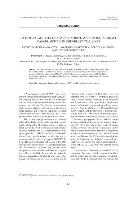
<strong>CYTOTOXIC</strong> <strong>ACTIVITY</strong> <strong>OF</strong> <strong>ε</strong>-<strong>AM<strong>IN</strong>OCAPROYLAM<strong>IN</strong>O</strong> <strong>ACIDS</strong> <strong>IN</strong> BREAST<br />
CANCER MCF-7 AND FIBROBLAST CELL L<strong>IN</strong>ES<br />
KRYSTYNA MIDURA-NOWACZEK 1 *, AGNIESZKA MARKOWSKA 1 , DOROTA KRAJEWSKA 1<br />
and S£AWOMIR WO£CZY—SKI 2<br />
1 Department of Organic Chemistry, Medical University of Bia≥ystok, 1 KiliÒski St.,<br />
15-223 Bia≥ystok, Poland<br />
2 Department of Gynecological Endocrinology, Medical University of Bia≥ystok, 24A Sk≥odowska-Curie St.,<br />
15-276 Bia≥ystok, Poland<br />
Abstract: The effect of H-EACA-L-Cys(S-Bzl)-OH, H-EACA-L-Leu-OH, H-EACA-L-Nle-OH and EACA on<br />
the viability of MCF-7 and fibroblast cells was examined. The antibacterial activity of these compounds was<br />
also tested. H-EACA-L-Leu-OH and H-EACA-L-Nle-OH showed cytotoxic activity against MCF-7 and fibroblast<br />
cell lines, particularly in the highest studied 20 mM concentration. None of the examined dipeptides showed<br />
antibacterial activity.<br />
Keywords: <strong>ε</strong>-aminocaproylamino acids, cytotoxity, MCF-7, fibroblast cells, antibacterial activity<br />
* Corresponding author: e-mail: krystyna.midura-nowaczek@umwb.edu.pl<br />
201<br />
Plasmin, a key enzyme of fibrinolysis plays an<br />
important role in a variety of biological processes<br />
such as wound healing, tissue repair, cell migration<br />
and is also important in pathological phenomena<br />
such as inflammation, tumor cell growth and metastasis<br />
(5). Plasmin inhibitors, eg. N α -acetyl-L-lysine<br />
methyl ester (6) and the derivative of dipeptide containing<br />
AMCHA ñ YO-2 (trans-aminomethylcyclohexanecarbonyl-(O-picolyl)-tyrosine<br />
octylamide)<br />
(7, 8) possess proapoptocic action. YO-2 exerts an<br />
apoptosis-inducing effect on various human cell cultures<br />
(9, 10), it also inhibits the growth of human<br />
tumors, decreases the amount of metastasis and it<br />
can be a potential antitumor agent (9). Antibacterial<br />
activity of some ω-aminoacyl derivatives of amino<br />
acids is well known (11, 12). <strong>ε</strong>-Aminocaproyl derivatives<br />
of L-histidine and L-lysine possessed antistaphylococcal<br />
activity but only <strong>ε</strong>-aminocaproyl-Llysine<br />
(H-EACA-L-Lys-OH) was antifibrinolytic<br />
agent.<br />
It seems to be interesting to check if EACA<br />
containing dipeptides which possessed antifbrinolytic<br />
activity can also show antitumor or antibacterial<br />
activity. In the present paper we examine the
202 KRYSTYNA MIDURA-NOWACZEK et al.<br />
influence of H-EACA-L-Cys(S-Bzl)-OH, H-EACA-<br />
L-Leu-OH, H-EACA-L-Nle-OH and EACA on the<br />
viability of MCF-7 and fibroblast cells.<br />
Antibacterial activity of these compounds was also<br />
examined and compared with the activity of chloramphenicol.<br />
EXPERIMENTAL<br />
Reagents<br />
The examined compounds were synthesized in<br />
the Department of Organic Chemistry of University<br />
of Bia≥ystok (1). 3-(4,5-Dimethylthiazol-2-yl)-2,5diphenyltetrazolium<br />
bromide (MTT) was purchased<br />
from Sigma. Dulbeccoís minimal essential medium<br />
(DMEM) and fetal bovine serum (FBS) used in cell<br />
culture were products of Gibco (USA). Penicillin<br />
and streptomycin were obtained from Quality<br />
Biological Inc. (USA).<br />
Fibroblast cultures<br />
Normal human skin fibroblasts were maintained<br />
in DMEM supplemented with 10% FBS, 50<br />
U/mL penicillin, 50 µg/mL streptomycin at 37 O C in<br />
5% CO 2 in an incubator. The cells were used<br />
between the 12 th and 14 th passages. The fibroblasts<br />
were subcultivated by trypsinization. Subconfluent<br />
cells from Costar Flasks were detached with 0.05%<br />
trypsin and 0.02% ethylenediaminetetraacetic acid<br />
(EDTA) in calcium-free phosphate-buffered saline<br />
(PBS). Cells were counted in hemocytometers and<br />
cultured at 1◊ . 10 5 cells per well in 2 mL of growth<br />
medium.<br />
MCF-7 cultures<br />
Breast cancer MCF-7 cells were maintained in<br />
DMEM supplemented with 10% FBS, 50 µg/mL<br />
penicillin, 50 µg/mL streptomycin at 37 O C in 5%<br />
CO 2 in an incubator. Cells were cultured in Costar<br />
flasks and subconfluent cells were detached with<br />
0.05% trypsin, 0.02% EDTA in calcium-free phosphate-buffered<br />
saline, counted in hemocytometers<br />
and inculated at 5◊10 5 cells per well of six-well<br />
plates (Nunc, Wiesbaden, Germany) in 2 mL of<br />
growth medium. Cells reached about 80% of confluence<br />
at day 3 after inoculation and in most cases<br />
such cells were used for the assays.<br />
Cell viability assay<br />
The assay was performed according to the<br />
method of Carmichael using MTT (13). Confluent<br />
cells, cultured for 48 h with various concentrations<br />
of studied compounds in 6-well plates, were washed<br />
three times with PBS and then incubated for 4 h in<br />
1 mL of MTT solution (0.5 mg/mL of PBS) at 37 O C<br />
in 5% CO 2 in an incubator. The medium was<br />
removed and 1 mL of 0.1 M HCl in absolute isopropanol<br />
was added to attached cells. Absorbance of<br />
Table 1. Cytotoxic activity of dipeptides, expressed as percentage of the survivability MCF-7 mammal tumor cells.<br />
Concentration of<br />
compound (mM)<br />
EACA<br />
H-EACA-<br />
Nle-OH<br />
H-EACA-<br />
LeuOH<br />
H-EACA-<br />
Cys(S-Bzl)-OH<br />
0 100 100 100 100<br />
0.02 98 ± 0.63 98 ± 0.63 97 ± 0.63 98 ± 0.63<br />
0.2 98 ± 0.63 95 ± 1.09 97 ± 0.63 98 ± 0.63<br />
2 98 ± 0.63 70 ± 1.67 80 ± 1.06 98 ± 0.63<br />
20 95 ± 1.09 0 0 80 ± 1.09<br />
Table 2. Cytotoxic activity of dipeptides, expressed as percentage of the survivability of fibroblast cell lines.<br />
Concentration of<br />
compound (mM)<br />
EACA<br />
H-EACA-<br />
Nle-OH<br />
H-EACA-<br />
LeuOH<br />
H-EACA-<br />
Cys(S-Bzl)-OH<br />
0 100 100 100 100<br />
0.02 100 100 95 ± 1.09 100<br />
0.2 100 85 ± 1.72 80 ± 1.09 100<br />
2 100 80 ± 1.09 50 ± 1.26 100<br />
20 95 ± 1.09 15 ± 1.66 5 ± 0.63 90 ± 0.89
Cytotoxic activity of <strong>ε</strong>-aminocaproylamino acids in breast cancer MCF-7 and fibroblast cell lines 203<br />
Table 3. Minimum inhibitory concentrations (µg/mL) of <strong>ε</strong>-aminocaproylamino acids and EACA (chloramphenicol was used as a standard<br />
compound).<br />
Microorganism Chloramphenicol EACA<br />
Compound<br />
H-EACA- H-EAC- H-EACA-<br />
Staphylococcus<br />
(D-threo-) Nle-OH Leu-OH Cys(S-Bzl)-OH<br />
aureus<br />
ATTC 6538<br />
0.5 > 200 > 200 > 200 > 200<br />
Micrococcus<br />
luteus<br />
ATCC 9391<br />
0.5 > 200 > 200 > 200 > 200<br />
Bacillus<br />
subtilis<br />
ATCC 6633<br />
Pseudomonas<br />
0.5 > 200 > 200 > 200 > 200<br />
aeruginosa<br />
ATCC 27853<br />
Escherichia<br />
> 200 > 200 > 200 > 200 > 200<br />
coli<br />
ATCC 29922<br />
1.0 > 200 > 200 > 200 > 200<br />
converted dye in living cells was measured at a<br />
wavelength of 570 nm. Cell viability of breast cancer<br />
MCF-7 and fibroblast cells cultured in the presence<br />
of examined compounds was calculated as a<br />
percent of control cells ± SD. The results are given<br />
in Tables 1 and 2.<br />
Antibacterial assay<br />
The assay was performed according to the literature<br />
method (14). The mother solution of the<br />
compound in ethanol, of the concentration of 10<br />
mg/mL was twice diluted by the Mueller-Hinton<br />
broth. Two more dilutions of the solutions with the<br />
broth were performed in order to obtain working<br />
solutions of the concentrations from 512 µg/mL to<br />
0.5 µg/mL. Fifty µL of each working solution was<br />
placed in the plate cavities for microcoagulation. To<br />
each portion of the solution 50 µL of a suspension of<br />
microorganisms in the broth, containing 2◊10 5 of<br />
cells in 1 µL was added. The obtained mixtures were<br />
incubated for 18 h at 37 O C. After 18 h, it was established<br />
whether the growth of microorganisms had<br />
been halted in the colonies. The results are given in<br />
Table 3.<br />
RESULTS AND DISCUSSION<br />
The two examined dipeptides: H-EACA-L-<br />
Leu-OH and H-EACA-L-Nle-OH show antitumor<br />
properties against MCF-7 cells, particularly in the<br />
largest studied 20 mM concentration (Table 1). Both<br />
compounds also show the cytotoxic activities<br />
against cells of fibroblasts (Table 2). In the case of<br />
H-EACA-L-Leu-OH, this effect was observed also<br />
at lower 2mM concentration, in which this compound<br />
reduces the survivability of fibroblast cells by<br />
50%. In the case of EACA and H-EACA-L-Cys(S-<br />
Bzl)-OH, the cytotoxity in the both tests practically<br />
was not observed. Only at the maximum 20 mM<br />
concentration, the last dipeptide showed a weak<br />
cytotoxic effect against MCF-7 breast cells.<br />
According to the results obtained, it is possible to<br />
suggest that the compounds with specific antifibrinolytic<br />
activity (EACA and H-EACA-L-Cys(S-<br />
Bzl)-OH) do not show cytotoxic activity against<br />
MCF-7 and fibroblast cells (Table 1, 2).<br />
Antitumor activity was observed only in the<br />
case of the antifibrinolytic dipeptides which are also<br />
the weak inhibitors of proteolytic and amidolytic<br />
activities of plasmin (1). These compounds influence<br />
not only the lysine binding sites but also the<br />
active centre of the enzyme. The relationship<br />
between antitumor activity and plasmin inhibition<br />
was observed earlier in the case of YO compounds<br />
(8, 15) but the correlation was not clear. Poor plasmin<br />
inhibitors did not effect apoptosis induction, but<br />
not every potent active centre directed inhibitor of<br />
this enzyme showed proapoptocic activity.<br />
Additionally, the apoptosis induction was not<br />
observed in the case of classic plasmin inhibitors<br />
such as diisopropylfluorophosphates or leupeptin<br />
(15). The problem of the possible relationship
204 KRYSTYNA MIDURA-NOWACZEK et al.<br />
between an inhibition of plasmin and anticancer<br />
activity needs further investigation. The examined<br />
antifibrinolytics did not showed antibacterial activity<br />
(Table 3) and probably there is no relathionship<br />
between these activities. Described earlier (12) antistaphylococcal<br />
activity of antifibrinolytic dipeptide:<br />
H-EACA-L-Lys-OH is probably rather connected<br />
with the fact that some of lysine derivatives (16) can<br />
show antibacterial properties.<br />
REFERENCES<br />
1. Midura-Nowaczek K., Bruzgo I., Dubis E.,<br />
Roszkowska-Jakimiec W., Worowski K.:<br />
Pharmazie 51, 775 (1996).<br />
2. Maciejewska D., Midura-Nowaczek K., Wawer<br />
I.: J. Mol. Struct. 604, 269 (2002).<br />
3. Bruzgo I., Tomasiak M., Stelmach H., Midura-<br />
Nowaczek K.: Acta Biochim. Pol. 51, 73<br />
(2004).<br />
4. Bruzgo I., Midura-Nowaczek K., Bruzgo M.,<br />
KaczyÒska J., Roszkowska-Jakimiec W., Acta<br />
Pol. Pharm. Drug Res. 63, 149 (2006).<br />
5. Syrovets T., Simmet T.: Cell. Mol. Life Sci. 61,<br />
873 (2004).<br />
6. George S.J., Johnson J.L., Smith M.A.,<br />
Angelini G.D., Jackson C.L.: J. Vasc. Res. 42,<br />
247(2005).<br />
7. Enomoto R., Sugahara C., Tsuda Y., Okada Y.<br />
Lee E.: Ann. N.Y. Acad. Sci. 1030, 622 (2004).<br />
8. Enomoto R., Sugahara C., Komai T., Suzuki C.,<br />
Kinoshita N., et al.: Biochim. Biophys. Acta<br />
1674, 291 (2004).<br />
9. Szende B., Okada Y., Tsuda Y., Horvath A.,<br />
Bˆkˆnyi G., et al.: In Vivo 16, 281 (2002).<br />
10. Okada Y., Tsuda Y., Wanaka K., Tada M.,<br />
Okamoto U., et al.: Bioorg. Med. Chem. Lett.<br />
10, 2217 (2000).<br />
11. Fuji A., Tanaka K., Tsuchiya Y., Cook E. S.: J.<br />
Med. Chem. 14, 354 (1971).<br />
12. Fuji A., Tanaka K., Cook E. S., J. Med.<br />
Chem.15, 378 (1972).<br />
13. Carmichael J., De Graff W., Gazdar A., Minna<br />
J., Mitchell J.: Cancer Res. 47, 936 (1987).<br />
14. Jorgensen J.H., Turnidge J.D., Washington<br />
J.A.: Antibacterial susceptibility tests; dilution<br />
and disk diffusion methods. in: Manual of<br />
Clinical Microbiology (7 th ed.). Murray P.R.,<br />
Baron E.J., Pfaller M.A., Tenover F.C., Yolken<br />
R.H. Eds., pp. 1526-1543, ASM Press,<br />
Washington 1999.<br />
15. Lee E., Enomoto R., Takemura K., Tsuda Y.,<br />
Okada Y.: Biochem. Pharmacol. 63, 1315<br />
(2002).<br />
16. Ryge T.S., Hansen P.R.: Bioorg. Med. Chem.<br />
14, 4444 (2006).<br />
Received: 03. 03. 2009


