Supplement 45 Ultrasound Protocol Support - Dicom - NEMA
Supplement 45 Ultrasound Protocol Support - Dicom - NEMA
Supplement 45 Ultrasound Protocol Support - Dicom - NEMA
Create successful ePaper yourself
Turn your PDF publications into a flip-book with our unique Google optimized e-Paper software.
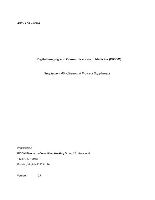
ASE • ACR • <strong>NEMA</strong><br />
Prepared by:<br />
Digital Imaging and Communications in Medicine (DICOM)<br />
<strong>Supplement</strong> <strong>45</strong>: <strong>Ultrasound</strong> <strong>Protocol</strong> <strong>Supplement</strong><br />
DICOM Standards Committee, Working Group 12 <strong>Ultrasound</strong><br />
1300 N. 17 th Street<br />
Rosslyn, Virginia 22209 USA<br />
Version: 0.7
ii<br />
8 June, 1999<br />
ii
<strong>Supplement</strong> <strong>45</strong>: <strong>Ultrasound</strong> <strong>Protocol</strong><br />
Page i<br />
Foreword..........................................................................................................................................................iii<br />
Scope and Field of Application....................................................................................................................... iv<br />
A.1.4 Overview of the Composite IOD Module Content.....................................................................2<br />
A.6.4 US Image IOD Module Table ...................................................................................................4<br />
A.7.4 US Multi-Frame Image IOD Module Table...............................................................................6<br />
C.8.5.x US Series Module ........................................................................................................7<br />
C.8.5.y US <strong>Protocol</strong> Module .....................................................................................................9<br />
6. REGISTRY OF DICOM DATA ELEMENTS .......................................................................................15
DOCUMENT HISTORY<br />
ii<br />
Document<br />
Version<br />
Date Content<br />
0.1 3 June, 1998 New draft based on 26-27 February meeting output<br />
0.2 4 June, 1998 Added data elements to Part 6. Added note in C.8.5.y US<br />
<strong>Protocol</strong> Module<br />
0.3 8 June, 1998 Synchronized with <strong>Supplement</strong> 36. Added tables with<br />
preliminary picklists for microglossary inclusion.<br />
0.4 30 November, 1998 Added supplement number (<strong>45</strong>). Added DICOMDIR<br />
support for Part 11. Made new modules optional. Defined<br />
relationship of ultrasound protocols to DICOM Real-World<br />
Model.<br />
0.5 1 December, 1998 Clean up of Forward, Scope and Field of Application and<br />
page numbering.<br />
0.6 3 December, 1998 Clarify relationship of ultrasound protocols to DICOM<br />
Real-World Model.<br />
0.7 8 June, 1999 Updated with changes from 3/29/99 teleconference.<br />
Added informative SDM codes from ASE.<br />
OPEN ISSUES<br />
Finish defining picklists and context ID’s.<br />
Get element numbers for new attributes.<br />
ii
Foreword<br />
<strong>Supplement</strong> <strong>45</strong>: <strong>Ultrasound</strong> <strong>Protocol</strong><br />
Page iii<br />
This draft <strong>Supplement</strong> to the DICOM Standard was developed according to <strong>NEMA</strong> Procedures. This<br />
<strong>Supplement</strong> to the Standard is developed in liaison with other standardization organizations including CEN<br />
TC251 in Europe, MEDIS-DC and JIRA in Japan, with review also by other organizations including IEEE,<br />
HL7 and ANSI in the USA.<br />
The DICOM Standard is structured as a multi-part document using the guidelines established in the<br />
following document:<br />
- ISO/IEC Directives, 1989 Part 3 : Drafting and Presentation of International Standards.<br />
This document is a <strong>Supplement</strong> to the DICOM Standard. It is an extension to PS 3.3, 3.6 and 3.11 of the<br />
published DICOM Standard which consists of the following parts:<br />
PS 3.1 - Introduction and Overview<br />
PS 3.2 - Conformance<br />
PS 3.3 - Information Object Definitions<br />
PS 3.4 - Service Class Specifications<br />
PS 3.5 - Data Structures and Encoding<br />
PS 3.6 - Data Dictionary<br />
PS 3.7 - Message Exchange<br />
PS 3.8 - Network Communication <strong>Support</strong> for Message Exchange<br />
PS 3.9 - Point-to-Point Communication <strong>Support</strong> for Message Exchange<br />
PS 3.10 Media Storage and File Format for Data Interchange<br />
PS 3.11 Media Storage Application Profiles<br />
PS 3.12 Media Formats and Physical Media for Data Interchange<br />
PS 3.13 Print Management Point-to-Point Communication <strong>Support</strong><br />
PS 3.14 Gray-scale Display Function Standard<br />
These parts are related but independent documents.
iv<br />
Scope and Field of Application<br />
This <strong>Supplement</strong> to the DICOM Standard specifies changes to the DICOM Image Information Object<br />
Definition (IOD) for <strong>Ultrasound</strong> (US) and <strong>Ultrasound</strong> Multi-frame (USMF) Images. It specifies the new<br />
modules necessary to identify images acquired as part of an ultrasound acquisition protocol.<br />
Since this document proposes changes to existing Parts of DICOM the reader should have a working<br />
understanding of the Standard.<br />
This proposed <strong>Supplement</strong> includes a number of Addenda to existing Parts of DICOM:<br />
1. PS 3.3 Addenda (Extension to Annex A and Annex C)<br />
2. PS 3.6 Addenda (Extension to Section 6)<br />
3. PS 3.11 Addenda (Extension to Annex C)<br />
iv
Changes to:<br />
<strong>NEMA</strong> Standards Publication PS 3.3-1998<br />
<strong>Supplement</strong> <strong>45</strong>: <strong>Ultrasound</strong> <strong>Protocol</strong><br />
Page 1<br />
Digital Imaging and Communications in Medicine (DICOM)<br />
Part 3: Information Object Definitions
3.X <strong>Ultrasound</strong><br />
The following definitions are commonly used in the specification of the <strong>Ultrasound</strong> IOD’s.<br />
3.X.1 Acquisition <strong>Protocol</strong><br />
An Acquisition <strong>Protocol</strong> is a Procedure Type that defines the particular ultrasound images that will be<br />
acquired.<br />
Note: These can be well known or locale specific.<br />
3.X.2 Staged <strong>Protocol</strong><br />
A Staged <strong>Protocol</strong> is an Acquisition <strong>Protocol</strong> that is repeated at more than one specific instance of time.<br />
Note: These instances commonly occur due to simple events. The introduction of contrast media, drugs or stress<br />
to the patient are typical events.<br />
Item: Add to PS 3.3 Table A.1-1<br />
A.1.4 Overview of the Composite IOD Module Content<br />
Table A.1-1 provides an overview of the Modules used throughout the Composite IODs. This table is for<br />
informative purposes only. It is based on the IOD definitions found in the remaining sections of Annex A<br />
which are normative.<br />
Table A.1-1<br />
COMPOSITE INFORMATION OBJECT MODULES OVERVIEW<br />
IODs<br />
Modules<br />
US USmf<br />
Patient M M<br />
Patient<br />
Summary<br />
General<br />
Study<br />
M M<br />
Patient study U U<br />
Study Content<br />
General<br />
Series<br />
CR Series<br />
NM Series<br />
M M<br />
US Series U U<br />
Frame Of<br />
Reference<br />
U S Frame of<br />
Ref.<br />
General<br />
Equipment<br />
SC Equipment<br />
U U<br />
C C<br />
M M<br />
2
General<br />
Image<br />
Image Plane<br />
M* M<br />
Image Pixel M* M<br />
NM Image<br />
Pixel<br />
Pallet Color<br />
L o oku p Ta b le<br />
Contrast/<br />
Bolus<br />
C C<br />
C* C<br />
Cine M<br />
Multi-frame M<br />
NM Multiframe<br />
Frame<br />
Pointers<br />
Mask<br />
Display<br />
Shutter<br />
Device<br />
Therapy<br />
CR Image<br />
CT Image<br />
MR Image<br />
NM Image<br />
NM Isotope<br />
NM Detector<br />
NM TOMO<br />
Acquisition<br />
NM Multi-<br />
Gated<br />
Acquisition<br />
NM Phase<br />
NM<br />
Re co n str u ct io<br />
n<br />
US Region<br />
Calibration<br />
U* U<br />
US Image M* M<br />
US <strong>Protocol</strong> U U<br />
SC Image<br />
X-Ray Image<br />
<strong>Supplement</strong> <strong>45</strong>: <strong>Ultrasound</strong> <strong>Protocol</strong><br />
Page 3
X-Ray<br />
Acquisition<br />
X-Ray<br />
Collimator<br />
X-Ray Table<br />
XRF<br />
Positioner<br />
XRF Tomo<br />
Acquisition<br />
XA Positioner<br />
Bi-Plane<br />
Sequence<br />
Bi-Plane<br />
Image<br />
Overlay<br />
Identification<br />
Overlay Plane U*<br />
Multi-frame<br />
Overlay<br />
Bi-Plane<br />
Overlay<br />
Curve<br />
Identification<br />
M* M*<br />
Curve M* M*<br />
Audio U U<br />
Modality LUT<br />
VOI LUT U* U<br />
LUT<br />
Identification<br />
SOP<br />
Common<br />
M* M*<br />
Item: Add to PS 3.3 Table A.6.4-1 - for clarity, those modules that are changed from the existing standard<br />
or new are shown in Bold and Underlined<br />
A.6.4 US Image IOD Module Table<br />
Table A.6.4-1<br />
US IMAGE IOD MODULES<br />
IE Module Reference Usage<br />
4
<strong>Supplement</strong> <strong>45</strong>: <strong>Ultrasound</strong> <strong>Protocol</strong><br />
Page 5<br />
Patient Patient C.7.1.1 M<br />
Study General Study C.7.2.1 M<br />
Patient Study C.7.2.2 U<br />
Series General Series C.7.3.1 M<br />
Frame of<br />
Reference<br />
US Series C.8.5.x U – Suggested if image is<br />
part of an acquisition<br />
protocol<br />
Frame of Reference C.7.4.1 U<br />
US Frame of Reference C.8.5.4 C - Required if images are<br />
spatially related<br />
Equipment General Equipment C.7.5.1 M<br />
Image General Image C.7.6.1 M<br />
(See A.6.4.1) Image Pixel C.7.6.3 M<br />
Contrast/bolus C.7.6.4 C - Required if contrast<br />
media was used in this<br />
image<br />
Palette Color Lookup table C.7.9 C - Required if Photometic<br />
Interpretation (0028,0004)<br />
has a value of PALETTE<br />
COLOR<br />
US Region Calibration C.8.5.5 U<br />
US Image C.8.5.6 M<br />
US Staged <strong>Protocol</strong> C.8.5.y U – Suggested if image is<br />
part of a staged protocol<br />
Overplay Plane C.9.2 U<br />
VOI LUT C.11.2 U<br />
SOP Common C.12.1 M<br />
Curve Curve Identification C.10.1 M<br />
(See A.6.4.1) Curve C.10.2 M<br />
Audio C.10.3 U<br />
SOP Common C.12.1 M<br />
A.6.4.1 Mutually Exclusive IEs<br />
The Image and Curve IEs are mutually exclusive. Each SOP Instance using this IOD shall contain exactly<br />
one of these IEs.<br />
A.6.5 US Image IOD Relationship to the DICOM Real-World Model<br />
<strong>Ultrasound</strong> images that make use of the US Series & <strong>Protocol</strong> Modules have a more specific<br />
relationship to the DICOM Real-World Model than non-protocol images. An ultrasound acquisition<br />
protocol is considered one Requested Procedure and has one Procedure Plan. The Procedure<br />
Plan will have one or more Action Items. An ultrasound acquisition protocol will typically consist
of one Scheduled Procedure Step containing one or more Action Items. Assuming the acquisition<br />
protocol was successfully completed, this will result in one Modality Performed Procedure Step.<br />
Therefore, all images that are part of this particular instance of an ultrasound acquisition protocol<br />
will have the same Procedure Code Sequence (0032,1064). It is recommended that all the images<br />
that are part of this particular instance of an ultrasound acquisition protocol have the same<br />
Performed Procedure Step ID. In an environment with information system connectivity, the<br />
Procedure Code Sequence can be used to identify all the images that were acquired as part of that<br />
protocol. If no information system connectivity is available, then Performed Procedure Step ID<br />
may be used to identify all the images that were acquired as part of that protocol.<br />
It is recommended that each stage should map to a separate series.<br />
Item: Add to PS 3.3 Table A.7.4-1 - for clarity, those modules that are changed from the existing standard<br />
or new are shown in Bold and Underlined<br />
A.7.4 US Multi-Frame Image IOD Module Table<br />
Table A.7.4-1<br />
US MULTI-FRAME IMAGE IOD MODULES<br />
IE Module Reference Usage<br />
Patient Patient C.7.1.1 M<br />
Study General Study C.7.2.1 M<br />
Patient Study C.7.2.2 U<br />
Series General Series C.7.3.1 M<br />
Frame of<br />
Reference<br />
US Series C.8.5.x U – Suggested if image is<br />
part of an acquisition<br />
protocol<br />
Frame of Reference C.7.4.1 U<br />
US Frame of Reference C.8.5.4 C - Required if images are<br />
spatially related<br />
Equipment General Equipment C.7.5.1 M<br />
Image General Image C.7.6.1 M<br />
(See A.7.4.1) Image Pixel C.7.6.3 M<br />
Contrast/bolus C.7.6.4 C - Required if contrast<br />
media was used in this<br />
image.<br />
Cine C.7.6.5 M<br />
Multi-frame C.7.6.6 M<br />
Palette Color Lookup Table C.7.9 C - Required if Photometric<br />
Interpretation (0028,0004)<br />
has a value of PALETTE<br />
COLOR<br />
6
<strong>Supplement</strong> <strong>45</strong>: <strong>Ultrasound</strong> <strong>Protocol</strong><br />
Page 7<br />
US Region Calibration C.8.5.5 U<br />
US Image C.8.5.6 M<br />
US Staged <strong>Protocol</strong> C.8.5.y U – Suggested if image is<br />
part of a staged protocol<br />
VOI LUT C.11.2 U<br />
SOP Common C.12.1 M<br />
Curve Curve Identification C.10.1 M<br />
(see A.7.4.1) Curve C.10.2 M<br />
Audio C.10.3 U<br />
SOP Common C.12.1 M<br />
A.7.4.1 Mutually Exclusive IEs<br />
The Image and Curve IEs are mutually exclusive. Each SOP Instance using this IOD shall contain exactly<br />
one of these IEs.<br />
A.7.5 US Multi-Frame Image IOD Relationship to the DICOM Real-World Model<br />
<strong>Ultrasound</strong> images that make use of the US Series & <strong>Protocol</strong> Modules have a more specific<br />
relationship to the DICOM Real-World Model than non-protocol images. An ultrasound acquisition<br />
protocol is considered one Requested Procedure and has one Procedure Plan. The Procedure<br />
Plan will have one or more Action Items. An ultrasound acquisition protocol will typically consist<br />
of one Scheduled Procedure Step containing one or more Action Items. Assuming the acquisition<br />
protocol was successfully completed, this will result in one Modality Performed Procedure Step.<br />
Therefore, all images that are part of this particular instance of an ultrasound acquisition protocol<br />
will have the same Procedure Code Sequence (0032,1064). It is recommended that all the images<br />
that are part of this particular instance of an ultrasound acquisition protocol have the same<br />
Performed Procedure Step ID. In an environment with information system connectivity, the<br />
Procedure Code Sequence can be used to identify all the images that were acquired as part of that<br />
protocol. If no information system connectivity is available, then Performed Procedure Step ID<br />
may be used to identify all the images that were acquired as part of that protocol.<br />
It is recommended that each stage should map to a separate series.<br />
Item: Add to PS 3.3 New Sections C.8.5.x and C.8.5.y<br />
C.8.5.x US Series Module<br />
Table C.8-x contains IOD attributes that describe an ultrasound series.
Table C.8-x<br />
US SERIES MODULE ATTRIBUTES<br />
Attribute Name Tag Type Attribute Description<br />
<strong>Protocol</strong> Name (0018,1030) 1C User-defined description of the conditions<br />
under which the Series was performed.<br />
Required if <strong>Protocol</strong> Type Sequence<br />
(0018,xxxx) has no value.<br />
<strong>Protocol</strong> Type Sequence (0018,xxxx) 2 Sequence of one or more Items that<br />
identifies the protocol used to acquire this<br />
image.<br />
See section C.8.5.x.1.y<br />
>Include ‘Code Sequence Macro‘ Table 8.8-1 Baseline Context ID is ???.<br />
><strong>Protocol</strong> Type Modifier Sequence (0018,xxxx) 3 Sequence of one or more Items that<br />
modifies the primary protocol type used to<br />
acquire this image.<br />
See section C.8.5.x.1.y<br />
>>Include ‘Code Sequence Macro‘ Table 8.8-1 Baseline Context ID is ???.<br />
Performed Procedure Step ID (0040,0253) 1C Identification of that Procedure that has<br />
been carried out within this step. Required if<br />
image was acquired in an acquisition<br />
protocol.<br />
C.8.5.x.1 US Series Attributes Descriptions<br />
C.8.5.x.1.y <strong>Protocol</strong> Type Sequence<br />
These Data Elements are intended to replace <strong>Protocol</strong> Name (0018,1030) Data Element.<br />
<strong>Protocol</strong> Type Sequence (0018,xxxx) describes the acquisition protocol used to acquire this image.<br />
The Coding Scheme Designator (0008,0102) shall be SNM3.<br />
The Code Value (0008,0100) shall be drawn from the SNOMED DICOM Microglossary Context ID XXX.<br />
Note: The existence of this attribute means that this image is part of an acquisition protocol.<br />
Note: The following will not be included in the standard.<br />
Code Meaning<br />
Code Value<br />
(0008,0104)<br />
(0008,0100)<br />
Transesophageal echocardiography P5-B3002<br />
Transthoracic echocardiography P5-B3003<br />
Epicardial echocardiography P5-B3004<br />
Intravascular echocardiography P5-B3005<br />
Intracardiac echocardiography P5-B3006<br />
Limited M-mode only echocardiography P5-B301F<br />
Limited Doppler only echocardiography P5-B303F<br />
Exercise stress echocardiography P5-B3050<br />
Maximal stress echocardiography P5-B3051<br />
8
Submaximal stress echocardiography P5-B3052<br />
Treadmill exercise stress echocardiography P5-B3053<br />
Bruce treadmill stress echocardiography P5-B3054<br />
Modified Bruce treadmill stress echocardiography P5-B3055<br />
Naughton treadmill stress echocardiography P5-B3056<br />
Bicycle exercise stress echocardiography P5-B3058<br />
Echocardiography with administered drug stress P5-B3060<br />
Dobutamine stress echocardiography P5-B3061<br />
High dose dobutamine stress echocardiography P5-B3062<br />
Low dose dobutamine stress echocardiography P5-B3063<br />
Arbutamine stress echocardiography P5-B3065<br />
Dipyridamole stress echocardiography P5-B3066<br />
Cardiac pacing echocardiography P5-B3070<br />
Adult echocardiography P5-B3081<br />
Pediatric echocardiography P5-B3082<br />
Intraoperative echocardiography P5-B3083<br />
Upright echocardiography P5-B3084<br />
Supine echocardiography P5-B3085<br />
Contrast echocardiography P5-B3090<br />
Contrast left ventricular opacification echocardiography P5-B3091<br />
Contrast perfusion echocardiography P5-B3092<br />
Contrast Doppler enhancement echocardiography P5-B3093<br />
2D complete echocardiography P5-B3191<br />
Limited 2D only echocardiography P5-B3192<br />
Fetal echocardiography P5-B8215<br />
<strong>Supplement</strong> <strong>45</strong>: <strong>Ultrasound</strong> <strong>Protocol</strong><br />
Page 9<br />
<strong>Protocol</strong> Type Modifier Sequence (0018,xxxx) modifies the primary acquisition protocol stage when the<br />
image was acquired.<br />
The Coding Scheme Designator (0008,0102) shall be SNM3.<br />
The Code Value (0008,0100) shall be drawn from the SNOMED DICOM Microglossary Context ID XXX.<br />
C.8.5.y US Staged <strong>Protocol</strong> Module<br />
Table C.8-y contains IOD attributes that describe an ultrasound staged protocol.<br />
Table C.8-y<br />
US STAGED PROTOCOL MODULE ATTRIBUTES<br />
Attribute Name Tag Type Attribute Description<br />
Number of Stages (0008,2124) 2 Number of Stages in this protocol.<br />
Number of Views in Stage (0008,212A) 2 Number of views in this Stage.
Stage Name (0008,2120) 1C A Stage is a particular time slice of a<br />
protocol in which a set of images are<br />
collected. The names can be free form text.<br />
Recommended text for Stress Echo stage<br />
names are:<br />
PRE-EXERCISE,<br />
POST-EXERCISE,<br />
PEAK-EXERCISE,<br />
RECOVERY,<br />
BASELINE,<br />
LOW DOSE,<br />
PEAK DOSE<br />
Required if Stage Type Sequence<br />
(0008,xxxx) has no value.<br />
Stage Type Sequence (0008,xxxx) 2 Sequence of one or more items that<br />
identifies the acquisition protocol stage<br />
when the image was acquired.<br />
See Section 8.5.y.1.1<br />
>Include ‘Code Sequence Macro‘ Table 8.8-1 Baseline Context ID is ???.<br />
>Stage Type Modifier Sequence (0008,xxxx) 3 Sequence of one or more Items that<br />
modifies the primary stage type in this<br />
image.<br />
See Section 8.5.y.1.1<br />
>>Include ‘Code Sequence Macro‘ Table 8.8-1 Baseline Context ID is ???.<br />
Stage Number (0008,2122) 1 A number that identifies the Stage. Stage<br />
Number starts at one.<br />
See Section C.8.5.y.1.4<br />
View Number (0008,2128) 2 A number that identifies the View. Can be<br />
empty if View Number is unknown or if<br />
known, View Number starts at one.<br />
See Section C.8.5.y.1.5<br />
Transducer Position Sequence (0008,2240) 2 Sequence of one or more Items that<br />
identifies the transducer position used in this<br />
image.<br />
See section C.8.5.y.1.2.<br />
>Include ‘Code Sequence Macro‘ Table 8.8-1 Baseline Context ID is 4.<br />
> Transducer Position Modifier<br />
Sequence<br />
(0008,2242) 3 Sequence of one or more Items that<br />
modifies the primary transducer position of<br />
interest in this image.<br />
See Section C.8.5.y.1.2.<br />
>>Include ‘Code Sequence Macro‘ Table 8.8-1 Baseline Context ID is 5.<br />
Transducer Orientation Sequence (0008,2244) 2 Sequence of one or more Items that<br />
identifies the Transducer Orientation used in<br />
this image.<br />
See section C.8.5.y.1.3.<br />
10
Include ‘Code Sequence Macro‘ Table 8.8-1 Baseline Context ID is 6.<br />
> Transducer Orientation Modifier<br />
Sequence<br />
<strong>Supplement</strong> <strong>45</strong>: <strong>Ultrasound</strong> <strong>Protocol</strong><br />
Page 11<br />
(0008,2246) 3 Sequence of one or more Items that<br />
modifies the primary Transducer Orientation<br />
of interest in this image.<br />
See Section C.8.5.y.1.3<br />
>>Include ‘Code Sequence Macro‘ Table 8.8-1 Baseline Context ID is 7.<br />
Note: Images that are part of a staged acquisition protocol should be identified by using Stage Type Sequence<br />
(0008,xxxx), Transducer Position Sequence (0008, 2240) and Transducer Orientation Sequence<br />
(0008,2244).<br />
Stage Number (0008,2122), View Number (0008,2128), Number of Stages (0008,2124), Number of Views in<br />
Stage (0008,212A) and Stage Name (0008, 2120) are maintained for backwards compatibility but may<br />
produce ambiguous results when used to identify a staged acquisition protocol.<br />
C.8.5.y.1 US Staged <strong>Protocol</strong> Attribute Descriptions<br />
C.8.5.y.1.1 Stage Type Sequence<br />
These Data Elements are intended to replace Stage Name (0008,2120) Data Element.<br />
Stage Type Sequence (0008,xxxx) describes the acquisition protocol stage when the image was<br />
acquired.<br />
The Coding Scheme Designator (0008,0102) shall be SNM3.<br />
The Code Value (0008,0100) shall be drawn from the SNOMED DICOM Microglossary Context ID XXX<br />
Note: The existence of this attribute means that this image is part of an staged protocol.<br />
Note: The following will not be included in the standard.<br />
The following terms must be defined and a new Context ID created for them:<br />
These codes have been defined by SNOMED and are available. There is some debate that SNOMED<br />
requires a licensing fee, so these terms need to be defined in such a way that is agreeable to Working<br />
Group 6.<br />
Code Meaning<br />
(0008,0104)<br />
Code Value<br />
(0008,0100)<br />
Image acquisition procedure P5-01000<br />
Image acquisition after administration of contrast agent P5-01101<br />
Image acquisition during cardiac pacing P5-01103<br />
Image acquisition at user-defined cardiac pacing rate P5-01104<br />
Image acquisition during hand grip maneuver P5-01111<br />
Image acquisition during Valsalva P5-01112<br />
Image acquisition during postural maneuver P5-01113<br />
Pre-procedure image acquisition P5-01120<br />
Preoperative image acquisition P5-01121<br />
Intra-procedure image acquisition P5-01130<br />
Intra-operative image acquisition P5-01131<br />
Post-procedure image acquisition P5-01140<br />
Post-operative image acquisition P5-01141<br />
Image acquisition following first cardiopulmonary bypass P5-01142
Image acquisition following second cardiopulmonary<br />
P5-01143<br />
bypass Image acquisition following third cardiopulmonary bypass P5-01144<br />
Image acquisition during stress procedure P5-01200<br />
Image acquisition at baseline P5-01201<br />
Pre-stress image acquisition P5-01202<br />
Mid-stress image acquisition P5-01203<br />
Peak-stress image acquisition P5-01204<br />
Image acquisition during recovery P5-01205<br />
Image acquisition after drug administration P5-01300<br />
Image acquisition at user-defined dobutamine dose P5-01310<br />
Image acquisition at low-dose dobutamine P5-01311<br />
Image acquisition at mid-dose dobutamine P5-01312<br />
Image acquisition at peak dose dobutamine P5-01313<br />
Image acquisition at dobutamine 5 mcg/kg/min P5-01314<br />
Image acquisition at dobutamine 10 mcg/kg/min P5-01315<br />
Image acquisition at dobutamine 20 mcg/kg/min P5-01316<br />
Image acquisition at dobutamine 30 mcg/kg/min P5-01317<br />
Image acquisition at dobutamine 40 mcg/kg/min P5-01318<br />
Image acquisition at dobutamine 50 mcg/kg/min P5-01319<br />
Image at dobutamine 40 mcg/kg/min plus atropine P5-0131A<br />
Image acquisition at dobutamine 50 mcg/kg/min plus<br />
P5-0131B<br />
atropine Image acquisition at peak Arbutamine dose P5-01323<br />
Image acquisition at peak dipyridamole P5-01333<br />
Image acquisition after nitroglycerin P5-01341<br />
Image acquisition after amyl nitrite P5-01342<br />
Image acquisition after adenosine P5-01343<br />
Stage Type Modifier Sequence (0008,xxxx) modifies the primary acquisition protocol stage when the<br />
image was acquired.<br />
The Coding Scheme Designator (0008,0102) shall be SNM3.<br />
The Code Value (0008,0100) shall be drawn from the SNOMED DICOM Microglossary Context ID XXX.<br />
C.8.5.y.1.2 Transducer Position Sequence<br />
Transducer Position Sequence (0008,2240) identifies the transducer position used in this image.<br />
The Coding Scheme Designator (0008,0102) shall be SNM3.<br />
The Code Value (0008,0100) shall be drawn from the SNOMED DICOM Microglossary Context ID 4.<br />
Transducer Position Modifier Sequence (0008,2242) modifies the transducer position used in this image.<br />
The Coding Scheme Designator (0008,0102) shall be SNM3.<br />
The Code Value (0008,0100) shall be drawn from the SNOMED DICOM Microglossary Context ID 5.<br />
12
C.8.5.y.1.3 Transducer Orientation Sequence<br />
<strong>Supplement</strong> <strong>45</strong>: <strong>Ultrasound</strong> <strong>Protocol</strong><br />
Page 13<br />
Transducer Orientation Sequence (0008,2244) identifies the transducer orientation used in this image.<br />
The Coding Scheme Designator (0008,0102) shall be SNM3.<br />
The Code Value (0008,0100) shall be drawn from the SNOMED DICOM Microglossary Context ID 6.<br />
Transducer Orientation Modifier Sequence (0008,2246) modifies the transducer orientation used in this<br />
image.<br />
The Coding Scheme Designator (0008,0102) shall be SNM3.<br />
The Code Value (0008,0100) shall be drawn from the SNOMED DICOM Microglossary Context ID 7.<br />
C.8.5.y.1.4 Stages<br />
The Number of Stages (0008,2124) shall be the total number of stages performed in this instance of the<br />
protocol. Stage Number (0008,2122) shall be the number that indicates the default order in which the<br />
stages are to be presented. Stage Number shall start from 1.<br />
C.8.5.y.1.5 Views<br />
The View Number (0008,2128) shall be used identify the same view across stages and defines a default<br />
order for presentation within and across stages. If a particular view appears in one stage but not in a<br />
subsequent stage, then that View Number will not be used during that subsequent stage. View Number<br />
shall start from 1. The Number of Views in Stage (0008,212A) shall be used to indicate how many unique<br />
View Numbers exist for this stage.<br />
The addition of modification of the value for this attribute shall not by itself force a SOP Instance UID<br />
change.
Changes to:<br />
<strong>NEMA</strong> Standards Publication PS 3.6-1998<br />
Digital Imaging and Communications in Medicine (DICOM)<br />
Part 6: Data Dictionary<br />
14
6. REGISTRY OF DICOM DATA ELEMENTS<br />
Add the following Data Elements to PS 3.6, Section 6.<br />
<strong>Supplement</strong> <strong>45</strong>: <strong>Ultrasound</strong> <strong>Protocol</strong><br />
Page 15<br />
Tag Name VR VM<br />
(0008,xxxx) Stage Type Sequence SQ 1<br />
(0008,xxxx) Stage Type Modifier Sequence SQ 1<br />
(0018,xxxx) <strong>Protocol</strong> Type Sequence SQ 1<br />
(0018,xxxx) <strong>Protocol</strong> Type Modifier Sequence SQ 1
Changes to:<br />
<strong>NEMA</strong> Standards Publication PS 3.11-1998<br />
Digital Imaging and Communications in Medicine (DICOM)<br />
Part 11: Media Storage Application Profiles<br />
16
2<br />
4<br />
6<br />
<strong>Supplement</strong> <strong>45</strong>: <strong>Ultrasound</strong> <strong>Protocol</strong><br />
Page 17<br />
Item: Add to PS 3.11 Section C.3.3.1 - for clarity, that text which is removed from the existing standard is<br />
shown as Strikethrough and that text which is changed or new is shown in Underlined<br />
C.3.3.1 Additional Keys<br />
File Set Creators and Updaters are only required to generate mandatory elements specified in PS 3.10.<br />
8 Table C.3-4 specifies the additional associated keys that shall be supported. Refer to the Basic Directory<br />
IOD in PS 3.3. At each directory record level any additional data elements can be added as keys, but is<br />
10 not required by File Set Readers to be able to use them as keys.<br />
12<br />
Table C.3-4<br />
STD-US-XX-XX-XXXX ADDITIONAL DICOMDIR KEYS<br />
Key Attribute Tag Directory<br />
Record Type<br />
<strong>Protocol</strong> Type<br />
Sequence<br />
Type Notes<br />
(0018,xxxx) SERIES 2C Sequence of one or more Items<br />
that identifies the protocol used to<br />
acquire this image. Required if US<br />
Series module is included.<br />
>Include ‘Code Sequence Macro‘ Table 8.8-1 Baseline Context ID is ???.<br />
><strong>Protocol</strong> Type<br />
Modifier Sequence<br />
See Part 3.3 Section C.8.5.x.1.y<br />
(0018,xxxx) SERIES 3 Sequence of one or more Items<br />
that modifies the primary protocol<br />
type used to acquire this image.<br />
>>Include ‘Code Sequence Macro‘ Table 8.8-1 Baseline Context ID is ???.<br />
Performed<br />
Procedure Step ID<br />
See Part 3.3 Section C.8.5.x.1.y<br />
(0040,0253) SERIES 1C Identification of that Procedure that<br />
has been carried out within this<br />
step. Required if series was part of<br />
a performed procedure step.<br />
Number of Stages (0008,2124) IMAGE 2C Number of Stages in this protocol.<br />
Required if Stage Type Sequence<br />
(0008,xxxx) is present.<br />
Number of Views in<br />
Stage<br />
See Part 3.3 Section C.8.5.y.1.4<br />
(0008,212A) IMAGE 2C Number of unique views in this<br />
Stage. Required if Stage Type<br />
Sequence (0008,xxxx) is present.<br />
See Part 3.3 Section C.8.5.y.1.5
Stage Type<br />
Sequence<br />
(0008,xxxx) IMAGE 2C Sequence of one or more items that<br />
identifies the acquisition protocol<br />
stage when the image was<br />
acquired. Required US <strong>Protocol</strong><br />
module is present.<br />
>Include ‘Code Sequence Macro‘ Table 8.8-1 Baseline Context ID is ???.<br />
>Stage Type<br />
Modifier Sequence<br />
See Part 3.3 Section 8.5.y.1.1<br />
(0008,xxxx) IMAGE 3 Sequence of one or more Items<br />
that modifies the primary stage type<br />
in this image.<br />
>>Include ‘Code Sequence Macro‘ Table 8.8-1 Baseline Context ID is ???.<br />
See Part 3.3 Section 8.5.y.1.1<br />
Stage Number (0008,2122) IMAGE 1C A number that identifies the Stage.<br />
Stage Number starts at one.<br />
Required if Stage Type Sequence<br />
(0008,xxxx) is present.<br />
See Part 3.3 Section C.8.5.y.1.4<br />
View Number (0008,2128) IMAGE 1C A number that identifies the View.<br />
View Number starts at one.<br />
Required if Stage Type Sequence<br />
(0008,xxxx) is present.<br />
Transducer<br />
Position Sequence<br />
See Part 3.3 Section C.8.5.y.1.5<br />
(0008,2240) IMAGE 2C Sequence of one or more Items<br />
that identifies the transducer<br />
position used in this image.<br />
Required if Stage Type Sequence<br />
(0008,xxxx) is present.<br />
>Include ‘Code Sequence Macro‘ Table 8.8-1 Baseline Context ID is 4.<br />
> Transducer<br />
Position Modifier<br />
Sequence<br />
See Part 3.3 Section C.8.5.y.1.2.<br />
(0008,2242) IMAGE 3 Sequence of one or more Items<br />
that modifies the primary<br />
transducer position of interest in<br />
this image.<br />
>>Include ‘Code Sequence Macro‘ Table 8.8-1 Baseline Context ID is 5.<br />
Transducer<br />
Orientation<br />
Sequence<br />
See Part 3.3 Section C.8.5.y.1.2.<br />
(0008,2244) IMAGE 2C Sequence of one or more Items<br />
that identifies the Transducer<br />
Orientation used in this image.<br />
Required if Stage Type Sequence<br />
(0008,xxxx) is present.<br />
>Include ‘Code Sequence Macro‘ Table 8.8-1 Baseline Context ID is 6.<br />
See Part 3.3 Section C.8.5.y.1.3.<br />
18
2<br />
> Transducer<br />
Orientation Modifier<br />
Sequence<br />
<strong>Supplement</strong> <strong>45</strong>: <strong>Ultrasound</strong> <strong>Protocol</strong><br />
Page 19<br />
(0008,2246) IMAGE 3 Sequence of one or more Items<br />
that modifies the primary<br />
Transducer Orientation of interest<br />
in this image.<br />
>>Include ‘Code Sequence Macro‘ Table 8.8-1 Baseline Context ID is 7.<br />
See Part 3.3 Section C.8.5.y.1.3


