Cytidine 5′-Monophosphate (CMP)-Induced ... - ResearchGate
Cytidine 5′-Monophosphate (CMP)-Induced ... - ResearchGate
Cytidine 5′-Monophosphate (CMP)-Induced ... - ResearchGate
Create successful ePaper yourself
Turn your PDF publications into a flip-book with our unique Google optimized e-Paper software.
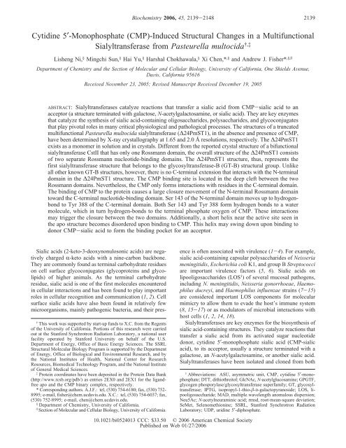
<strong>Cytidine</strong> <strong>5′</strong>-<strong>Monophosphate</strong> (<strong>CMP</strong>)-<strong>Induced</strong> Structural Changes in a Multifunctional<br />
Sialyltransferase from Pasteurella multocida †,‡<br />
Lisheng Ni, § Mingchi Sun, § Hai Yu, § Harshal Chokhawala, § Xi Chen,* ,§ and Andrew J. Fisher* ,§,|<br />
Department of Chemistry and the Section of Molecular and Cellular Biology, UniVersity of California, One Shields AVenue,<br />
DaVis, California 95616<br />
ReceiVed NoVember 23, 2005; ReVised Manuscript ReceiVed December 19, 2005<br />
ABSTRACT: Sialyltransferases catalyze reactions that transfer a sialic acid from <strong>CMP</strong>-sialic acid to an<br />
acceptor (a structure terminated with galactose, N-acetylgalactosamine, or sialic acid). They are key enzymes<br />
that catalyze the synthesis of sialic acid-containing oligosaccharides, polysaccharides, and glycoconjugates<br />
that play pivotal roles in many critical physiological and pathological processes. The structures of a truncated<br />
multifunctional Pasteurella multocida sialyltransferase (∆24PmST1), in the absence and presence of <strong>CMP</strong>,<br />
have been determined by X-ray crystallography at 1.65 and 2.0 Å resolutions, respectively. The ∆24PmST1<br />
exists as a monomer in solution and in crystals. Different from the reported crystal structure of a bifunctional<br />
sialyltransferase CstII that has only one Rossmann domain, the overall structure of the ∆24PmST1 consists<br />
of two separate Rossmann nucleotide-binding domains. The ∆24PmST1 structure, thus, represents the<br />
first sialyltransferase structure that belongs to the glycosyltransferase-B (GT-B) structural group. Unlike<br />
all other known GT-B structures, however, there is no C-terminal extension that interacts with the N-terminal<br />
domain in the ∆24PmST1 structure. The <strong>CMP</strong> binding site is located in the deep cleft between the two<br />
Rossmann domains. Nevertheless, the <strong>CMP</strong> only forms interactions with residues in the C-terminal domain.<br />
The binding of <strong>CMP</strong> to the protein causes a large closure movement of the N-terminal Rossmann domain<br />
toward the C-terminal nucleotide-binding domain. Ser 143 of the N-terminal domain moves up to hydrogenbond<br />
to Tyr 388 of the C-terminal domain. Both Ser 143 and Tyr 388 form hydrogen bonds to a water<br />
molecule, which in turn hydrogen-bonds to the terminal phosphate oxygen of <strong>CMP</strong>. These interactions<br />
may trigger the closure between the two domains. Additionally, a short helix near the active site seen in<br />
the apo structure becomes disordered upon binding to <strong>CMP</strong>. This helix may swing down upon binding to<br />
donor <strong>CMP</strong>-sialic acid to form the binding pocket for an acceptor.<br />
Sialic acids (2-keto-3-deoxynonulosonic acids) are negatively<br />
charged R-keto acids with a nine-carbon backbone.<br />
They are commonly found as terminal carbohydrate residues<br />
on cell surface glycoconjugates (glycoproteins and glycolipids)<br />
of higher animals. As the terminal carbohydrate<br />
residue, sialic acid is one of the first molecules encountered<br />
in cellular interactions and has been found to play important<br />
roles in cellular recognition and communication (1, 2). Cell<br />
surface sialic acids have also been found in relatively few<br />
microorganisms, mainly pathogenic bacteria, and their pres-<br />
† This work was supported by start-up funds to X.C. from the Regents<br />
of the University of California. Portions of this research were carried<br />
out at the Stanford Synchrotron Radiation Laboratory, a national user<br />
facility operated by Stanford University on behalf of the U.S.<br />
Department of Energy, Office of Basic Energy Sciences. The SSRL<br />
Structural Molecular Biology Program is supported by the Department<br />
of Energy, Office of Biological and Environmental Research, and by<br />
the National Institutes of Health, National Center for Research<br />
Resources, Biomedical Technology Program, and the National Institute<br />
of General Medical Sciences.<br />
‡ Protein coordinates have been deposited in the Protein Data Bank<br />
(http://www.rcsb.org/pdb/) as entries 2EX0 and 2EX1 for the ligandfree<br />
apo and the <strong>CMP</strong> binary complex, respectively.<br />
* Corresponding authors. A.J.F.: tel, (530) 754-6180; fax, (530) 752-<br />
8995; e-mail, fisher@chem.ucdavis.edu. X.C.: tel, (530) 754-6037; fax,<br />
(530) 752-8995; e-mail, chen@chem.ucdavis.edu.<br />
§ Department of Chemistry, University of California.<br />
| Section of Molecular and Cellular Biology, University of California.<br />
Biochemistry 2006, 45, 2139-2148<br />
2139<br />
ence is often associated with virulence (1-4). For example,<br />
sialic acid-containing capsular polysaccharides of Neisseria<br />
meningitidis, Escherichia coli K1, and group B Streptococci<br />
are important virulence factors (5, 6). Sialic acids on<br />
lipooligosaccharides (LOS 1 ) of several mucosal pathogens,<br />
including N. meningitidis, Neisseria gonorrhoeae, Haemophilus<br />
ducreyi, and Haemophilus influenzae strains (7-15)<br />
are considered important LOS components for molecular<br />
mimicry to allow them to evade the host’s immune system<br />
(8, 15-17) or as modulators of microbial interactions with<br />
host cells (1, 2, 14, 18).<br />
Sialyltransferases are key enzymes for the biosynthesis of<br />
sialic acid-containing structures. They catalyze reactions that<br />
transfer a sialic acid from its activated sugar nucleotide<br />
donor, cytidine <strong>5′</strong>-monophosphate sialic acid (<strong>CMP</strong>-sialic<br />
acid), to its acceptor, usually a structure terminated with a<br />
galactose, an N-acetylgalactosamine, or another sialic acid.<br />
Sialyltransferases have been isolated and cloned from both<br />
1 Abbreviations: ASU, asymmetric unit, <strong>CMP</strong>, cytidine <strong>5′</strong>-monophosphate;<br />
DTT, dithiothreitol; GlcNAc, N-acetylglucosamine; GPGTF,<br />
glycogen phosporylase/glycosyltransferase superfamily; GT, glycosyltransferase;<br />
IPTG, isopropyl-1-thio--D-galactopyranoside; LOS, lipooligosaccharide;<br />
MAD, multiple wavelength anomalous dispersion;<br />
Neu5Ac; N-acetylneuraminic acid; rmsd, root-mean-square deviation;<br />
SeMet, Selenomethionine; SSRL, Stanford Synchrotron Radiation<br />
Laboratory; UDP, uridine <strong>5′</strong>-diphosphate.<br />
10.1021/bi0524013 CCC: $33.50 © 2006 American Chemical Society<br />
Published on Web 01/27/2006
2140 Biochemistry, Vol. 45, No. 7, 2006 Ni et al.<br />
Scheme 1: Multiple Functions of ∆24PmST1<br />
eukaryotes and bacteria (3, 13, 19-29). It has been<br />
demonstrated that sialyltransferase activity is crucial for<br />
bacterial pathogenicity (1, 2). Bacterial sialyltransferases,<br />
therefore, are potential drug targets in fighting against<br />
bacterial infection. Despite important biological roles of<br />
sialyltransferases and great interests in sialyltransferase<br />
studies, little is known about the molecular architecture and<br />
mechanism of these enzymes (30). The only reported<br />
sialyltransferase structure was for a recombinant bifunctional<br />
sialyltransferase, CstII, from Campylobacter jejuni strain<br />
OH4384, a highly prevalent food-borne pathogen (31).<br />
Previously, we reported the cloning and characterization<br />
of a C-terminal His6-tagged N-terminus truncated multifunctional<br />
sialyltransferase from Pasteurella multocida strain<br />
P-1059 (32). In this construct, the N-terminal 24 residues of<br />
the full-length protein comprising a putative membranespanning<br />
helix were removed. The recombinant protein was<br />
previously named tPm0188Ph for truncated Pm0188 Protein<br />
homolog. Here, we will use ∆24PmST1 (P. multocida<br />
sialyltransferase 1 with an N-terminal 24 amino acid residues<br />
deletion) to represent the construct. The recombinant protein<br />
was highly active and was expressed in E. coli in high yields.<br />
Four types of functions have been found for the ∆24PmST1<br />
(Scheme 1) (32). It is (a) an R2,3-sialyltransferase with an<br />
optimal activity at pH 7.5-9.0, (b) an R2,6-sialyltransferase<br />
with an optimal activity at pH 4.5-6.0, (c) an R2,3-sialidase<br />
with an optimal pH of 5.0-5.5, and (d) an R2,3-transsialidase<br />
with an optimal pH of 5.5-6.5. It is unusual that<br />
a single enzyme possesses so many different functions.<br />
Therefore, it is of extreme interest to elucidate the molecular<br />
structure of this protein to help understand its mechanisms<br />
of catalysis.<br />
We report herein the crystal structures of the multifunctional<br />
sialyltransferase ∆24PmST1 in the absence and the<br />
presence of <strong>CMP</strong> at 1.65 and 2.0 Å resolutions, respectively.<br />
The overall structure of ∆24PmST1 consists of two separate<br />
Rossmann nucleotide-binding domains that forms a deep<br />
nucleotide-binding cleft between the two domains. Comparison<br />
of two structures shows a large conformational<br />
change of the enzyme upon binding to <strong>CMP</strong>. This study<br />
elucidates a new class of sialyltransferase structure that<br />
differs greatly from that of CstII.<br />
EXPERIMENTAL PROCEDURES<br />
Expression and Purification of Pasteurella multocida<br />
Multifunctional Sialyltransferase ∆24PmST1. These procedures<br />
were performed as previously reported (32). Briefly,<br />
the sialyltransferase gene was cloned excluding the predicted<br />
N-terminal membrane anchor comprising the first 24 residues.<br />
E. coli BL21 (DE3) harboring the recombinant plasmid<br />
in a pET23a(+) vector was grown in LB-rich medium (10<br />
g/L tryptone, 5 g/L yeast extract, and 10 g/L NaCl)<br />
containing 100 µg/mL ampicillin until the OD600nm reached<br />
0.8-1.0. The expression of the protein was induced by<br />
adding 0.1 mM IPTG (isopropyl-1-thio--D-galactopyranoside)<br />
followed by incubation at 37 °C for 3 h with vigorous<br />
shaking at 250 rpm in a C25KC incubator shaker (New<br />
Brunswick Scientific, Edison, NJ). The bacterial cells were<br />
harvested by centrifugation at 4 °C in a Sorvall Legend RT<br />
centrifuge with a swinging bucket rotor at 3696g for 60 min.<br />
The cell pellet was resuspended in 20 mL/L cell culture in<br />
lysis buffer (pH 8.0, 100 mM Tris-HCl containing 0.1%<br />
Triton X-100). Lysozyme (1 mg/L culture) and DNaseI (50<br />
µg/L culture) were then added to the cell suspension. After<br />
the mixture was incubated at 37 °C for 50 min with vigorous<br />
shaking (250 rpm), the cell lysate was separated from<br />
inclusion bodies and other cellular debris by centrifugation<br />
(Sorvall RC-5B centrifuge with a S5-34 rotor) at 7000g for<br />
30 min. The enzyme was purified directly from the lysate<br />
using a Ni 2+ -NTA affinity column. All purification procedures<br />
were performed on ice or at 4 °C. After loading the<br />
cell lysate, the Ni 2+ column was washed with 6 column vol<br />
of binding buffer (5 mM imidazole, 0.5 M NaCl, and 20<br />
mM Tris-HCl, pH 7.5), followed by 8 column vol of washing<br />
buffer (20 mM imidazole, 0.5 M NaCl, and 20 mM Tris-<br />
HCl, pH 7.5). The protein was eluted with 8 column vol of<br />
elute buffer (200 mM imidazole, 0.5 M NaCl, and 20 mM<br />
Tris-HCl, pH 7.5) and detected by a UV/Vis spectrometer<br />
at 280 nm using a Smart-Spec 3000 Spectrophotometer (Bio-<br />
Rad, Hercules, CA). Fractions containing the purified enzyme<br />
were collected and stored at 4 °C.<br />
Preparation of Selenomethionyl ∆24PmST1. Selenomethionine-containing<br />
P. multocida (Pm) sialyltransferase<br />
was prepared as reported with modifications (33). Briefly,<br />
recombinant E. coli BL21 (DE3) were grown in modified<br />
M9 minimal media (6.78 g/L Na2HPO4, 3.00 g/L KH2PO4,<br />
0.50 g/L NaCl, 1.24 g/L (NH4)2SO4, 2 mM MgSO4, 0.1 mM<br />
CaCl2, 4 g/L glucose, trace amount of ZnSO4, MnSO4, H3-<br />
BO3, NaMoO4, CoCl2, KI, CuSO4, FeSO4, and H2SO4) (34)<br />
containing 400 µg/mL ampicillin until the OD600nm reached<br />
0.6-0.8. A sterile mixture of amino acids (100 mg/L of<br />
L-lysine, L-phenylalanine, and L-threonine; 50 mg/L of<br />
L-isoleucine, L-leucine, and L-valine; and 60 mg/L of<br />
L-selenomenthionine) was then added. The expression of the<br />
protein was induced 15 min later by addition of 0.1 mM<br />
IPTG and incubation at 37 °C for 6 h with vigorous shaking<br />
at 250 rpm. The procedures for lysate preparation and protein<br />
purification were the same as described above except that<br />
0.2 mM of dithiothreitol (DTT) was added to the lysate and<br />
all the buffers used for the purification.<br />
Crystallization of ∆24PmST1. Purified enzyme was dialyzed<br />
in Tris-HCl buffer (20 mM, pH 7.5) and concentrated<br />
to 10 mg/mL for crystallization. Native protein was crystallized<br />
by hanging drop vapor diffusion using 3-µL drops of<br />
protein mixed with an equal volume of reservoir buffer (26%<br />
poly(ethylene glycol) 3350, 100 mM HEPES, pH ) 7.5, 100<br />
mM NaCl, and 0.4% Triton X-100). The crystals grew to<br />
full size of 0.3 mm × 0.3 mm × 0.05 mm after 2 weeks.<br />
The binary <strong>CMP</strong> crystals were grown under identical<br />
conditions except for addition of 2 mM <strong>CMP</strong>-N-acetylneuraminic<br />
acid (<strong>CMP</strong>-Neu5Ac). Structure determination<br />
indicated that the <strong>CMP</strong>-Neu5Ac was hydrolyzed and the<br />
N-acetylneuraminic acid was removed, resulting in only <strong>CMP</strong><br />
binding to the enzyme (see below). The SeMet-derivatized
Pm Sialyltransferase Crystal Structures Biochemistry, Vol. 45, No. 7, 2006 2141<br />
Table 1: Data Collection, Phasing, and Refinement Statistics<br />
Se<br />
peak<br />
Se<br />
remote<br />
protein was crystallized under identical conditions as for the<br />
native protein. All crystals were transferred to Paratone-N<br />
and frozen in a stream of nitrogen to -173 °C for data<br />
collection.<br />
Data Collection and Phase Determination. X-ray data<br />
from all three crystals (Native, SeMet, and <strong>CMP</strong>-bound) were<br />
collected on beam line 9-2 at Stanford Synchrotron Radiation<br />
Laboratory (SSRL). Although all three crystals were grown<br />
under identical conditions, the three crystallized in space<br />
group P21 but with different cell parameters and crystal<br />
packing (Table 1). The data were indexed and integrated with<br />
DENZO and were reduced with SCALEPACK (35). Complete<br />
data sets were collected to 1.65, 1.8, and 2.0 Å<br />
resolution for the native, SeMet, and <strong>CMP</strong> complex, respectively.<br />
For the native crystals, the Matthews coefficient VM<br />
(36) was calculated to be 2.0 Å 3 /Da assuming two monomers<br />
per asymmetric unit (ASU), which corresponds to a solvent<br />
content in the crystal of ∼38%. The SeMet crystals had one<br />
monomer per ASU with a Matthews coefficient of VM )<br />
2.1 Å 3 /Da and a solvent content of ∼41%, while the <strong>CMP</strong><br />
complex also had one monomer per ASU (VM ) 2.7 Å 3 /Da,<br />
solvent content of ∼55%).<br />
Multiwavelength anomalous dispersion (MAD) data were<br />
collected on beamline 9-2 at SSRL. Excellent 1.8 Å<br />
resolution data sets were collected at three wavelengths<br />
Se<br />
inflection native<br />
<strong>CMP</strong><br />
complex<br />
x-ray source SSRL SSRL SSRL SSRL SSRL<br />
BL 9-2 BL 9-2 BL 9-2 BL 9-2 BL 9-2<br />
wavelength 0.97879 Å 0.91838 Å 0.97931 Å 0.9800 Å 0.97931 Å<br />
resolution 1.80 (1.86-1.80) Å 1.80 (1.86-1.80) Å 1.80 (1.86-1.80) Å 1.65 (1.69-1.65) Å 2.00 (2.05-2.00) Å<br />
space group<br />
cell parameters<br />
P21<br />
a ) 52.72<br />
P21<br />
a ) 63.72<br />
P21<br />
a ) 61.25<br />
b ) 61.54 b ) 61.28 b ) 64.57<br />
c ) 65.05 c ) 96.65 c ) 64.84<br />
) 111.79° ) 101.56° ) 98.62°<br />
monomers/ASU 1 2 1<br />
VM (Å3 /Da); % solvent 2.1; 41% 2.0; 38% 2.7; 55%<br />
no. of reflections 241355 253767 251778 280823 126172<br />
no. unique 35255 35320 35728 86273 33403<br />
Rmerge a (%) 7.2 (20.8) 6.6 (20.2) 6.4 (21.3) 5.2 (30.8) 4.5 (17.0)<br />
mean (I)/σ(I) 24.9 (7.4) 23.5 (7.3) 23.4 (7.2) 15.1 (2.5) 17.8 (5.4)<br />
completeness (%) 99.6 (99.9) 99.6 (100) 99.5 (99.3) 98.1 (82.1) 98.3 (97.7)<br />
no. of Se sites 6<br />
figure of merit (solve/resolve) 0.66/0.74<br />
Refinement Statistics<br />
resolution (Å) 30-1.65 64.15-2.00<br />
no. reflections (F g 0) 86273 33403<br />
R-factorb 19.3 19.2<br />
R-freeb 21.9 22.1<br />
rms bond length 0.005 0.006<br />
rms bond angles 1.22 1.24<br />
Ramachandran Plot Statistics<br />
residuesc 710 343<br />
most favorable region (%) 90.6 91.5<br />
allowed region (%) 9.0 7.3<br />
generously allowed region (%) 0.3 0.9<br />
disallowed (%) 0.1 0.3<br />
Asymmetric Unit Content<br />
nonhydrogen protein atoms 6365 3110<br />
water 841 398<br />
<strong>CMP</strong>s (atoms) 1 (21)<br />
a Rmerge ) [∑h∑i|Ih - Ihi|/∑h∑iIhi] where Ih is the mean of Ihi observations of reflection h. Numbers in parentheses represent highest resolution<br />
shell. b R-factor and Rfree ) ∑||Fobs| - |Fcalc||/∑|Fobs| ×100 for 95% of recorded data (R-factor) or 5% of data (Rfree). Numbers in parentheses<br />
represent highest resolution shell. c Number of nonproline and nonglycine residues used for calculation.<br />
corresponding to the Selenium absorption edge (peak and<br />
inflection point) as well as a high-energy remote wavelength<br />
(Table 1). The three data sets were input into the program<br />
SOLVE (37), which was able to find 6 of the 7 selenium<br />
sites. The overall Figure of Merit (FOM) from SOLVE was<br />
0.66. The phases were improved by solvent flattening using<br />
the program RESOLVE (38, 39), which improved the FOM<br />
to 0.74 for all data to 1.8 Å resolution. The SeMet data with<br />
phases were then input into the automated protein modelbuilding<br />
program ARP/wARP (40), which was able to build<br />
the entire protein structure with the exception of residues<br />
98-107 and 145-154, with a final R-factor of 20.2%. This<br />
1.8 Å resolution structure was used to solve the structure of<br />
native sialyltransferase using the 1.65 Å resolution data by<br />
molecular replacement using the program AMoRe (41). The<br />
SeMet structure was also used as a phasing model to solve<br />
the binary <strong>CMP</strong> structure by molecular replacement. However,<br />
initial molecular replacement attempts were not successful<br />
using the complete enzyme structure. A solution,<br />
however, was achieved by breaking the apo-enzyme structure<br />
into two separate search domains, which corresponded to<br />
the two separate Rossmann domains. The solution revealed<br />
a significant closure between the two domains upon binding<br />
to <strong>CMP</strong> (see below).
2142 Biochemistry, Vol. 45, No. 7, 2006 Ni et al.<br />
FIGURE 1: Stereoview ribbon drawing of the ligand-free ∆24PmST1 structure. The N-terminal domain is colored with blue R-helices and<br />
green -strands. The C-terminal domain is colored with salmon helices and teal -strands. Intervening loops are colored tan in both domains.<br />
The secondary structural elements are labeled. The R-helices are numbered after the -strand number that precedes it, and loops between<br />
-strands that have multiple helices are sequentially labeled with letters a, b, etc. All figures except Figure 2C were generated with the<br />
program PyMol (http://www.pymol.org).<br />
Model Building and Refinement. Model building was<br />
carried out with the molecular graphics program O (42).<br />
Energy minimization-coupled coordinate and B-factor refinement<br />
were carried out with the program CNS (43) using 95%<br />
of the data to the respective maximum resolution. During<br />
each stage of refinement, the R-free and R-factor decreased.<br />
Noncrystallographic restraints for the native ligand-free<br />
structure were released during later stages of refinement<br />
resulting in a lower R-free. The quality of the model was<br />
checked with the program PROCHECK (44, 45), and results<br />
are summarized in Table 1. Only Ala 219 falls in the<br />
disallowed region of the Ramachandran plot in all three<br />
models (two apo molecules in ASU and one binary structure).<br />
The electron density map clearly defines the same conformation<br />
in all three models ( ≈ 45°, ψ ≈ 120°). The reason<br />
for the unfavorable main-chain angles is that Ala 219 falls<br />
in a tight turn between R6b and 7, which then proceeds<br />
into the C-terminal domain. The native and the <strong>CMP</strong>-bound<br />
atomic coordinates have been deposited in the protein data<br />
bank, accession numbers 2EX0 and 2EX1, respectively.<br />
RESULTS AND DISCUSSION<br />
OVerall NatiVe Structure. The sialyltransferase from P.<br />
multocida is likely anchored into the membrane by a<br />
predicted membrane-spanning domain consisting of an<br />
N-terminal 24 residue stretch of basic and hydrophobic<br />
residues (32). Other sialyltransferases are also predicted to<br />
be anchored to a membrane, through either an N-terminal<br />
or a C-terminal tail (31, 46, 47). A soluble construct was<br />
made by deleting the first 24 amino acid residues and<br />
replacing Ser 25 with the Met start codon (∆24PmST1),<br />
which was expressed and crystallized. The monomer is made<br />
up of two separate nonidentical R//R domains whose<br />
structures and topologies are similar to the Rossmann<br />
nucleotide-binding domain (48). There is a deep cleft that<br />
forms between the two domains, which comprises the <strong>CMP</strong><br />
binding site (see below). Thus, the PmST1 structure belongs<br />
to the glycosyltransferases B (GT-B) structural group (46,<br />
49-53), which has more diversity in its substrates and<br />
products compared to the glycosyltransferases A family. The<br />
PmST1 structure, therefore, differs from the only other<br />
sialyltransferase structure reported to date, the CstII sialyltransferase<br />
from C. jejuni (31). The tetrameric CstII structure<br />
belongs to glycosyltransferases-A (GT-A) or GT-A-like<br />
structural group (46, 49-54). In the GT-A structures, each<br />
monomer consists of a single Rossmann domain and a<br />
smaller lid-like structure that folds over the active site (31).<br />
However, CstII lacks the DXD motif found in GT-A<br />
structures, which is known to coordinate divalent cations<br />
involved in binding to the sugar-nucleotide diphosphate<br />
moiety (31, 55, 56).<br />
A ribbon diagram of the native ligand-free ∆24PmST1<br />
structure is shown in Figure 1. The N-terminal Rossmannlike<br />
domain (residues 25-246) consists of a central sevenstranded<br />
parallel -sheet (3, 2, 1, 4, 5, 6, and 7)<br />
flanked by four R-helices on the backside (as viewed in<br />
Figure 1, R1, R2, R7a, and R7b) and seven R-helices on the<br />
front side (R3, R4, R5a, R5b, R5c, R6a, and R6b). The<br />
smaller C-terminal domain (residues 247-416) is also a<br />
R//R fold with a central six-stranded parallel -sheet (10,<br />
9, 8, 11, 12, and 13) sandwiched by two R-helices<br />
on one side (top in Figure 1, R8 and R9) and five R-helices<br />
on the other side (R7c, R10, R11, R12a, and R12b). This<br />
domain also has a topology of the Rossmann nucleotidebinding<br />
domain. The genomic sequence of wild-type PmST1<br />
ends at Leu 412, but the first four residues of the His-tag<br />
linker (413-416) of the recombinant ∆24PmST1 are visible<br />
in the electron density map and were modeled in extended<br />
conformation over the top of the structure opposite to the<br />
N-terminal domain.<br />
The quaternary structure of ∆24PmST1 consists of a<br />
monomer with overall dimensions of ∼73 Å × 54 Å × 45
Pm Sialyltransferase Crystal Structures Biochemistry, Vol. 45, No. 7, 2006 2143<br />
Å. Even though the native ligand-free enzyme crystallized<br />
with two monomers in the asymmetric unit, inspection of<br />
crystal packing revealed there is little interaction between<br />
the two monomers or with other symmetry-related molecules.<br />
Size exclusion chromatography also suggested a monomeric<br />
arrangement in solution (data not shown). There is very little<br />
structural difference between the two monomers in the<br />
crystallographic asymmetric unit. The two monomers superimpose<br />
with a root-mean-squared deviation (rmsd) of 0.75<br />
Å for 390 equivalent R-carbons. The largest structural<br />
difference is in the loop between R5b and R5c. Residues<br />
Thr 174-Lys 178 in the B monomer are pulled away from<br />
the N-terminal core slightly to make a lattice contact with a<br />
symmetry-related B monomer. The R-carbon of Gly 175<br />
shifts 5.0 Å between the two monomers. Superposition of<br />
the individual domains independently resulted in a slightly<br />
lower rmsd of 0.64 Å for both respective domains, suggesting<br />
a minor difference in the orientation angle between the two<br />
domains and mobility between the two domains (see <strong>CMP</strong><br />
binding below).<br />
<strong>CMP</strong> Binary Structure. To understand the structural<br />
requirements for donor substrate binding and obtain clues<br />
for catalytic activity, ∆24PmST1 was cocrystallized with the<br />
sialyltransferase donor <strong>CMP</strong>-Neu5Ac. Inspection of the<br />
resultant electron density map, however, only revealed the<br />
<strong>CMP</strong> moiety with several well-ordered water molecules<br />
binding in the deep cleft between the two nucleotide-binding<br />
domains. It is likely that during the time required for<br />
crystallization, the enzyme hydrolyzed off the Neu5Ac<br />
moiety from <strong>CMP</strong>-Neu5Ac and only <strong>CMP</strong> was left bound<br />
to the enzyme. This phenomenon is very similar to that<br />
reported for CstII, for which crystallization of the CstII<br />
sialyltransferase in the presence of <strong>CMP</strong>-Neu5Ac also<br />
resulted in only observing <strong>CMP</strong> in the active site (31).<br />
The initial <strong>CMP</strong> structure could not be solved by molecular<br />
replacement using the SeMet ligand-free structure as a search<br />
model. Nevertheless, a molecular replacement solution was<br />
found by dividing the ligand-free structure into two separate<br />
search models, each corresponding to an individual Rossmann<br />
domain. Indeed, inspection of the solution revealed a<br />
significant closure of the two domains over the <strong>CMP</strong> binding<br />
pocket in the deep cleft (Figure 2A) (see below), explaining<br />
the failure of the single-model molecular replacement search.<br />
In the final refined structure, residues Met 25-Lys 375 and<br />
Asn 385-Leu 412 are well-ordered in the electron density<br />
map. The stretch of nine disordered residues, Gln 376-Leu<br />
384, corresponding to helix R12a, could be involved in<br />
acceptor binding (see below).<br />
<strong>CMP</strong> makes extensive hydrogen bonds with the C-terminal<br />
nucleotide-binding domain of the enzyme, and no contacts<br />
are made with the N-terminal domain. A total of 12 hydrogen<br />
bonds develop between the enzyme C-terminal domain and<br />
atoms from the base, ribose, and phosphate of <strong>CMP</strong> (Figures<br />
2B,C). Additionally, three ordered water molecules are in<br />
hydrogen-bonding distance to each of the terminal phosphate<br />
oxygens (Figures 2B,C). The specificity for the cytosine base<br />
comes from two hydrogen bonds between N4 of the base<br />
and the main-chain carbonyl oxygens of Gly 266 and Lys<br />
309. In comparison, a uracil (or a thymine) base contains a<br />
deprotonated carbonyl O4, which is incapable of serving as<br />
a hydrogen donor to the main-chain carbonyl oxygens.<br />
Additionally, N of Lys 309 donates a hydrogen bond to<br />
unprotonated N3 of cytosine, while in uracil (or thymine),<br />
N3 is protonated and would be unable to accept this hydrogen<br />
bond. The O2 in the cytosine ring accepts hydrogen bonds<br />
from N of Lys 309 and main-chain N of Phe 337. There is<br />
insufficient space to accommodate a purine base. Additionally,<br />
unlike that in the CstII sialyltransferase structure, there<br />
is no aromatic stacking of the cytosine ring with any amino<br />
acid residues in ∆24PmST1.<br />
Interactions between ∆24PmST1 and the ribose-phosphate<br />
moiety of <strong>CMP</strong> display some resemblance to UDP<br />
binding in UDP-GlcNAc 2-epimerase (57), its closest<br />
structural homologue (see below). In ∆24PmST1, the carboxyl<br />
group of Glu 338 makes a parallel bidentate hydrogenbond<br />
pair with both hydroxyl groups (2′ and 3′) of the ribose<br />
in <strong>CMP</strong>, reminiscent of Glu 296 in UDP-GlcNAc 2-epimerase.<br />
In ∆24PmST1, the ribose ring is in a slight C2′-exo<br />
conformation, but almost planar, while in UDP-GlcNAc<br />
2-epimerase, the sugar pucker is more pronounced and in<br />
the C2′-endo-C3′-exo conformation. The phosphate moiety<br />
of <strong>CMP</strong> is positioned at the positive dipole moment at the<br />
N-terminus of helix R11, similar to the position of the<br />
-phosphate of UDP in UDP-GlcNAc 2-epimerase, which<br />
lies off the N-terminus of the topologically same helix. In<br />
both enzymes, the phosphate makes extensive interactions<br />
with both protein and solvent. In ∆24PmST1, each terminal<br />
phosphate oxygen hydrogen-bonds to both a solvent molecule<br />
and a hydrophilic side chain of the enzyme. <strong>CMP</strong> phosphate<br />
oxygen O1P hydrogen-bonds to Nɛ2 of His 311 and Wat<br />
29. O2P hydrogen-bonds to both Oγ and N from Ser 356<br />
and Wat 1. And finally, O3P hydrogen-bonds to Oγ of Ser<br />
355 and Wat 11 (Figure 2B,C). His 311 and Ser 355 are<br />
analogous to His 213 and Ser 290 in UDP-GlcNAc<br />
2-epimerase, which make similar hydrogen bonds to the UDP<br />
-phosphate. These residues, His 311, Ser 355, and Ser 356<br />
in ∆24PmST1, could play catalytic roles in one or more of<br />
the measured activities of this enzyme. In particular, His 311,<br />
which stabilizes the phosphate, may serve as a general acid<br />
and protonate the departing phosphate oxygen of <strong>CMP</strong>. This<br />
histidine is comparable to His 213 in UDP-GlcNAc 2-epimerase,<br />
which hydrogen-bonds to a -phosphate oxygen of<br />
UDP and is hypothesized to serve as a general acid (57).<br />
Apo and Binary Structure Comparison. Comparison of the<br />
ligand-free and the binary <strong>CMP</strong>-enzyme structures reveals<br />
a large N-terminal domain movement upon the enzyme<br />
binding to <strong>CMP</strong>. However, it is possible that the domain<br />
shift occurred during or after <strong>CMP</strong>-Neu5Ac binding or<br />
hydrolysis. This domain closure is a result of substrate or<br />
<strong>CMP</strong> binding and not of crystal packing, because comparison<br />
of the apo SeMet crystal structure to the apo native enzyme<br />
(natural amino acids) structure revealed little structural<br />
differences and an rmsd of only 0.486 and 0.345 Å (A and<br />
B subunits, respectively) between the two apo forms, which<br />
pack in diverse crystal arrangements.<br />
Since the <strong>CMP</strong> moiety only contacts the C-terminal<br />
domain, the C-terminal domain was thus used as a fixed point<br />
for superposition and comparison of the <strong>CMP</strong>-bound and<br />
the native apo structures. When the two structures are<br />
overlaid at their C-terminal domain, there is a substantial<br />
rotation of the N-terminal domain upon binding <strong>CMP</strong> (Figure<br />
3A). The N-terminal domain rotates ∼23° up toward the<br />
<strong>CMP</strong> binding site to close up the cleft between the two<br />
Rossmann domains. This results in a movement of >18 Å
2144 Biochemistry, Vol. 45, No. 7, 2006 Ni et al.<br />
FIGURE 2: Binary <strong>CMP</strong>-bound ∆24PmST1 structure. (A) Stereo-ribbon diagram revealing the <strong>CMP</strong>-bound (stick model with white-colored<br />
carbons) in the deep cleft between the two Rossmann domains. Coloring of the protein is the same as in Figure 1. Residues 375 and 385<br />
mark the region of helix R12a, which becomes disordered upon binding <strong>CMP</strong>. (B) Close-up stereoview of the <strong>CMP</strong> binding site. The <strong>CMP</strong><br />
is drawn as a stick model with white-colored carbon atoms (nitrogen, oxygen, and phosphorus atoms are colored blue, red, and orange,<br />
respectively). The amino acid side chains of residues that interact with <strong>CMP</strong> are also drawn as a stick model with tan-colored carbon atoms.<br />
Three water molecules that hydrogen-bond to terminal phosphate oxygens in <strong>CMP</strong> are represented as small red spheres. Many other wellordered<br />
solvent molecules in the active site are not shown for clarity. Potential hydrogen bonds are drawn as green dashes. Electron density<br />
corresponding to a simulated-annealing omit map is drawn in gray mesh around the <strong>CMP</strong> moiety contoured at 5σ. (C) Flatten two-dimensional<br />
representation showing the interactions between <strong>CMP</strong> and protein or solvent atoms. Covalent bonds are drawn as solid purple lines for<br />
<strong>CMP</strong> and tan lines for protein residues. Hydrogen bonds are drawn as green dashed lines with numbers corresponding to the distances in<br />
angstroms. van der Waals contacts are represented by hashed red lines around an atom, which point to the direction of the corresponding<br />
atom (also with red hashed lines) with which it interacts. Panel C was generated with the program LIGPLOT (68).
Pm Sialyltransferase Crystal Structures Biochemistry, Vol. 45, No. 7, 2006 2145<br />
FIGURE 3: Conformational changes in ∆24PmST1 upon binding to <strong>CMP</strong>. (A) Stereo ribbon drawing of the ligand-free apo structure drawn<br />
in tan and the <strong>CMP</strong>-bound binary structure drawn in green with <strong>CMP</strong> drawn as stick model with white-colored carbons. The structures<br />
were superimposed at the C-terminal <strong>CMP</strong> binding domain to illustrate the large conformational change upon binding to <strong>CMP</strong>. The C-terminal<br />
domains superimpose with an rmsd value of 2.41 Å for 156 equivalent R-carbons. The N-terminal domain rotates ∼23° resulting in a<br />
movement of >18 Å for some loops in a direction shown by the arrow. (B) Close-up stereoview of the active site showing the movement<br />
of ∆24PmST1 upon binding to <strong>CMP</strong>. The <strong>CMP</strong> binary structure is drawn in green and the ligand-free apo structure is drawn semitransparent<br />
in tan color. Tyr 388 in helix R12b moves up and flips around to hydrogen-bond to Ser 143 and Wat 1 (green sphere). Upon binding to<br />
<strong>CMP</strong>, helix R11 moves slightly away from Tyr 388 to accommodate this large movement. Ser 143 in the N-terminal domain moves 5.2 Å<br />
to hydrogen-bond to both Tyr 388 and Wat 1. This interaction could stabilize the conformational change seen in the <strong>CMP</strong>-bound structure.<br />
Residues 375-385 correspond to the disordered helix R12a in the <strong>CMP</strong>-bound state, which could be involved in sugar binding that would<br />
likely occur to the right of the <strong>CMP</strong> phosphate as shown.<br />
for some residues at the distal end of the N-terminal domain<br />
(Figure 3A). The pivot point is Asp 249 in the loop linking<br />
the two domains.<br />
Comparison of the individual N- or C-terminal domains<br />
reveals that significant structural changes have also occurred<br />
within the C-terminal domain upon binding <strong>CMP</strong>, but little<br />
difference is seen in the N-terminal domain. The rmsd values<br />
for overlaying the C-terminal domains between the <strong>CMP</strong>bound<br />
and the apo structures are 2.41 and 2.48 Å for A and<br />
B apo subunits, respectively (156 equivalent R-carbons). In<br />
contrast, the rmsd values for the superposition of the<br />
N-terminal domains alone are only 0.55 and 0.72 Å for the<br />
A and B apo subunits, respectively (223 equivalent R-carbons).<br />
The significant differences observed when comparing the<br />
two C-terminal domain structures center around helix R12a<br />
in the C-terminal domain. This helix, which is well-defined<br />
in the ligand-free structure, becomes completely disordered<br />
in the <strong>CMP</strong>-bound structure, indicating its mobility in the<br />
latter (Figure 3B). The conformational flexibility of this helix<br />
suggests that it may swing down and interact with sugar<br />
substrates. The R12a helix contains many hydrophilic<br />
residues (including three lysines, an aspartate, a glutamate,<br />
a serine, an asparagine, and a glutamine), which may be<br />
involved in binding to the sialic acid residue in donor <strong>CMP</strong>sialic<br />
acid and/or the acceptor sugar and possibly contribute<br />
residues for catalysis. This is reminiscent of the CstII<br />
sialyltransferase structures in which a small loop was<br />
disordered in the <strong>CMP</strong> complex but became ordered upon<br />
binding to a <strong>CMP</strong>-sialic acid analogue (31).<br />
In addition to the disorder of helix R12a, helix R12b rotates<br />
up away from the N-terminal domain (Figure 3B). This<br />
rotation is due to the flipping of Tyr 388 in helix R12b. In<br />
the <strong>CMP</strong> structure, the phenolic oxygen (Oη) of Tyr 388<br />
hydrogen-bonds to ordered water molecule Wat 1, which in<br />
turn hydrogen-bonds to the terminal phosphate oxygen of
2146 Biochemistry, Vol. 45, No. 7, 2006 Ni et al.<br />
<strong>CMP</strong>. In the apo structure, there would be insufficient room<br />
for this flipping because of the position of helix R11. When<br />
<strong>CMP</strong> binds to the enzyme, the phosphate in <strong>CMP</strong> binds to<br />
Ser 355 and Ser 356 at the N-terminal end of helix R11 and<br />
pulls it slightly away from helix R12b, thus, creating room<br />
for Tyr 388 to flip. Other significant C-terminal domain<br />
structural differences include the movement of 13, which<br />
can no longer hydrogen-bond with 12 because of the<br />
rotation of helix R12b. Additionally, helix R8 also rotates<br />
down toward the bound <strong>CMP</strong>. This movement is likely<br />
facilitated by the hydrogen bonding of main-chain carbonyl<br />
oxygen of Gly 266 (in the 8-R8 loop) to the N4 of the<br />
<strong>CMP</strong> cytosine base.<br />
One must investigate the nature of the closing of the two<br />
Rossmann domains in ∆24PmST1 upon its binding to <strong>CMP</strong>,<br />
since it does not interact with the N-terminal domain<br />
(residues 25-246). Therefore, what induces the N-terminal<br />
domain to move? Inspection of the <strong>CMP</strong>-bound structure<br />
reveals that Ser 143 of helix R5a moves up 5.2 Å in the<br />
<strong>CMP</strong>-bound structure where the Oγ hydrogen-bonds to both<br />
Water 1 and the Oη of Tyr 388. Water 1 in turn hydrogenbonds<br />
to the terminal phosphate oxygen in the <strong>CMP</strong> moiety.<br />
The side chain of Tyr 388 flips ∼180°, and the CR moves<br />
5.5 Å causing helix R12b to shift up upon <strong>CMP</strong> binding<br />
(Figure 3B). Therefore, it is likely that this water molecule<br />
may occupy the same position of a glycosidic oxygen in<br />
<strong>CMP</strong>-Neu5Ac. A similar conformational change, thus, may<br />
be induced in ∆24PmST1 upon its binding to <strong>CMP</strong>-<br />
Neu5Ac.<br />
Comparison to Other GT-B Structures. A search of the<br />
PDB database for other structures with similar folds revealed<br />
that the closest match was bacterial UDP-N-acetylglucosamine<br />
2-epimerase (57) (PDB ID, 1f6d). The DALI search<br />
resulted in a Z score of 10.3 (58). The rmsd between the<br />
two enzymes is 3.8 Å based on the structural alignment of<br />
248 R-carbons. Despite the structural similarity, these two<br />
enzymes only share 9% sequence identity. Both structures<br />
belong to the glycosyltransferase group B (GT-B) structural<br />
family and have very similar topologies. Both have an<br />
N-terminal Rossmann nucleotide-binding domain consisting<br />
of a central seven-stranded parallel -sheet sandwiched<br />
between R-helices and a C-terminal six-stranded nucleotidebinding<br />
domain. However, the ∆24PmST1 structure contains<br />
additional helices inserted between some of the -strands.<br />
While the UDP-GlcNAc 2-epimerase structure only has a<br />
single helix between each -strand except for the interdomain<br />
linkage, the ∆24PmST1 has three helices between 5 and<br />
6 (R5a, R5b, and R5c), two helices between 6 and 7<br />
(R6a and R6b), and two helices between 12 and 13 (R12a<br />
and R12b). Another major divergence in topology between<br />
these two structures is that the bacterial epimerase contains<br />
a C-terminal helix that extends out from the C-terminal<br />
nucleotide-binding domain and interacts extensively with the<br />
N-terminal domain (57). This interaction, which is uniquely<br />
absent in the ∆24PmST1 structure, appears to be common<br />
to all other members of the GT-B family whose structures<br />
are known (49, 59). In ∆24PmST1, the absence of this<br />
C-terminal helix tethering to the N-terminal domain may<br />
allow for greater flexibility of the substrate binding pocket<br />
between the two Rossmann domains to accommodate a wider<br />
variety of substrates and catalytic activities as measured for<br />
this enzyme (32).<br />
The second highest structural similarity DALI score<br />
(Z score ) 9.3) corresponds to the structure of the UDPglucosyltransferase<br />
GtfB that modifies the heptapeptide<br />
aglycone in biosynthesis of vancomycin group antibiotics<br />
(60) (PDB ID, 1iir). The rmsd between this structure and<br />
that of ∆24PmST1 is 4.9 Å for 260 equivalent R-carbons,<br />
for which it shares only 6% identity in sequence. GtfB also<br />
contains two domains with similar topologies, but GtfB binds<br />
UDP-glucose. However, since this structure was determined<br />
in the absence of nucleotide, it is difficult to compare the<br />
active sites. Given that these enzymes have similar structures<br />
and bind sugar nucleotides, an evolutionary link is suggested<br />
between this family of sialyltransferases and the bacterial<br />
epimerase and glucosyltransferase.<br />
The DALI search did not identify any significant correlation<br />
between the P. multocida sialyltransferase reported here<br />
and the only other sialyltransferase whose structure is known,<br />
CstII from C. jejuni (31). This was expected, since the CstII<br />
structure belongs to the GT-A structural family, which<br />
consists of a single Rossmann nucleotide-binding domain<br />
with a small lid inserted in the center of the Rossmann<br />
domain (between 4 and 5) involved in substrate binding<br />
(31). It was a surprise, though, that even DALI searches using<br />
each individual Rossmann domain of the ∆24PmST1 structure<br />
did not result in any homology with a Z score greater<br />
than 2.0 to the CstII structure.<br />
In conclusion, the crystal structure of ∆24PmST1 represents<br />
the first GT-B structure type sialyltransferase and a<br />
new member of the GT-B glycosyltransferase family. Despite<br />
their significant structure similarity, GT-B-type glycosyltransferases<br />
have great sequence diversity and dramatic<br />
substrate differentiation (49). For example, T4 phage -glucosyltransferase<br />
(BGT) (50, 61) transfers glucose from<br />
UDP-glucose (donor substrate) to hydroxymethylcytosine<br />
on duplex DNA (acceptor). E. coli MurG, another GT-B<br />
glycosyltransferase, transfers GlcNAc from UDP-GlcNAc<br />
(donor) to the C4 position of a lipid-linked N-acetylmuramyl<br />
peptide (acceptor) (46, 62). Additionally, GtfB transfers<br />
glucose from UDP-glucose to heptapeptide aglycone in the<br />
biosynthesis of vancomycin group antibiotics (60). Thus, the<br />
donor substrates of the GT-B glycosyltransferases diverge<br />
from UDP-glucose, UDP-GlcNAc, to <strong>CMP</strong>-sialic acid.<br />
The acceptor substrates vary dramatically from duplex DNA,<br />
lipid-linked glycopeptide, peptide, to oligosaccharide. A<br />
common feature shared among these GT-B-type glycosyltransferases<br />
is that they belong to microbial or bacteriophage<br />
inverting type glycosyltransferases (63). Because of their<br />
structural similarity to GT-B glycosyltransferases, both<br />
UDP-GlcNAc 2-epimerase (57) and glycogen phosphorylase<br />
(64-66) have been categorized together with GT-B glycosyltransferases<br />
into a superfamily named GPGTF (glycogen<br />
phosphorylase/glycosyltransferase) superfamily (67). There<br />
is excellent structural conservation between protein members<br />
in this superfamily, particularly in the C-terminal nucleotidebinding<br />
domain. The GPGTF topology, with a large cleft<br />
between the two Rossmann domains, appears to be welladapted<br />
to accommodate substrate molecules of varied sizes<br />
and diverse structures.<br />
REFERENCES<br />
1. Angata, T., and Varki, A. (2002) Chemical diversity in the sialic<br />
acids and related R-keto acids: an evolutionary perspective, Chem.<br />
ReV. 102, 439-469.
Pm Sialyltransferase Crystal Structures Biochemistry, Vol. 45, No. 7, 2006 2147<br />
2. Schauer, R. (2000) Achievements and challenges of sialic acid<br />
research, Glycoconjugate J. 17, 485-499.<br />
3. Gilbert, M., Brisson, J. R., Karwaski, M. F., Michniewicz, J.,<br />
Cunningham, A. M., Wu, Y., Young, N. M., and Wakarchuk, W.<br />
W. (2000) Biosynthesis of ganglioside mimics in Campylobacter<br />
jejuni OH4384. Identification of the glycosyltransferase genes,<br />
enzymatic synthesis of model compounds, and characterization<br />
of nanomole amounts by 600-MHz (1)H and (13)C NMR analysis,<br />
J. Biol. Chem. 275, 3896-3906.<br />
4. Smith, H., Parsons, N. J., and Cole, J. A. (1995) Sialylation of<br />
neisserial lipopolysaccharide: a major influence on pathogenicity,<br />
Microb. Pathog. 19, 365-377.<br />
5. Weisgerber, C., Hansen, A., and Frosch, M. (1991) Complete<br />
nucleotide and deduced protein sequence of <strong>CMP</strong>-NeuAc: polyalpha-2,8<br />
sialosyl sialyltransferase of Escherichia coli K1, Glycobiology<br />
1, 357-365.<br />
6. Frosch, M., Edwards, U., Bousset, K., Krausse, B., and Weisgerber,<br />
C. (1991) Evidence for a common molecular origin of the capsule<br />
gene loci in gram-negative bacteria expressing group II capsular<br />
polysaccharides, Mol. Microbiol. 5, 1251-1263.<br />
7. Vimr, E., Lichtensteiger, C., and Steenbergen, S. (2000) Sialic<br />
acid metabolism’s dual function in Haemophilus influenzae, Mol.<br />
Microbiol. 36, 1113-1123.<br />
8. Vimr, E., and Lichtensteiger, C. (2002) To sialylate, or not to<br />
sialylate: that is the question, Trends Microbiol. 10, 254-257.<br />
9. Schilling, B., Goon, S., Samuels, N. M., Gaucher, S. P., Leary, J.<br />
A., Bertozzi, C. R., and Gibson, B. W. (2001) Biosynthesis of<br />
sialylated lipooligosaccharides in Haemophilus ducreyi is dependent<br />
on exogenous sialic acid and not mannosamine. Incorporation<br />
studies using N-acylmannosamine analogues, N-glycolylneuraminic<br />
acid, and 13 C-labeled N-acetylneuraminic acid, Biochemistry<br />
40, 12666-12677.<br />
10. Mansson, M., Bauer, S. H., Hood, D. W., Richards, J. C., Moxon,<br />
E. R., and Schweda, E. K. (2001) A new structural type for<br />
Haemophilus influenzae lipopolysaccharide. Structural analysis of<br />
the lipopolysaccharide from nontypeable Haemophilus influenzae<br />
strain 486, Eur. J. Biochem. 268, 2148-2159.<br />
11. Mandrell, R. E., McLaughlin, R., Aba Kwaik, Y., Lesse, A.,<br />
Yamasaki, R., Gibson, B., Spinola, S. M., and Apicella, M. A.<br />
(1992) Lipooligosaccharides (LOS) of some Haemophilus species<br />
mimic human glycosphingolipids, and some LOS are sialylated,<br />
Infect. Immun. 60, 1322-1328.<br />
12. Hood, D. W., Makepeace, K., Deadman, M. E., Rest, R. F.,<br />
Thibault, P., Martin, A., Richards, J. C., and Moxon, E. R. (1999)<br />
Sialic acid in the lipopolysaccharide of Haemophilus influenzae:<br />
strain distribution, influence on serum resistance and structural<br />
characterization, Mol. Microbiol. 33, 679-692.<br />
13. Hood, D. W., Cox, A. D., Gilbert, M., Makepeace, K., Walsh, S.,<br />
Deadman, M. E., Cody, A., Martin, A., Mansson, M., Schweda,<br />
E. K., Brisson, J. R., Richards, J. C., Moxon, E. R., and<br />
Wakarchuk, W. W. (2001) Identification of a lipopolysaccharide<br />
alpha-2,3-sialyltransferase from Haemophilus influenzae, Mol.<br />
Microbiol. 39, 341-350.<br />
14. Bouchet, V., Hood, D. W., Li, J., Brisson, J. R., Randle, G. A.,<br />
Martin, A., Li, Z., Goldstein, R., Schweda, E. K., Pelton, S. I.,<br />
Richards, J. C., and Moxon, E. R. (2003) Host-derived sialic acid<br />
is incorporated into Haemophilus influenzae lipopolysaccharide<br />
and is a major virulence factor in experimental otitis media, Proc.<br />
Natl. Acad. Sci. U.S.A. 100, 8898-8903.<br />
15. Mandrell, R. E., Griffiss, J. M., and Macher, B. A. (1988)<br />
Lipooligosaccharides (LOS) of Neisseria gonorrhoeae and Neisseria<br />
meningitidis have components that are immunochemically<br />
similar to precursors of human blood group antigens. Carbohydrate<br />
sequence specificity of the mouse monoclonal antibodies that<br />
recognize crossreacting antigens on LOS and human erythrocytes,<br />
J. Exp. Med. 168, 107-126.<br />
16. Campagnari, A. A., Spinola, S. M., Lesse, A. J., Kwaik, Y. A.,<br />
Mandrell, R. E., and Apicella, M. A. (1990) Lipooligosaccharide<br />
epitopes shared among gram-negative non-enteric mucosal pathogens,<br />
Microb. Pathog. 8, 353-362.<br />
17. Mandrell, R. E., and Apicella, M. A. (1993) Lipo-oligosaccharides<br />
(LOS) of mucosal pathogens: molecular mimicry and hostmodification<br />
of LOS, Immunobiology 187, 382-402.<br />
18. Harvey, H. A., Porat, N., Campbell, C. A., Jennings, M., Gibson,<br />
B. W., Phillips, N. J., Apicella, M. A., and Blake, M. S. (2000)<br />
Gonococcal lipooligosaccharide is a ligand for the asialoglycoprotein<br />
receptor on human sperm, Mol. Microbiol. 36, 1059-<br />
1070.<br />
19. Tsuji, S., Datta, A. K., and Paulson, J. C. (1996) Systematic<br />
nomenclature for sialyltransferases, Glycobiology 6, v-vii.<br />
20. Tsuji, S. (1996) Molecular cloning and functional analysis of<br />
sialyltransferases, J. Biochem. (Tokyo) 120, 1-13.<br />
21. Harduin-Lepers, A., Recchi, M. A., and Delannoy, P. (1995) 1994,<br />
the year of sialyltransferases, Glycobiology 5, 741-758.<br />
22. Wakarchuk, W. W., Watson, D., St Michael, F., Li, J., Wu, Y.,<br />
Brisson, J. R., Young, N. M., and Gilbert, M. (2001) Dependence<br />
of the bi-functional nature of a sialyltransferase from Neisseria<br />
meningitidis on a single amino acid substitution, J. Biol. Chem.<br />
276, 12785-12790.<br />
23. Steenbergen, S. M., Wrona, T. J., and Vimr, E. R. (1992)<br />
Functional analysis of the sialyltransferase complexes in Escherichia<br />
coli K1 and K92, J. Bacteriol. 174, 1099-1108.<br />
24. Yamamoto, T., Nakashizuka, M., Kodama, H., Kajihara, Y., and<br />
Terada, I. (1996) Purification and characterization of a marine<br />
bacterial beta-galactoside alpha 2,6-sialyltransferase from Photobacterium<br />
damsela JT0160, J. Biochem. (Tokyo) 120, 104-110.<br />
25. Gilbert, M., Watson, D. C., Cunningham, A. M., Jennings, M. P.,<br />
Young, N. M., and Wakarchuk, W. W. (1996) Cloning of the<br />
lipooligosaccharide alpha-2,3-sialyltransferase from the bacterial<br />
pathogens Neisseria meningitidis and Neisseria gonorrhoeae, J.<br />
Biol. Chem. 271, 28271-28276.<br />
26. Bozue, J. A., Tullius, M. V., Wang, J., Gibson, B. W., and Munson,<br />
R. S., Jr. (1999) Haemophilus ducreyi produces a novel sialyltransferase.<br />
Identification of the sialyltransferase gene and construction<br />
of mutants deficient in the production of the sialic acidcontaining<br />
glycoform of the lipooligosaccharide, J. Biol. Chem.<br />
274, 4106-4114.<br />
27. Harduin-Lepers, A., Vallejo-Ruiz, V., Krzewinski-Recchi, M. A.,<br />
Samyn-Petit, B., Julien, S., and Delannoy, P. (2001) The human<br />
sialyltransferase family, Biochimie 83, 727-737.<br />
28. Datta, A. K., and Paulson, J. C. (1997) Sialylmotifs of sialyltransferases,<br />
Indian J. Biochem. Biophys. 34, 157-165.<br />
29. Nakayama, J., Angata, K., Ong, E., Katsuyama, T., and Fukuda,<br />
M. (1998) Polysialic acid, a unique glycan that is developmentally<br />
regulated by two polysialyltransferases, PST and STX, in the<br />
central nervous system: from biosynthesis to function, Pathol.<br />
Int. 48, 665-677.<br />
30. Williams, S. J., and Davies, G. J. (2001) Protein-carbohydrate<br />
interactions: learning lessons from nature, Trends Biotechnol. 19,<br />
356-362.<br />
31. Chiu, C. P., Watts, A. G., Lairson, L. L., Gilbert, M., Lim, D.,<br />
Wakarchuk, W. W., Withers, S. G., and Strynadka, N. C. (2004)<br />
Structural analysis of the sialyltransferase CstII from Campylobacter<br />
jejuni in complex with a substrate analog. Nat. Struct. Mol.<br />
Biol. 11, 163-170.<br />
32. Yu, H., Chokhawala, H., Karpel, R., Yu, H., Wu, B., Zhang, J.,<br />
Zhang, Y., Jia, Q., and Chen, X. (2005) A multifunctional<br />
Pasteurella multocida sialyltransferase: a powerful tool for the<br />
synthesis of sialoside libraries, J. Am. Chem. Soc. 127, 17618-<br />
17619.<br />
33. Doublie, S. (1997) Preparation of selenomethionyl proteins for<br />
phase determination, in Methods in Enzymology, (Carter, C. W.<br />
J., and Sweet, R. M., Eds.) Vol. 276, pp 523-530, Academic<br />
Press, New York.<br />
34. Sambrook, J. R., and Russell, D. W. (2001) Molecular cloning:<br />
A laboratory manual, 3rd ed., Cold Spring Harbor Laboratory<br />
Press, Cold Spring Harbor, New York.<br />
35. Otwinowski, Z., and Minor, W. (1997) Processing of X-ray<br />
diffraction data collected in oscillation mode, in Methods in<br />
Enzymology, (Carter, C. W. J., and Sweet, R. M., Eds.) Vol. 276,<br />
pp 307-326, Academic Press, New York.<br />
36. Matthews, B. W. (1968) Solvent content of protein crystals, J.<br />
Mol. Biol. 33, 491-497.<br />
37. Terwilliger, T. C., and Berendzen, J. (1999) Automated structure<br />
solution for MIR and MAD, Acta Crystallogr. D55, 849-861.<br />
38. Terwilliger, T. C. (1999) Reciprocal-space solvent flattening, Acta<br />
Crystallogr. D55, 1863-1871.<br />
39. Terwilliger, T. C. (2000) Maximum likelihood density modification,<br />
Acta Crystallogr. D56, 965-972.<br />
40. Perrakis, A., Morris, R., and Lamzin, V. S. (1999) Automated<br />
protein model building combined with iterative structure refinement,<br />
Nat. Struct. Biol. 6, 458-463.<br />
41. Navaza, J. (1993) On the computation of the fast rotation function,<br />
Acta Crystallogr. D49, 588-591.<br />
42. Jones, T. A., Zou, J. Y., Cowan, S. W., and Kjeldgaard. (1991)<br />
Improved methods for building protein models in electron density
2148 Biochemistry, Vol. 45, No. 7, 2006 Ni et al.<br />
maps and the location of errors in these models, Acta Crystallogr.<br />
A47 (Pt. 2), 110-119.<br />
43. Brunger, A. T., Adams, P. D., Clore, G. M., DeLano, W. L., Gros,<br />
P., Grosse-Kunstleve, R. W., Jiang, J. S., Kuszewski, J., Nilges,<br />
M., Pannu, N. S., Read, R. J., Rice, L. M., Simonson, T., and<br />
Warren, G. L. (1998) Crystallography & NMR system: a new<br />
software suite for macromolecular structure determination, Acta<br />
Crystallogr., Sect. D 54 (Pt. 5), 905-921.<br />
44. Ramakrishnan, C., and Ramachandran, G. N. (1965) Stereochemical<br />
criteria for polypeptide and protein chain conformations. II.<br />
Allowed conformations for a pair of peptide units, Biophys. J. 5,<br />
909-933.<br />
45. Laskowski, R. A., Macarthur, M. W., Moss, D. S., and Thornton,<br />
J. M. (1993) Prochecksa program to check the stereochemical<br />
quality of protein structures, J. Appl. Crystallogr. 26, 283-291.<br />
46. Ha, S., Walker, D., Shi, Y., and Walker, S. (2000) The 1.9 Å<br />
crystal structure of Escherichia coli MurG, a membrane-associated<br />
glycosyltransferase involved in peptidoglycan biosynthesis, Protein<br />
Sci. 9, 1045-1052.<br />
47. Hodson, N., Griffiths, G., Cook, N., Pourhossein, M., Gottfridson,<br />
E., Lind, T., Lidholt, K., and Roberts, I. S. (2000) Identification<br />
that KfiA, a protein essential for the biosynthesis of the Escherichia<br />
coli K5 capsular polysaccharide, is an alpha-UDP-GlcNAc<br />
glycosyltransferase. The formation of a membrane-associated K5<br />
biosynthetic complex requires KfiA, KfiB, and KfiC, J. Biol.<br />
Chem. 275, 27311-27315.<br />
48. Rossmann, M. G., Moras, D., and Olsen, K. W. (1974) Chemical<br />
and biological evolution of nucleotide-binding protein, Nature 250,<br />
194-199.<br />
49. Hu, Y., and Walker, S. (2002) Remarkable structural similarities<br />
between diverse glycosyltransferases, Chem. Biol. 9, 1287-1296.<br />
50. Vrielink, A., Ruger, W., Driessen, H. P., and Freemont, P. S.<br />
(1994) Crystal structure of the DNA modifying enzyme betaglucosyltransferase<br />
in the presence and absence of the substrate<br />
uridine diphosphoglucose, EMBO J. 13, 3413-3422.<br />
51. Gibson, R. P., Turkenburg, J. P., Charnock, S. J., Lloyd, R., and<br />
Davies, G. J. (2002) Insights into trehalose synthesis provided by<br />
the structure of the retaining glucosyltransferase OtsA, Chem. Biol.<br />
9, 1337-1346.<br />
52. Bourne, Y., and Henrissat, B. (2001) Glycoside hydrolases and<br />
glycosyltransferases: families and functional modules, Curr. Opin.<br />
Struct. Biol. 11, 593-600.<br />
53. Unligil, U. M., and Rini, J. M. (2000) Glycosyltransferase structure<br />
and mechanism, Curr. Opin. Struct. Biol. 10, 510-517.<br />
54. Breton, C., Snajdrova, L., Jeanneau, C., Koea, J., and Imberty, A.<br />
(2005) Structures and mechanisms of glycosyltransferases superfamily,<br />
Glycobiology, Epub ahead of print, PMID 16037492.<br />
55. Hagen, F. K., Hazes, B., Raffo, R., deSa, D., and Tabak, L. A.<br />
(1999) Structure-function analysis of the UDP-N-acetyl-Dgalactosamine:<br />
polypeptide N-acetylgalactosaminyltransferase.<br />
Essential residues lie in a predicted active site cleft resembling a<br />
lactose repressor fold, J. Biol. Chem. 274, 6797-6803.<br />
56. Wiggins, C. A., and Munro, S. (1998) Activity of the yeast MNN1<br />
alpha-1,3-mannosyltransferase requires a motif conserved in many<br />
other families of glycosyltransferases, Proc. Natl. Acad. Sci. U.S.A.<br />
95, 7945-7950.<br />
57. Campbell, R. E., Mosimann, S. C., Tanner, M. E., and Strynadka,<br />
N. C. (2000) The structure of UDP-N-acetylglucosamine 2-epimerase<br />
reveals homology to phosphoglycosyl transferases, Biochemistry<br />
39, 14993-15001.<br />
58. Holm, L., and Sander, C. (1993) Protein structure comparison by<br />
alignment of distance matrices, J. Mol. Biol. 233, 123-138.<br />
59. Unligil, U. M., Zhou, S., Yuwaraj, S., Sarkar, M., Schachter, H.,<br />
and Rini, J. M. (2000) X-ray crystal structure of rabbit Nacetylglucosaminyltransferase<br />
I: catalytic mechanism and a new<br />
protein superfamily, EMBO J. 19, 5269-5280.<br />
60. Mulichak, A. M., Losey, H. C., Walsh, C. T., and Garavito, R.<br />
M. (2001) Structure of the UDP-glucosyltransferase GtfB that<br />
modifies the heptapeptide aglycone in the biosynthesis of vancomycin<br />
group antibiotics, Structure (London) 9, 547-557.<br />
61. Morera, S., Imberty, A., Aschke-Sonnenborn, U., Ruger, W., and<br />
Freemont, P. S. (1999) T4 phage beta-glucosyltransferase: substrate<br />
binding and proposed catalytic mechanism, J. Mol. Biol.<br />
292, 717-730.<br />
62. Hu, Y., Chen, L., Ha, S., Gross, B., Falcone, B., Walker, D.,<br />
Mokhtarzadeh, M., and Walker, S. (2003) Crystal structure of the<br />
MurG:UDP-GlcNAc complex reveals common structural principles<br />
of a superfamily of glycosyltransferases, Proc. Natl. Acad.<br />
Sci. U.S.A. 100, 845-849.<br />
63. Qasba, P. K., Ramakrishnan, B., and Boeggeman, E. (2005)<br />
Substrate-induced conformational changes in glycosyltransferases,<br />
Trends Biochem. Sci. 30, 53-62.<br />
64. Mitchell, E. P., Withers, S. G., Ermert, P., Vasella, A. T., Garman,<br />
E. F., Oikonomakos, N. G., and Johnson, L. N. (1996) Ternary<br />
complex crystal structures of glycogen phosphorylase with the<br />
transition state analogue nojirimycin tetrazole and phosphate in<br />
the T and R states, Biochemistry 35, 7341-7355.<br />
65. Artymiuk, P. J., Rice, D. W., Poirrette, A. R., and Willett, P. (1995)<br />
beta-Glucosyltransferase and phosphorylase reveal their common<br />
theme, Nat. Struct. Biol. 2, 117-120.<br />
66. Holm, L., and Sander, C. (1995) Evolutionary link between<br />
glycogen phosphorylase and a DNA modifying enzyme, EMBO<br />
J. 14, 1287-1293.<br />
67. Wrabl, J. O., and Grishin, N. V. (2001) Homology between<br />
O-linked GlcNAc transferases and proteins of the glycogen<br />
phosphorylase superfamily, J. Mol. Biol. 314, 365-374.<br />
68. Wallace, A. C., Laskowski, R. A., and Thornton, J. M. (1995)<br />
LIGPLOT: a program to generate schematic diagrams of proteinligand<br />
interactions, Protein Eng. 8, 127-134.<br />
BI0524013


