Descargar PDF - Nefrología
Descargar PDF - Nefrología
Descargar PDF - Nefrología

Create successful ePaper yourself
Turn your PDF publications into a flip-book with our unique Google optimized e-Paper software.
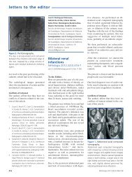
letters to the editor<br />
This is a representative case of a type 1<br />
cardiorenal syndrome (kidney involvement<br />
secondary to acute heart failure) in<br />
the context of a recently reported complex<br />
pathology, such as the Tako-Tsubo<br />
syndrome. It is a prerenal failure, in<br />
which an adequate recovery of renal function<br />
should be expected.<br />
While there are no good early markers<br />
available for kidney dysfunction, renal<br />
function must be closely monitored for<br />
any patient with an acute heart disease<br />
for an early diagnosis. If the cardiologist<br />
and the nephrologist work together in<br />
acute management of the treatment, patient<br />
prognosis could improve.<br />
1. Paixao AR, Balaji N, Ryzhikov A, Ghafouri K,<br />
Collins S. Left ventricular contraction patterns<br />
in patients with suspected acute coronary<br />
syndrome and normal coronary angiograms:<br />
a new look at the Takotsubo syndrome. Clin<br />
Cardiol 2011;34(1):45-50.<br />
2. Dote K, Sato H, Tateishi H, Uchida T, Ishihara<br />
M. Myocardial stunning due to simultaneous<br />
multivessel coronary spasms: a review of 5<br />
cases. J Cardiol 1991;21(2):203-14.<br />
3. Prasad A, Lerman A, Rihal CS. Apical<br />
ballooning syndrome (Tako-Tsubo or stress<br />
cardiomyopathy): a mimic of acute<br />
myocardial infarction. Am Heart J<br />
2008;155(3):408-17.<br />
4. Singh NK. Apical ballooning syndrome: the<br />
emerging evidence of a neurocardiogenic<br />
basis. Am Heart J 2008;156(3):e33.<br />
5. Previtali M, Repetto A, Camporotondo R,<br />
Citro R, Faggiano P. Clinical characteristics<br />
and outcome of left ventricular ballooning<br />
syndrome in a European population. Am J<br />
Cardiol 2011;107(1):120-5.<br />
D. Arroyo1 , N. Panizo1 , U. Verdalles1 ,<br />
M.E. Vázquez-Álvarez2 , D. Barraca1 ,<br />
B. Quiroga1 , J. Luño1 1 Nephrology Department. Gregorio Marañón<br />
University General Hospital. Madrid, Spain<br />
2 Cardiology Department. Gregorio Marañón<br />
University General Hospital. Madrid, Spain<br />
Correspondence: David Arroyo Rueda<br />
Servicio de <strong>Nefrología</strong>.<br />
Hospital General Universitario Gregorio Marañón.<br />
Dr. Esquerdo, 46. 28007 Madrid. Spain.<br />
dvdrry@gmail.com<br />
nayapanizo@gmail.com<br />
Chronic pulmonary<br />
bleeding as the first<br />
sign of microscopic<br />
polyangiitis associated<br />
with autoimmune<br />
thyroiditis<br />
Nefrologia 2011;31(4):494-5<br />
doi:10.3265/Nefrologia.pre2011.Apr.10841<br />
To the Editor,<br />
Microscopic polyangiitis (MPA) is defined<br />
as a non granulomatous, anti-neutrophil<br />
cytoplasmic antibodies (ANCA)positive<br />
systemic necrotising vasculitis<br />
which affects small vessels. Kidneys and<br />
lungs are the most frequently main affected<br />
organs. 1 Pulmonary bleeding in MPA is<br />
usually acute or even fulminant. 2,3 Pulmonary<br />
bleeding as part of chronic MPA<br />
is not described as often. Autoimmune<br />
thyroiditis is associated with glomerulopathies,<br />
especially with membranous<br />
nephropathy. 4<br />
We describe a patient with chronic pulmonary<br />
bleeding as a sign of MPA; the examination<br />
also showed that she presented<br />
with autoimmune thyroiditis.<br />
A 54-year-old woman under examination<br />
for a year and a half in another hospital for<br />
iron deficiency anaemia of several years.<br />
She was being treated with orally-administered<br />
iron and needed transfusions. They<br />
found that she had renal failure, proteinuria<br />
and microhaematuria during this examination,<br />
and she was referred to us. Patient<br />
history: arterial hypertension for a<br />
year. Clinically, she presented with important<br />
asthenia and medium-effort dyspnoea.<br />
Physical examination: she did not<br />
present with fever, blood pressure was<br />
140/96mm Hg, and had no oedemas on<br />
lower limbs. The analytical study showed:<br />
haemoglobin: 8.6g/dl; iron: 33.5µg/dl; total<br />
iron-binding capacity (TIBC):<br />
264µg/dl; ferritin: 137ng/ml; urea:<br />
67mg/dl; creatinine: 1.50mg/dl; estimated<br />
glomerular filtration rate (eGFR) (according<br />
to Modification of Diet in Renal Disease<br />
[MDRD4]): 38.46ml/min/1.73m 2 ;<br />
uric acid: 7.19mg/dl; total cholesterol:<br />
233mg/dl (LDL: 163; HDL: 33.7);<br />
triglycerides: 178mg/dl. Normal/negative<br />
immunoglobulins, antinuclear antibodies<br />
(ANA), anti-DNA, rheumatoid factor<br />
(RF), anti-glomerular basement membrane<br />
antibody treatment (anti-MBG) and<br />
anti-transglutaminase antibodies (ATA);<br />
positive myeloperoxidase (MPO)-ANCA,<br />
negative PR3-ANCA. High resolution<br />
electrophoresis (HRE) of serum: no monoclonal<br />
band. Thyroid-stimulating hormone<br />
(TSH): 326mIU/l (0.350-4.940),<br />
free thyroxine (T4): 0.11ng/dl (0.7-1.48);<br />
anti-thyroid peroxidase antibodies (anti-<br />
TPO) >1000IU/ml. Negative Mantoux<br />
test. Negative hepatitis B surface antigens<br />
(HbsAg), hepatitis B anti-core antibodies<br />
(anti-HBc), hepatitis C antibodies (anti-<br />
HCV) and human immunodeficiency<br />
virus (HIV) tests. Cytomegalovirus<br />
(CMV) serology test and Epstein-Barr<br />
virus (EBV): previous exposure. Urine<br />
showed a proteinuria of 1.3g/24hr (moderately<br />
selective glomerular proteinuria),<br />
sediment with 60-100 red blood cells<br />
(30%-50% dysmorphic) and hyalinegranular<br />
casts. Urine culture: negative.<br />
Chest X-ray and electrocardiogram<br />
(ECG) were normal. Abdominal ultrasound:<br />
kidneys of a normal size with<br />
slight cortical thinning. Chest computerised<br />
tomography (CT) showed images<br />
of ‘ground-glass opacity’ on both sides in<br />
the middle and lower fields and a bronchoscopy<br />
with a bronchoalveolar lavage<br />
was indicated, showing 80%<br />
siderophores. The malignant cell cytology<br />
and cell cultures were negative. The patient<br />
was interviewed, revealing that she<br />
sporadically presented with a slight, darkcoloured<br />
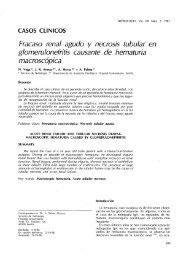
expectoration. The renal biopsy<br />
(Figure 1) showed a focal and segmental<br />
necrotising glomerulonephritis, and approximately<br />
a third of the glomeruli were<br />
sclerotic, interstitial fibrosis and tubular<br />
atrophy in 30% of the cylinder. The immunofluorescence<br />
study showed slight<br />
deposits of IgM, C3 and C4 on capillary<br />
walls and the mesangium. Given the pulmonary<br />
bleeding, the positive ANCA, and<br />
the focal necrotising glomerulonephritis<br />
with scarce immune deposits, MPAwas diagnosed,<br />
associated with autoimmune thyroiditis-induced<br />
hypothyroidism. Treatment<br />
with prednisone at 50mg/day was started for<br />
8 weeks, gradually reducing the dosage, and<br />
six 750-mg cyclophosphamide boli, which<br />
494 Nefrologia 2011;31(4):489-504