

Physiological and molecular determinants of embryo implantation
Physiological and molecular determinants of embryo implantation
Physiological and molecular determinants of embryo implantation

You also want an ePaper? Increase the reach of your titles
YUMPU automatically turns print PDFs into web optimized ePapers that Google loves.
16 S. Zhang et al. / Molecular Aspects <strong>of</strong> Medicine xxx (2013) xxx–xxx<br />
5.4. Uterine gl<strong>and</strong>s for <strong>embryo</strong> <strong>implantation</strong><br />
The initial development <strong>of</strong> the female reproductive tract occurs during <strong>embryo</strong>genesis soon after gastrulation (Kobayashi<br />
<strong>and</strong> Behringer, 2003; Orvis <strong>and</strong> Behringer, 2007; Spencer et al., 2012). In laboratory rodents, domestic animals <strong>and</strong> humans,<br />
the uterus is not fully developed or differentiated at birth; thus, postnatal uterine morphogenesis is a critical event for normal<br />
uterine functions in adult life (Gray et al., 2001a). Uterine gl<strong>and</strong>s are thought to be the principal source <strong>of</strong> uterine secretions<br />
that are required for blastocyst survival <strong>and</strong> <strong>implantation</strong>, as well as the establishment <strong>and</strong> maintenance <strong>of</strong> pregnancy<br />
(Bazer, 1975). Using the uterine gl<strong>and</strong> knockout ewe model, it has been demonstrated that uterine gl<strong>and</strong> <strong>and</strong> its secretion<br />
during early pregnancy are essential for conceptus survival (Gray et al., 2001a,b,c). Molecular <strong>and</strong> genetic studies have demonstrated<br />
that growth factors, cytokine factors <strong>and</strong> potential histotrophic factors produced by uterine gl<strong>and</strong>s are able to regulate<br />
the endometrial function.<br />
5.4.1. Uterine adenogenesis<br />
Coordinated development <strong>of</strong> the endometrial gl<strong>and</strong>s from the luminal epithelium, known as uterine adenogenesis, is a<br />
predominant event during postnatal uterine morphogenesis. The process <strong>of</strong> adenogenesis involves three typical steps, budding,<br />
penetration <strong>and</strong> branching. The gl<strong>and</strong>ular epithelium derived from the luminal epithelium differentiates <strong>and</strong> buds, followed<br />
by the penetration <strong>of</strong> the gl<strong>and</strong>ular tubes into the uterine stroma, <strong>and</strong> then the gl<strong>and</strong>ular elements keep branching<br />
<strong>and</strong> coiling to exp<strong>and</strong> throughout the endometrial stroma (Bartol et al., 1999, 1993). In mice, epithelial invaginations or<br />
gl<strong>and</strong> buds appear at postnatal day (PND) 5, nascent endometrial gl<strong>and</strong>s are present at PND 7, <strong>and</strong> complex gl<strong>and</strong>s extending<br />
from the luminal epithelium into the endometrial stroma are established by PND 12 (Branham et al., 1985; Brody <strong>and</strong> Cunha,<br />
1989). In contrast, endometrial gl<strong>and</strong> development in humans begins during fetal development within the womb, continues<br />
from birth to age 6, <strong>and</strong> is fully developed by puberty (K<strong>of</strong>f, 1933).<br />
The process <strong>of</strong> uterine adenogenesis involves intrinsic epithelial–stromal interactions <strong>and</strong> is precisely orchestrated by<br />
multifactorial gene networks (Baker et al., 1996; Miller <strong>and</strong> Sassoon, 1998; Reardon et al., 2012). Exposure <strong>of</strong> mice to native<br />
estrogen, estrogen analogs or progesterone during critical developmental periods inhibits endometrial adenogenesis by disrupting<br />
the normal expression <strong>of</strong> morphoregulatory genes (Cooke et al., 2012; Filant et al., 2012; Hayashi et al., 2011; Miller<br />
et al., 1998; Stewart et al., 2011; Talbi et al., 2006). Ablation <strong>of</strong> these key genes in mice reduces or even eliminates uterine<br />
gl<strong>and</strong>s (Table 1). Interestingly, abnormalities <strong>of</strong> uterine gl<strong>and</strong>s are <strong>of</strong>ten accompanied by defects in the uterine stroma <strong>and</strong><br />
luminal epithelium, leading to <strong>implantation</strong> failure. For example, uterine conditional depletion <strong>of</strong> Dicer, an RNase III endonuclease<br />
that is essential for the biogenesis <strong>of</strong> microRNAs <strong>and</strong> small interfering RNAs, leads to female sterility <strong>and</strong> underdeveloped<br />
small uteri (Hawkins et al., 2012). The most prominent features <strong>of</strong> Dicer-deficient uteri are a complete lack <strong>of</strong> uterine<br />
gl<strong>and</strong>s <strong>and</strong> enhanced stromal apoptosis with failure to proliferate in response to progesterone (Hawkins et al., 2012). Females<br />
with conditional knockout <strong>of</strong> prohibitin 2 (also known as repressor <strong>of</strong> estrogen receptor activity, Rea) in the uterus<br />
show <strong>implantation</strong> <strong>and</strong> decidualization failure due to altered endometrial gl<strong>and</strong> morphogenesis (Park et al., 2012b). In addition,<br />
Wnt5a plays a crucial role in the generation <strong>of</strong> uterine gl<strong>and</strong>s <strong>and</strong> is required for cellular <strong>and</strong> <strong>molecular</strong> responses to<br />
exogenous estrogen (Mericskay et al., 2004). During postnatal uterine development, Wnt5a participates in a regulatory loop<br />
with other uterine patterning genes, including Wnt7a, Hoxa10 <strong>and</strong> Hoxa11 (Mericskay et al., 2004). Mice with uterine-specific<br />
ablation <strong>of</strong> Wnt4, Wnt7a or E-cadherin show defective <strong>implantation</strong> associated with a reduction or absence <strong>of</strong> uterine<br />
gl<strong>and</strong>s (Dunlap et al., 2011; Franco et al., 2011a; Hayashi et al., 2011).<br />
5.4.2. Gl<strong>and</strong>ular–luminal epithelial interaction<br />
Of the products secreted by uterine gl<strong>and</strong>s, LIF is one <strong>of</strong> the most important molecules. It has been well documented that<br />
membrane-associated LIFR <strong>and</strong> gp130 are required for LIF signaling (Layton et al., 1994; Narazaki et al., 1994). In mice, the<br />
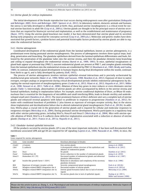
Table 1<br />
Genes required for uterine adenogenesis.<br />
Gene Gene encoded Knockout phenotype in females Ref.<br />
Ctnnb1 b-Catenin Few uterine gl<strong>and</strong>s Jeong et al. (2009)<br />
Cdh1 E-cadherin A disorganized cellular structure <strong>of</strong> the epithelium <strong>and</strong><br />
ablation <strong>of</strong> endometrial gl<strong>and</strong>s<br />
Reardon et al. (2012)<br />
Dicer Dicer No gl<strong>and</strong>s Hawkins et al. (2012)<br />
Foxa2 Forkhead box A2 Severe reduction in the number <strong>of</strong> endometrial gl<strong>and</strong>s Jeong et al. (2010)<br />
Hoxa11 Homeobox A11 (TF) Decreased number <strong>of</strong> endometrial gl<strong>and</strong>s <strong>and</strong> abnormal<br />
gl<strong>and</strong>ular differentiation<br />
Gendron et al. (1997)<br />
Igf1 Insulin-like growth factor 1 (IGF1) Reduction in abundance <strong>and</strong> complexity <strong>of</strong> the secretory<br />
gl<strong>and</strong>ular elements<br />
Baker et al. (1996)<br />
REA Repressor <strong>of</strong> estrogen receptor activity Severely compromised gl<strong>and</strong> morphogenesis Park et al. (2012b)<br />
Wnt4 Wingless-related MMTV integration site 4 Decreased number <strong>of</strong> uterine gl<strong>and</strong>s Franco et al. (2011a)<br />
Wnt5a Wingless-related MMTV integration site 5a Uterine gl<strong>and</strong> formation failure due to lack <strong>of</strong> stromal<br />
signals<br />
Mericskay et al. (2004)<br />
Wnt7a Wingless-related MMTV integration site 7a No uterine gl<strong>and</strong>s due to lack <strong>of</strong> epithelial signals Dunlap et al. (2011),<br />
Miller <strong>and</strong> Sassoon (1998)<br />
Please cite this article in press as: Zhang, S., et al. <strong>Physiological</strong> <strong>and</strong> <strong>molecular</strong> <strong>determinants</strong> <strong>of</strong> <strong>embryo</strong> <strong>implantation</strong>. Molecular Aspects <strong>of</strong><br />
Medicine (2013), http://dx.doi.org/10.1016/j.mam.2012.12.011