Pigmented lesion clinic - Goals and Objectives - Dermatology
Pigmented lesion clinic - Goals and Objectives - Dermatology
Pigmented lesion clinic - Goals and Objectives - Dermatology

Create successful ePaper yourself
Turn your PDF publications into a flip-book with our unique Google optimized e-Paper software.
<strong>Goals</strong> <strong>and</strong> <strong>Objectives</strong> <strong>Pigmented</strong> Lesion Clinic<br />
First-Year Residents:<br />
PIGMENTED LESION CLINIC<br />
RES C<br />
As a dermatologist you will become an expert on skin cancer, as well as a the main<br />
<strong>clinic</strong>al advisor for patients at risk. You have to be able to perform a thorough total<br />
body investigation to identify potential skin cancers <strong>clinic</strong>ally, <strong>and</strong> to decide whether a<br />
<strong>lesion</strong> will require total excision or further follow-up. Dermoscopy is a great tool for the<br />
diagnosis of early melanoma <strong>and</strong> other pigmented <strong>and</strong> non-pigmented tumors of the<br />
skin.<br />
This means you will have to:<br />
1. Recognize the <strong>clinic</strong>al features of common nevi, atypical nevi, melanoma, <strong>and</strong> other<br />
pigmented skin <strong>lesion</strong>s (pigmented BCC, seborrhoeic keratoses, angiomas, etc.)<br />
2. Learn the basic terminology used to describe pigmented <strong>lesion</strong>s examined with a<br />
dermoscope<br />
3. Learn the dermoscopic algorithm to differentiate between melanocytic <strong>and</strong> nonmelanocytic<br />
pigmented <strong>lesion</strong>s<br />
4. Learn one of the dermoscopic algorithms to diagnose melanoma <strong>and</strong> be able to use<br />
it in your daily routine (see readings below)<br />
Required Readings for First-Year Residents:<br />
1. Thompson JF et al. Cutaneous melanoma. Lancet 365:687-701, 2005<br />
2. Braun RP et al. Dermoscopy of pigmented skin <strong>lesion</strong>s. J Am Acad Dermatol:109-21,<br />
2005<br />
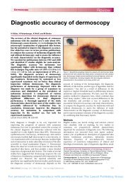
3. Kittler H et al. Diagnostic accuracy of dermoscopy. Lancet Oncol 3:159-65, 2002<br />
4. Curtin JA et al: Distinct sets of genetic alterations in melanoma. N Engl J Med<br />
353:2135-47, 2005<br />
5. Nachbar F, Stolz W et al. The ABCD rule of dermatoscopy. High prospective value in<br />
the diagnosis of doubtful melanocytic skin <strong>lesion</strong>s. J Am Acad Dermatol. 30:551-9,<br />
1994<br />
or<br />
Argenziano G et al. Epiluminescence microscopy for the diagnosis of doubtful<br />
melanocytic skin <strong>lesion</strong>s. Comparison of the ABCD rule of dermatoscopy <strong>and</strong> a new<br />
7-point checklist based on pattern analysis. Arch Dermatol. 134:1563-70, 1998<br />
or<br />
Menzies SW et al. A sensitivity <strong>and</strong> specificity analysis of the surface microscopy<br />
features of invasive melanoma. Melanoma Res. 6:55-62, 1996
<strong>Goals</strong> <strong>and</strong> <strong>Objectives</strong> <strong>Pigmented</strong> Lesion Clinic<br />
Second-Year Residents:<br />
PIGMENTED LESION CLINIC<br />
RES C<br />
You will learn to identify patients at increased risk for melanoma or who already have<br />
melanoma, <strong>and</strong> to manage, educate, encourage, <strong>and</strong> lead them. By improving your<br />
dermoscopy skills, <strong>and</strong> using short-term <strong>and</strong> long-term follow-up, you will improve<br />
early detection of melanoma <strong>and</strong> other skin cancers, <strong>and</strong> reduce unnecessary<br />
excisions, i.e. improve sensitivity <strong>and</strong> specificity of your melanoma diagnosis.<br />
In more detail, you will:<br />
1. Be able <strong>and</strong> confident in identifying <strong>lesion</strong>s suspicious for melanoma, <strong>and</strong> other<br />
pigmented tumors of the skin using dermoscopy.<br />
2. Be familiar with dermoscopic patterns suggestive for melanoma <strong>and</strong> other pigmented<br />
<strong>lesion</strong>s, as well as with the concept of “featureless melanoma”.<br />
3. Be able to outline a patient’s melanoma risk integrating patient’s common <strong>and</strong><br />
atypical melanocytic nevus counts, skin type, history, family history, <strong>and</strong> other<br />
factors.<br />
4. Learn to manage patients at high risk for melanoma, including required surgery, <strong>and</strong><br />
short- <strong>and</strong> long-term follow-up using digital images. You will learn the advantages<br />
<strong>and</strong> disadvantages of shave biopsies versus complete excisions.<br />
5. Be able to raise patient awareness of melanocytic nevi <strong>and</strong> melanoma risk without<br />
creating fear. You will also be able to educate patients regarding skin selfexamination,<br />
sun-protection, <strong>and</strong> required follow-up visits.<br />
6. Learn dermoscopic features of acral <strong>and</strong> facial pigmented tumors.<br />
Required Second-Year Readings:<br />
1. Bauer J, Garbe C. Acquired melanocytic nevi as risk factor for melanoma<br />
development. A comprehensive review of epidemiological data. Pigment Cell Res.<br />
16:297-306, 2003<br />
2. Pehamberger H, et al. In vivo epiluminescence microscopy of pigmented skin<br />
<strong>lesion</strong>s. I. Pattern analysis of pigmented skin <strong>lesion</strong>s. J Am Acad Dermatol 17:571-<br />
83, 1987<br />
3. Steiner A, et al. In vivo epiluminescence microscopy of pigmented skin <strong>lesion</strong>s. II.<br />
Diagnosis of small pigmented skin <strong>lesion</strong>s <strong>and</strong> early detection of malignant<br />
melanoma. J Am Acad Dermatol 17:584-91, 1987<br />
4. Menzies SW. Cutaneous melanoma: making a <strong>clinic</strong>al diagnosis, present <strong>and</strong> future.<br />
Dermatol Ther. 19:32-9, 2006<br />
5. Kittler H et al. Identification of <strong>clinic</strong>ally featureless incipient melanoma using<br />
sequential dermoscopy imaging. Arch Dermatol. 142:1113-9, 2006<br />
6. Stolz W at al. Dermatoscopy for facial pigmented skin <strong>lesion</strong>s. Clin Dermatol. 20:276-<br />
8, 2002<br />
7. Saida et al. Dermoscopy for acral pigmented skin <strong>lesion</strong>s. Clin Dermatol. 20:279-85,<br />
2002