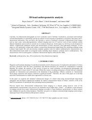
3D Tooth Shape from Radiographs using Thin-Plate Splines
3D Tooth Shape from Radiographs using Thin-Plate Splines
3D Tooth Shape from Radiographs using Thin-Plate Splines

Create successful ePaper yourself
Turn your PDF publications into a flip-book with our unique Google optimized e-Paper software.
Introduction<br />
<strong>3D</strong> <strong>Tooth</strong> <strong>Shape</strong> <strong>from</strong> <strong>Radiographs</strong><br />
<strong>using</strong> <strong>Thin</strong>-<strong>Plate</strong> <strong>Splines</strong><br />
Reyes ENCISO*°, John P. LEWIS°, Ulrich NEUMANN°<br />
and James MAH*<br />
*Craniofacial Virtual Reality Lab, DEN 312<br />
°Integrated Media Systems Center, EEB 131<br />
University of Southern California, Los Angeles, CA 90089, USA<br />
Abstract. Current methods to produce 3-dimensional tooth root models involve<br />
conversion <strong>from</strong> radiographic means (computed tomography) or creation <strong>using</strong><br />
computer-assisted design (CAD) software. The former lacks detail while the second<br />
is manually fabricated and can bear little resemblance to the original. <strong>Thin</strong>-plate<br />
splines have been used in morphometrics to define changes of shape between<br />
subjects of the same species [1]. Herein, we use thin-plate splines to deform a <strong>3D</strong><br />
geometric prior model of a tooth to match 2D patient radiographs, producing a “bestfit”<br />
patient specific <strong>3D</strong> geometric polygonal mesh of the tooth.<br />
Very accurate three-dimensional geometric crown models are now commercially available<br />
<strong>from</strong> different sources. Destructive scanning methods (Orthocad <strong>from</strong> Cadent) slice the<br />
dental impression of the patient as it is scanned producing a stack of images that are<br />
rendered to produce the final model. Laser systems are also used to directly image the stone<br />
model of patient’s dentition (e-Models <strong>from</strong> GeoDigm Corp.). A direct imaging method<br />
(OraScanner <strong>from</strong> OraMetrix GmbH) also exists wherein an intra-oral camera capable of<br />
generating highly accurate <strong>3D</strong> crown models is used after applying an opaquing agent to the<br />
teeth. However all these methods provide data on the tooth crowns only and nothing on root<br />
form. Therefore, researchers have explored <strong>using</strong> root information <strong>from</strong> computed<br />
tomography (CT) volumes with crown forms obtained <strong>using</strong> laser scanning methods[4][5].<br />
Registration of the data sets is accomplished <strong>using</strong> ceramic markers present in both images.<br />
In this paper we propose to use a patient-specific radiograph and a <strong>3D</strong> geometric<br />
prior model to produce a “best-fit” patient specific <strong>3D</strong> model of the whole tooth <strong>using</strong><br />
radial basis functions, previously used for volume morphing [3] and to fill in a hole on the<br />
skull and fabricate the implant [2]. The rationale for this approach is that 2D radiographs of<br />
dental patients are the norm, while there are applications being developed for modeling and<br />
treatment simulation that require <strong>3D</strong> tooth root information. CT imaging of dental patients<br />
is not routine and in most cases the risk cannot be justified.<br />
Of note, there are <strong>3D</strong> volumetric imaging devices developed specifically for<br />
dentistry recently released (NewTom <strong>from</strong> Aperio Services) and others being developed<br />
which feature a lower absorbed radiation dose but until these devices become widely used,<br />
2D radiographs are the only available source of root morphological information. Therefore,<br />
this information can be “fitted” to a geometric model to provide a reasonable shape of the<br />
tooth root.
1. Materials and Methods<br />
The input data for our interactive <strong>3D</strong> computer program is a 2D periapical radiograph of a<br />
tooth (Figure 1a) and a generic <strong>3D</strong> polygonal surface mesh model (courtesy of 3M Unitek)<br />
of the corresponding tooth (Figure 1b). These items are overlaid in close proximity prior to<br />
the fitting (Figure 1c).<br />
(a) (b) (c)<br />
Figure 1: (a) Periapical radiograph of the second bicuspid; (b) View of the <strong>3D</strong><br />
prior geometric tooth; (c) Both the radiograph and the <strong>3D</strong> model overlaid.<br />
In our system the user is asked to interactively indicate with the mouse<br />
corresponding landmarks of their choice on both the radiograph (indicated with a “x” in<br />
Figure 2a) and a view of the <strong>3D</strong> model (indicated with a “+” in Figure 2a). The computer<br />
program first computes the thin-plate coefficients <strong>from</strong> these landmarks for the X and<br />
(separately) Y displacements and then warps the image of the <strong>3D</strong> model (shaded in Figure<br />
2b) and the <strong>3D</strong> mesh (Figure 2c) in X and Y directions (depth information is kept constant)<br />
<strong>using</strong> the thin-plate interpolant. <strong>Thin</strong>-plate splines minimize a bending energy:<br />
2 2<br />
2<br />
( + 2 f + f ) dxdy , while interpolating values of a set of 2d points ( xi,<br />
yi)<br />
∫<br />
∫<br />
named landmarks. The thin-plate spline can be represented and solved <strong>using</strong> radial basis<br />
functions:<br />
f xx xy yy<br />
p<br />
ay y + ∑ wi<br />
⋅U<br />
1<br />
i i<br />
( ( x , y ) − ( x,<br />
y)<br />
)<br />
f ( x,<br />
y)<br />
= a1<br />
+ ax<br />
x +<br />
, with U<br />
2 ( r ) = r log( r)<br />
.<br />
(a) (b) (c)<br />
Figure 2: Landmarks interactively selected by the user in the <strong>3D</strong> prior model image (+)<br />
and the radiograph (x) are shown in (a). (b) and (c) show the overlaid image of the shell of<br />
the tooth <strong>3D</strong> model (before fitting) and the shaded warped image of the fitted <strong>3D</strong> model.
2. Results<br />
The input landmarks and resulting <strong>3D</strong> polygonal mesh can be seen in Figure 2. The prior<br />
and fitted <strong>3D</strong> models can be interactively rotated to any viewpoint <strong>using</strong> a virtual trackball<br />
interface (Figure 3).<br />
(a) (b) (c) (d)<br />
Figure 3: Frontal (a) and side (b) view of the prior geometric model <strong>from</strong> 3M Unitek.<br />
Frontal (c) and side (d)viewofthe<strong>3D</strong> fitted model <strong>using</strong> thin-plate splines.<br />
3. Novelty/Discussion<br />
This new technique uses computer graphics algorithms to combine current patient records<br />
in the form of two-dimensional radiographs with 3-D geometric prior models of teeth,<br />
producing a “best-fit” patient-specific model (including root information). The resultant <strong>3D</strong><br />
model is smooth and conserves the overall shape of the tooth, but is adapted in scale,<br />
orientation and shape to fit the X-ray picture and provide patient-specific information.<br />
Additional radiographs <strong>from</strong> known directions would provide additional depth<br />
information but these are generally not available. As such the selected 3d model provides a<br />
“best fit” in the presence of this unavailable information. Future research includes fitting<br />
the crown models obtained <strong>from</strong> clinical services and assessing geometric accuracy with<br />
micro-CT following extraction of the tooth for clinical reasons.<br />
Acknowledgements:<br />
The research has been funded in part by the Integrated Media Systems Center, a National<br />
Science Foundation Engineering Research Center, Cooperative Agreement No. EEC-<br />
9529152, Sun Microsystems and the American Association of Orthodontists Foundation.<br />
References<br />
[1] Bookstein FL: Principal warps: <strong>Thin</strong>-plate splines and the decomposition of deformations. IEEE<br />
Transactions on Pattern Analysis and Machine Intelligence 11:567-585, 1989<br />
[2] Carr JC, Fright WR, Beatson RK: Surface interpolation with radial basis functions for medical imaging.<br />
Medical Imaging, IEEE Transactions on 16:96-107, 1997<br />
[3] Fang S, Raghavan R, Richtsmeier JT. Volume Morphing Methods for Landmark Based <strong>3D</strong> Image<br />
Deformation, SPIE Int'l Symposium on Medical Imaging, 1996.<br />
[4] Nishii Y, Nojima K, Takane Y, Isshiki Y: Integration of the maxillofacial three-dimensional CT image<br />
and the three-dimensional dental surface image. The Journal of Japan Orthodontic Society 57:189-194,<br />
1998<br />
[5] Terai H, Shimahara M, Sakinaka Y, Tajima S: Accuracy of Integration of Dental Casts in Three-<br />
Dimensional Models. J Oral Maxillofac Surgery 57:662-665, 1999.