Soft body morphology, dissection and slide-preparation of Ostracoda
Soft body morphology, dissection and slide-preparation of Ostracoda
Soft body morphology, dissection and slide-preparation of Ostracoda

Create successful ePaper yourself
Turn your PDF publications into a flip-book with our unique Google optimized e-Paper software.
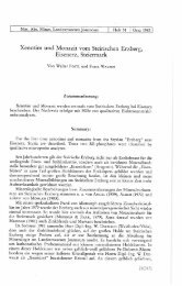
Joannea Geol. Paläont. 11: 327-343 (2011)<br />
<strong>S<strong>of</strong>t</strong> <strong>body</strong> <strong>morphology</strong>, <strong>dissection</strong> <strong>and</strong> <strong>slide</strong>-<strong>preparation</strong> <strong>of</strong> <strong>Ostracoda</strong>:<br />
a primer<br />
Tadeusz NAMIOTKO, Dan L. DANIELOPOL & Angel BALTANÁS<br />
Abstract: Most commonly used techniques for treating ostracod s<strong>of</strong>t <strong>body</strong> for taxonomical<br />
purposes with optical microscopy are described with emphasis on the order Podocopida.<br />
A variety <strong>of</strong> procedures for pre-treatment, storage, recovery <strong>of</strong> dried specimens,<br />
<strong>dissection</strong>, temporary <strong>and</strong> permanent mounting, <strong>and</strong> staining methods are presented<br />
<strong>and</strong> evaluated. General <strong>morphology</strong> <strong>and</strong> terminology <strong>of</strong> the ostracod appendages are<br />
also summarised.<br />
Key Words: <strong>Ostracoda</strong>; Dissection; Mounting; Staining; Appendages; Morphology.<br />
1. Introduction<br />
One <strong>of</strong> best diagnostic <strong>and</strong> most conspicuous trait <strong>of</strong> <strong>Ostracoda</strong> is a bivalved carapace<br />
that may completely envelop the whole animal <strong>body</strong> with limbs. Ostracodologists usually<br />
refer to calcified valves as “hard parts”, whereas to the appendages <strong>and</strong> other internal<br />
organs as „s<strong>of</strong>t parts”. The classification <strong>of</strong> Recent ostracods is based mainly on<br />
differences in the s<strong>of</strong>t part <strong>morphology</strong> which is ordinarily <strong>of</strong> little use by palaeontologists.<br />
However, considering the close functional relationship <strong>of</strong>ten existing between the<br />
s<strong>of</strong>t <strong>and</strong> hard parts, each expressing to a considerable extent the <strong>morphology</strong> <strong>of</strong> the<br />
other, even basic knowledge on the appendages without doubt should help palaeontologists<br />
to better underst<strong>and</strong> <strong>and</strong> interpret various features displayed in the valves <strong>of</strong> the<br />
fossil ostracods. Examining living material collected from modern habitats enables palaeontologists<br />
also to become familiar with intraspecific variability or ecology, which<br />
could greatly facilitate both taxonomical <strong>and</strong> palaeoenvironmental studies. Therefore<br />
the <strong>morphology</strong> <strong>and</strong> terminology <strong>of</strong> the s<strong>of</strong>t parts <strong>of</strong> ostracods is outlined (focusing on<br />
limbs) in this primer prior to the description <strong>of</strong> various laboratory techniques involved<br />
in the appendage <strong>preparation</strong> for optical microscopy. Analogously, one may advise a<br />
biologist (neontologist) to familiarise oneself with the details <strong>of</strong> the “hard parts” <strong>morphology</strong><br />
<strong>and</strong> some basic laboratory techniques used routinely in micropalaeontological<br />
work.<br />
327
Several laboratory techniques <strong>and</strong> chemicals used in the study <strong>of</strong> the s<strong>of</strong>t <strong>body</strong> <strong>of</strong><br />
ostracods are in many respects analogous to those applied to other aquatic animals <strong>of</strong><br />
similar size. However, the presence <strong>of</strong> the overdeveloped calcified carapace in ostracods,<br />
has proved necessary to develop a number <strong>of</strong> special methods for treating both<br />
Recent <strong>and</strong> fossil specimens. The bivalved nature <strong>of</strong> the carapace coupled with the<br />
small size <strong>of</strong> most ostracods appear also to be an important obstacle <strong>and</strong> the reason <strong>of</strong><br />
a disinclination to study this group by biologists, since acquiring the expertise to perform<br />
properly a full <strong>dissection</strong> can take sometimes several months.<br />
The present introductory text was intended to be used primarily by palaeontologists<br />
<strong>and</strong> novices working mainly with hard parts, thus it was deemed not necessary to<br />
include in it a detailed description <strong>of</strong> the techniques typically pertaining the study <strong>of</strong> the<br />
valves. Both <strong>preparation</strong> techniques <strong>and</strong> general <strong>morphology</strong> <strong>of</strong> the ostracod s<strong>of</strong>t <strong>body</strong><br />
is presented <strong>and</strong> exemplified by the order Podocopida (or subclass Podocopa; classification<br />
scheme according to HORNE et al. 2002) which is the most species-rich <strong>and</strong> diverse<br />
ostracod group at the present day as well as has the best fossil record (HORNE et<br />
al. 2002).<br />
For the practical knowledge concerning methods <strong>of</strong> sampling, sample processing,<br />
initial laboratory treatments <strong>and</strong> culturing <strong>of</strong> Recent ostracods the reader is referred to<br />
other publications in which such procedures are described in detail, <strong>of</strong> which the most<br />
recommendable are the following: BRONSTEIN (1947, Engl. transl. 1988); ATHERSUCH et<br />
al. (1989); HENDERSON (1990); GRIFFITHS & HOLMES (2000); MEISCH (2000) <strong>and</strong> DANIE-<br />
LOPOL et al. (2002), as well as those concerning mei<strong>of</strong>auna in general (e.g., HIGGINS &<br />
THIEL 1988; GALASSI et al. 2002; SOMMERFIELD et al. 2005 or GIERE 2009).<br />
2. General <strong>morphology</strong> <strong>and</strong> terminology <strong>of</strong> the s<strong>of</strong>t <strong>body</strong><br />
Although several authors have recently attempted to unify terminology pertaining ostracod<br />
limbs <strong>and</strong> their chaetotaxy (e.g., HARTMANN 1966–1989; DANIELOPOL 1978; BROOD-<br />
BAKKER & DANIELOPOL 1982; MARTENS 1987; MATZKE-KARASZ 1995; HARTMANN &<br />
GUILLAUME 1996; MEISCH 1996; COHEN et al. 1998; MARTENS 1998; MEISCH 2000;<br />
MADDOCKS 2000; HORNE 2005; KARANOVIC 2005; SMITH & TSUKAGOSHI 2005; SMITH et al.<br />
2005; TSUKAGOSHI et al. 2006; BOXSHALL et al. 2010), there is still lack <strong>of</strong> consensus on<br />
this matter in the literature <strong>and</strong> many confusion exists about limb homologies with<br />
other crustacean taxa <strong>and</strong> even within <strong>Ostracoda</strong>. Here we adopted terminology <strong>of</strong> the<br />
general limb <strong>morphology</strong> after HORNE et al. (2002).<br />
Ostracods have a short compact <strong>body</strong> (Fig. 1) with no true segmentation as <strong>of</strong>ten<br />
recognisable in other crustaceans. A faint constriction <strong>of</strong> the <strong>body</strong> usually just in front<br />
<strong>of</strong> the centre (indicated by a dashed-line in Fig. 1) marks the indistinct boundary between<br />
two main parts, the anterior head (cephalon) <strong>and</strong> the posterior trunk (consisting<br />
<strong>of</strong> the reduced thorax <strong>and</strong> the rudimentary abdomen). The later portion shows in a few<br />
328
taxa some external traces <strong>of</strong> segmentation, suggesting 4–7 (subclass Myodocopa) or<br />
10–11 (subclass Podocopa) barely discernible postcephalic segments (HORNE et al.<br />
2002).<br />
Ostracod limbs (or appendages), excepting the antennula (or first antenna), are<br />
considered to derive from a generalised ancestral crustacean appendage composed <strong>of</strong><br />
a basal protopod on which distally two rami are carried: an inner endopod (commonly<br />
larger) <strong>and</strong> an outer exopod (<strong>of</strong>ten strongly reduced). The rami (<strong>and</strong> sometimes protopod)<br />
are usually composed <strong>of</strong> a number <strong>of</strong> podomeres (articles or joints). From the protopod<br />
may arise medial endites <strong>and</strong> lateral exites (epipods if branchial in function).<br />
Adult ostracods possess up to eight pairs <strong>of</strong> functionally specialised limbs, including<br />
male copulatory appendages (HORNE et al. 2002), which is the fewest number <strong>of</strong> limbs<br />
<strong>of</strong> any crustaceans (BRUSCA & BRUSCA 2003).<br />
In Podocopa four pairs <strong>of</strong> limbs are considered to be clearly attached to the cephalon,<br />
which is untypical for crustaceans (for some details on a debate as to whether ostracods<br />
have four or five cephalic appendages see MEISCH (2000) or HORNE et al.<br />
(2002) <strong>and</strong> references therein). These are the following limbs from the anterior-most:<br />
antennula (A1), antenna (A2), m<strong>and</strong>ibula (Md) <strong>and</strong> maxillula (Mx1) (Fig. 1). First two<br />
pairs (<strong>and</strong> the eye) are attached to the pre-oral forehead, while m<strong>and</strong>ibula <strong>and</strong> maxillula<br />
are connected to the hypostome, constituting the ventral posterior portion <strong>of</strong> the<br />
cephalon, <strong>and</strong> forming the posterior edge <strong>of</strong> the mouth opening. The forehead, hypostome<br />
<strong>and</strong> upper lip (laying below the forehead <strong>and</strong> forming the anterior edge <strong>of</strong> the<br />
mouth) constitute parts <strong>of</strong> the complex chitinous framework <strong>of</strong> the cephalon. In the<br />
mouth region there are also food-rakes assisting with chewing food (MEISCH 2000).<br />
The antennula (A1, Fig. 1) is uniramous (BOXSHALL et al. 2010), composed <strong>of</strong> 5-8<br />
podomeres bearing a number <strong>of</strong> various setae <strong>and</strong> claws (up to c. 30 in Podocopa:<br />
MADDOCKS 2000; SMITH & TSUKAGOSHI 2005), <strong>and</strong> has locomotory (swimming, crawling<br />
<strong>and</strong>/or burrowing) <strong>and</strong> sensory functions (served by chemo-sensorial setae or aesthetascs).<br />
It is usually long, flexible <strong>and</strong> moving upward <strong>and</strong> back. The chaetotaxy <strong>of</strong> A1 (the<br />
number <strong>and</strong> arrangement <strong>of</strong> various setae) as well as the number <strong>of</strong> podomeres differ<br />
in the various groups, providing useful but yet not fully exploited diagnostic characters.<br />
It is advised here to follow the antennular podomere notation <strong>of</strong> SMITH & TSUKAGOSHI<br />
(2005), however, this notation can be applied successfully only if the ontogeny <strong>of</strong> A1<br />
<strong>of</strong> a given species is sufficiently known.<br />
The antenna (A2, Fig. 1) is biramous, however, the degree <strong>of</strong> development <strong>of</strong> the<br />
two rami, arising from usually one podomere-protopod, differs substantially in Myodocopa<br />
<strong>and</strong> Podocopa (HORNE et al. 2002). In the former subclass the exopod is well developed<br />
(with at least 9 podomeres), while the endopod is reduced. On the contrary, in<br />
Podocopa it is the endopod which is better-developed ramus (with 3–4 podomeres<br />
armed with various setae, aesthetascs <strong>and</strong> terminal claws), whereas the exopodite is<br />
rudimentary (in a form <strong>of</strong> a small scale with short seta(-e) or <strong>of</strong> a long spinneret seta,<br />
except the order Platycopida where the exopod is developed almost as strongly as the<br />
endopod). The antennae are the most important locomotory appendages for ostracods,<br />
329
Fig. 1: Morphology <strong>of</strong> a female <strong>of</strong> Cypris pubera O.F. MÜLLER as an example <strong>of</strong> a cypridoidean<br />
ostracod (Podocopa: Podocopida: Cypridoidea: Cyprididae). Right valve in internal lateral view,<br />
the whole <strong>body</strong> removed from the valves <strong>and</strong> dissected individual limbs: A1 – antennula, A2 –<br />
antenna, Md – m<strong>and</strong>ibula, Mx1 – maxillula, L5 – fifth limb (maxilliped), L6 – sixth limb (walking<br />
leg), L7 – seventh leg (cleaning leg), CR – caudal ramus.<br />
330
with long natatory setae in swimming forms <strong>and</strong>/or chelate claws for crawling <strong>and</strong> burrowing.<br />
The complex chaetotaxy <strong>of</strong> A2 (<strong>of</strong>ten sexually dimorphic) is a significant character<br />
in ostracod taxonomy, sometimes also allowing distinction <strong>of</strong> particular larval<br />
stages. It is strongly recommended to consult the A2 chaetotaxic schemes <strong>and</strong> terminology<br />
for the detailed taxonomical study (BROODBAKKER & DANIELOPOL 1982; MARTENS<br />
1987; MEISCH 2000 for the superfamily Cypridoidea, <strong>and</strong> ROSSETTI & MARTENS 1996,<br />
1998 for Darwinuloidea, as well as KAJI & TSUKAGOSHI 2010 for homology <strong>of</strong> the<br />
antennal chaetotaxy among all podocopid superfamilies).<br />
The m<strong>and</strong>ibula (Md, Fig. 1) functions mostly as a feeding organ <strong>and</strong> typically corresponds<br />
well to the general structure <strong>of</strong> the ancestral crustacean biramous appendage,<br />
having both rami developed in addition to the protopod (HORNE 2005). The protopod is<br />
composed <strong>of</strong> two podomeres, the large <strong>and</strong> heavily sclerotized coxa ventrally bearing<br />
strong endite (teeth), <strong>and</strong> the basis, which bears the exopodial branchial plate (<strong>of</strong>ten<br />
greatly reduced) <strong>and</strong> constitutes the first segment <strong>of</strong> a m<strong>and</strong>ibular palp, consisting also<br />
<strong>of</strong> the endopod. The endopodal part <strong>of</strong> the palp is armed with various setae, the<br />
number <strong>and</strong> structure <strong>of</strong> which proved to be important diagnostic traits for some groups<br />
(e.g., the subfamily C<strong>and</strong>oninae). For the terminology <strong>of</strong> the m<strong>and</strong>ibular chaetotaxy <strong>of</strong><br />
the superfamily Cypridoidea see BROODBAKKER & DANIELOPOL (1982).<br />
The maxillula (Mx1, Fig. 1) is the fourth pair <strong>of</strong> the cephalic limb, usually modified<br />
to a great extent, having masticatory <strong>and</strong> respiratory functions. In the subclass Podocopa<br />
Mx1 consists <strong>of</strong> a) the single-podomere protopod bearing three endites, b) the<br />
endopod constituting a palp with up to three podomeres <strong>and</strong> c) the exopod forming usually<br />
a well-developed, large branchial plate, <strong>morphology</strong> <strong>of</strong> which seems to <strong>of</strong>fer useful<br />
diagnostic <strong>and</strong> phylogenetical features (HORNE 2005; SMITH et al. 2005). Myodocopa<br />
do not possess large exopodial branchial plates, instead, they have them on the fifth<br />
limb, while on Mx1 small epipodial (arising from the protopod) branchial plates may be<br />
observed in some taxa (HORNE 2005).<br />
The four pairs <strong>of</strong> the head limbs are followed by three pairs <strong>of</strong> the trunk limbs, one<br />
pair <strong>of</strong> the male copulatory appendages <strong>and</strong> a pair <strong>of</strong> caudal rami (or furcae). The trunk<br />
limbs vary in structure <strong>and</strong> function among taxa.<br />
The fifth limb (L5, Fig. 1) differs in the structure in various taxa depending on the<br />
function it performs <strong>and</strong> is regarded important in classification in several groups. It may<br />
serve as a locomotory appendage (for instance in the superfamilies Cytheroidea or Bairdioidea)<br />
<strong>and</strong> then it has a form <strong>of</strong> a walking leg with a single protopod podomere <strong>and</strong><br />
up to four endopod podomeres (the distal one armed with a strong claw), while the<br />
exopod is reduced, represented usually just by one seta or rarely by a well-developed<br />
branchial plate (HORNE 2005). This limb may be also modified for feeding (e.g., in the<br />
superfamily Cypridoidea) <strong>and</strong> forms a maxilliped with a single protopod podomere (provided<br />
with setae used in feeding), a leg-like or palp-like endopod, <strong>and</strong> a small or totally<br />
lacking exopodial branchial plate. In males <strong>of</strong> Cypridoidea the endopod is transformed<br />
into an <strong>of</strong>ten unsymmetrical clasping organ used for holding the female during copulation.<br />
331
In several Myodocopa the fifth limb serves as a respiratory (<strong>and</strong>/or filter feeding)<br />
appendage as it bears a large epipodial branchial plate (HORNE 2005; COHEN et al.<br />
2007). Between or just in front <strong>of</strong> the fifth limbs the so-called brush-shaped organs,<br />
considered vestiges <strong>of</strong> an additional pair <strong>of</strong> appendages <strong>of</strong> an unknown function, are<br />
found in males <strong>of</strong> several representatives <strong>of</strong> the order Podocopida (e.g., the suborder<br />
Cytherocopina; HORNE et al. 2002).<br />
The sixths limb (L6, Fig. 1) in the majority <strong>of</strong> the representatives <strong>of</strong> the subclass<br />
Podocopa is a uniramous walking leg with the (usually undifferentiated) protopod <strong>and</strong><br />
up to four podomere long endopod, armed distally with a strong claw. In other taxa it<br />
may be a walking leg with an epipodial branchial plate (suborder Halocypridina), lamelliform<br />
(order Myodocopida), modified into claspers in males <strong>and</strong> being rudimentary in<br />
females (order Platycopida) or absent (suborder Cladocopina; COHEN et al. 1998,<br />
2007; HORNE et al. 2002; HORNE 2005).<br />
The seventh limb (L7, Fig. 1) in the Podocopa is either a walking leg similar to the<br />
L6 (as in e.g., the order Palaeocopida or the suborders Cytherocopina <strong>and</strong> Darwinulocopina<br />
<strong>of</strong> the order Podocopida) or a cleaning leg, directed upwards, <strong>of</strong>ten terminated<br />
with a set <strong>of</strong> complex pincers <strong>and</strong> used for removing foreign material from the interior<br />
<strong>of</strong> the valves (as in Cypridocopina) or it is completely lacking (as in Platycopida; HORNE<br />
et al. 2002). In the Myodocopa this limb has the cleaning function, being long, vermiform<br />
<strong>and</strong> flexible (as in the order Myodocopida) or it is greatly reduced or totally absent<br />
(as in the order Halocyprida; COHEN et al. 2007).<br />
The male copulatory appendages (or hemipenes), located in front <strong>of</strong> or attached<br />
to the caudal rami <strong>and</strong> usually paired, may be regarded as the transformation <strong>and</strong> integration<br />
<strong>of</strong> 3–5 pairs <strong>of</strong> thoracic appendages (MARTENS & HORNE 2009). These are <strong>of</strong>ten<br />
large <strong>and</strong> complex structures, varied in various taxa <strong>and</strong> considered very important<br />
taxonomic characters. The detailed internal structure <strong>of</strong> the copulatory organs is <strong>of</strong>ten<br />
very difficult to study <strong>and</strong> needs much practice to obtain satisfactory results. For more<br />
details on <strong>morphology</strong> <strong>and</strong> terminology pertaining to the hemipenis the reader is referred<br />
to MCGREGOR & KESLING (1969), DANIELOPOL (1969, 1978), MARTENS (1990,<br />
1998), MEISCH (2000), SMITH et al. (2006) <strong>and</strong> SMITH & KAMIYA (2007).<br />
The paired caudal rami or furcae (CR, Fig. 1) are attached to the posteroventral<br />
end <strong>of</strong> the ostracod <strong>body</strong>, however, their position relative to the anus differs fundamentally<br />
in Myodocopa <strong>and</strong> Podocopa. In the former subclass they are situated posterior,<br />
while in the latter – anterior to the anus (COHEN et al. 2007). When fully developed they<br />
are plate- or rod-shaped structures with setae <strong>and</strong>/or claws <strong>and</strong> have essentially a locomotory<br />
function (Myodocopa, Palaeocopida, Platycopida, most Podocopida). In some<br />
groups they are reduced to various extent, being represented in extreme case just by<br />
minute setae (as in e.g., Cytheroidea or Darwinuloidea). Caudal rami <strong>and</strong> their attachment<br />
are <strong>of</strong> systematic importance (HORNE et al. 2002; MEISCH 2007).<br />
332
3. Dissection, staining <strong>and</strong> mounting<br />
Although an experienced ostracodologist (zoologist) is sometimes able to identify ostracod<br />
specimens to the genus or species level just by the external <strong>morphology</strong> <strong>of</strong> the carapace<br />
<strong>and</strong> can dispense with the examination <strong>of</strong> the s<strong>of</strong>t <strong>body</strong>, for the accurate identification<br />
<strong>and</strong> description <strong>of</strong> specimens a complete <strong>dissection</strong> <strong>of</strong> the s<strong>of</strong>t parts is usually<br />
necessary. Without experience, dissecting appendages is a difficult task, <strong>and</strong> <strong>of</strong>ten, especially<br />
in case <strong>of</strong> small ostracods, it is not possible to avoid damage. Hence, if there<br />
is enough material for study, it is recommended for practice that an initiate should examine<br />
several specimens <strong>and</strong> select larger <strong>and</strong> well-preserved specimens with open or<br />
at least not tightly closed valves (Fig. 2).<br />
Fig. 2: If there is enough material for study,<br />
larger <strong>and</strong> well-preserved specimens with<br />
open valves should be selected for <strong>dissection</strong><br />
(indicated by an arrow).<br />
The first step is to open <strong>and</strong> disarticulate the valves, <strong>and</strong> then separate valves <strong>and</strong><br />
the s<strong>of</strong>t <strong>body</strong>. If live ostracods are being killed for <strong>dissection</strong>, it is vital to use dilute c.<br />
30 % ethyl alcohol, as this causes animals to die with more or less open valves, making<br />
removal <strong>of</strong> the s<strong>of</strong>t parts from the carapace easier. Alternatively, similar results may be<br />
obtained by killing animals by adding narcotics used for anaesthesia or euthanasia in<br />
veterinary practice, as e.g., urethane (or ethyl carbamate) (HENDERSON 1990) or MS-<br />
222 (or tricaine methanesulfonate) (SCHMIT & MEZQUITA 2010) but these may be toxic<br />
<strong>and</strong> the present authors have no experience with such chemical compounds. Details on<br />
other chemicals which may be potentially used in the ostracod studies <strong>and</strong> are commonly<br />
used as pre-preservation anaesthetising agents in other marine <strong>and</strong> fresh water<br />
animals when material must be preserved in a relaxed condition may be found in GREEN<br />
(2001).<br />
Prior to removal the animal s<strong>of</strong>t <strong>body</strong> from the carapace, the examination <strong>of</strong> the<br />
shape <strong>and</strong> general surface features as well as measuring <strong>of</strong> the complete carapace has<br />
to be done. Opening the valves <strong>of</strong> freshly killed or preserved specimens is best carried<br />
out on a st<strong>and</strong>ard 3 × 1 inch microscope glass <strong>slide</strong> in a drop <strong>of</strong> glycerine under a stereoscopic<br />
binocular microscope at a magnification <strong>of</strong> c. 20–60×. As finally the transmitted<br />
light <strong>and</strong> high magnification will be used for examining fine morphological de-<br />
333
tails <strong>of</strong> the dissected appendages, the glass <strong>slide</strong> should not be thicker than 1 mm <strong>and</strong><br />
the volume <strong>of</strong> the glycerine drop should be just enough to fill the area under a cover-slip<br />
(see also below). For larger specimens a depression <strong>slide</strong>, a watch glass or an embryo<br />
dish may be used to separate valves from the s<strong>of</strong>t <strong>body</strong>, <strong>and</strong> 96 % ethanol or water instead<br />
<strong>of</strong> glycerine as a dissecting medium, if valves are to be used for SEM or geochemical<br />
analyses. Two dissecting needles are necessary, the finest entomological pins (no.<br />
000) fitted in dissecting-needle h<strong>and</strong>les (or fixed just to any convenient grip, as e.g., a<br />
matchstick) are most suitable. Pasteur pipettes, small flexible forceps, fine brushes <strong>and</strong><br />
tiny wire loops may also be useful while dissecting, <strong>and</strong> it is important first to have all<br />
necessary materials on h<strong>and</strong> (Fig. 3).<br />
Fig. 3: A set <strong>of</strong> materials necessary for <strong>dissection</strong> <strong>and</strong> <strong>slide</strong>-<strong>preparation</strong> <strong>of</strong> ostracods: dissecting<br />
needles, pipettes, forceps, fine brushes, an embryo dish, nail varnish, mounting medium (Hydro-<br />
Matrix®), an aluminium holder for a glass <strong>slide</strong> <strong>and</strong>/or cover slips, a three-well embryo <strong>slide</strong>, a<br />
micropalaeontological <strong>slide</strong>, self-adhesive paper labels, a st<strong>and</strong>ard glass <strong>slide</strong>, depression glass<br />
<strong>slide</strong>s, cover slips <strong>and</strong> a small Petri dish.<br />
If the carapace valves are not tightly closed, the needles should be put between<br />
the valves allowing valves to be slightly open (Fig. 4A–C) so that one is able to insert<br />
one needle between one valve <strong>and</strong> the <strong>body</strong>, <strong>and</strong> holding the specimen in the place to<br />
pry this valve <strong>of</strong>f the <strong>body</strong> with the second needle, cutting the central adductor muscle<br />
<strong>and</strong> dorsal connection <strong>of</strong> the <strong>body</strong> to the valves (Fig. 4D–F). Finally, one should remove<br />
the <strong>body</strong> from the other valve freeing it also from the adductor muscles (Fig. 4G).<br />
Specimens with firmly closed valves may be opened up in a numbers <strong>of</strong> ways. The<br />
most straightforward way is to place one needle in the middle <strong>of</strong> the ventral margin <strong>of</strong><br />
334
Fig. 4: Successive stages <strong>of</strong> a separation <strong>of</strong> the valves <strong>and</strong> the s<strong>of</strong>t <strong>body</strong>. Consult text for details.<br />
a specimen immersed in a drop <strong>of</strong> glycerine on a glass <strong>slide</strong> <strong>and</strong> attempt to put a gentle<br />
pressure on its dorsal part by the second needle. If this action successfully causes the<br />
valves slightly open, the procedure described above can be followed. Sometimes, alcohol<br />
fixed specimens may open when simply transferred to water (F. VIEHBERG, pers.<br />
comm.). If unsuccessful, sometimes it is necessary to breach one valve a little or more<br />
(preferably in the middle <strong>of</strong> the ventral margin where the mutilation is the least severe)<br />
in order to prise the valves definitely apart. As a last resort (particularly when sufficient<br />
335
material is available for study), specimens with tightly closed valves (especially those<br />
minute <strong>and</strong> with ball-shaped carapaces) can be pressed when in a glycerine drop on a<br />
glass <strong>slide</strong> by a cover-slip to crush the carapace. The appendages (particularly those<br />
well-chitinized) may be farther examined either after removing the cover-slip <strong>and</strong> cleaning<br />
the broken valve remains <strong>of</strong>f the <strong>body</strong> or just as undissected smashed <strong>body</strong>.<br />
For stubborn specimens when there is enough material for study, other more complex<br />
procedures may be also employed which consist <strong>of</strong> repeated heating <strong>and</strong> cooling<br />
in water or gluing one valve to a <strong>slide</strong> <strong>and</strong> prising <strong>of</strong>f the other (see for details VAN MORK-<br />
HOVEN 1962; ATHERSUCH et al. 1989). The opening <strong>of</strong> the valves <strong>and</strong> removing the s<strong>of</strong>t<br />
<strong>body</strong> is one <strong>of</strong> the most frustrating part <strong>of</strong> ostracod <strong>preparation</strong> not only for a novice <strong>and</strong><br />
only much practice could provide satisfactory results.<br />
As both opening <strong>of</strong> valves <strong>and</strong> subsequent <strong>dissection</strong> <strong>of</strong> appendages sometimes<br />
result in damage <strong>and</strong> finally partial loss or unavailability <strong>of</strong> some specimens or parts<br />
there<strong>of</strong> for further studies, it is always recommended that whenever possible one<br />
should retain additional intact voucher specimens preserved in ethanol <strong>and</strong> deposit these<br />
in any recognisable collection for a verification <strong>of</strong> the identification or for a use in<br />
other studies.<br />
When the valves <strong>and</strong> s<strong>of</strong>t <strong>body</strong> are separated, the valves have to be removed from<br />
glycerine, transferred to a small petri-dish or better to a watch glass <strong>and</strong> washed thoroughly<br />
by immersing in distilled water or alcohol to get rid <strong>of</strong> the glycerine. Cleaned<br />
valves should then be dried in air <strong>and</strong> finally placed <strong>and</strong> mounted with a water-soluble<br />
gum tragacanth adhesive in labelled cardboard or plastic cavity <strong>slide</strong>s (micropalaeontological<br />
<strong>slide</strong>s) for storing (Fig. 5A). MEISCH (2000) advocates also to mount valves for<br />
permanent preservation in Euparal or glycerine jelly on a microscopic depression <strong>slide</strong>,<br />
which allows observation in transmitted light. The valves can also be stored in a vial <strong>of</strong><br />
70–80 % ethyl alcohol. However, sometimes in the long run both Euparal <strong>and</strong> alcohol<br />
cause some decalcification, thus not all workers are fully satisfied with this method.<br />
Decalcification <strong>of</strong> valves can be avoided or minimised when using ethanol which is buffered,<br />
absolute (analytically pure) or denatured by butanone or methyl ethyl ketone (F.<br />
VIEHBERG, pers. comm., later method according to B. SCHARF).<br />
Dissection <strong>of</strong> the s<strong>of</strong>t <strong>body</strong> parts is routinely performed from specimens wet preserved<br />
in alcohol (or formalin). If dried, the specimens may be sometimes effectively<br />
restored to the state suitable for studying appendages but it requires additional treatments.<br />
ATHERSUCH et al. (1989) recommend gradual moistening with water or immersing<br />
in 10 % aqueous solution <strong>of</strong> TSP (trisodium phosphate) for 24 hours, after which<br />
the specimens can be washed <strong>and</strong> returned to alcohol or further dissected (see also<br />
WECHSLER et al. 2001).<br />
In a day-to-day practice, the <strong>dissection</strong> <strong>of</strong> appendages is found easiest to be continued<br />
in glycerine on the same glass <strong>slide</strong> where in the first step valves were separated<br />
from the s<strong>of</strong>t <strong>body</strong>. If that step was done thoroughly, the compact unfragmented <strong>body</strong><br />
<strong>of</strong> an animal can be seen under a stereomicroscope at about 20–60 ×. If you are unfamiliar<br />
with the ostracod s<strong>of</strong>t <strong>body</strong> <strong>morphology</strong>, it is advisable first to orient the animal,<br />
336
<strong>and</strong> sketch the general shape <strong>and</strong> position <strong>of</strong> the appendages before separating them<br />
from the <strong>body</strong>. Although most ostracods show no external evidence <strong>of</strong> segmentation, a<br />
slight constriction <strong>of</strong> the <strong>body</strong> near the middle indicates the boundary between the anterior<br />
head <strong>and</strong> the posterior trunk (see above). It is recommended to start the <strong>dissection</strong><br />
<strong>of</strong> the appendages by dividing the <strong>body</strong> into these two parts inserting the needles<br />
in the middle <strong>of</strong> the dorsal side <strong>and</strong> cutting the <strong>body</strong> along the transverse dorsoventral<br />
axis between the fourth <strong>and</strong> fifth limbs (indicated by a dashed-line in Fig. 1). Then the<br />
halves <strong>of</strong> the <strong>body</strong> should be divided along the sagittal plane into the right <strong>and</strong> left portions,<br />
<strong>and</strong> subsequently all appendages gently removed with the needles. In some taxa<br />
(representatives <strong>of</strong> the superfamily Cypridoidea) the fourth <strong>and</strong> fifth limbs are attached<br />
<strong>and</strong> have to be first removed from the <strong>body</strong> together <strong>and</strong> then teased apart (see Fig. 1).<br />
Small limbs or reduced caudal rami typical for some taxa are, with a little practice, difficult<br />
to localise <strong>and</strong> dissect, therefore they sometimes are removed together with adjacent<br />
parts <strong>and</strong> not separated.<br />
Fig. 5: Cleaned valves placed <strong>and</strong> mounted in a<br />
labelled micropalaeontological <strong>slide</strong> (A). Covering<br />
dissected appendages by a cover-slip (B <strong>and</strong> C)<br />
<strong>and</strong> final sealing by a nail polish (D).<br />
337
Anatomical <strong>dissection</strong> <strong>of</strong> the hemipenes requires more practice <strong>and</strong> patience (some<br />
details may be found in DANIELOPOL 1982). Care should be taken that no air bubbles<br />
remain in glycerine or are attached to pieces <strong>of</strong> the <strong>body</strong> causing them to float at the<br />
glycerine surface.<br />
Once the appendages are dissected <strong>and</strong> placed in the centre <strong>of</strong> the glycerine drop,<br />
they are covered carefully with a 15 mm round or 18–20 mm square <strong>and</strong> 0.13–<br />
0.17 mm thick cover-slip as follows. Lower the cover-slip over the drop at an angle,<br />
with one edge touching the glass <strong>slide</strong> first (Fig. 5B). Allow the glycerine to spread<br />
slowly out between the glass <strong>slide</strong> <strong>and</strong> the cover-slip without applying pressure (Fig.<br />
5C). It takes some practice to determine just how much glycerine to use. If too much is<br />
placed on the <strong>slide</strong>, the cover-slip will float, creating a glycerine layer that is too thick<br />
<strong>and</strong> causing the appendages spread out to the edges <strong>of</strong> the cover-slip. If too little glycerine<br />
is used, the layer is too thin, not extending to the edges <strong>of</strong> the cover-slip <strong>and</strong><br />
appendages may be squashed. Finally, the <strong>preparation</strong> is sealed with a nail polish (Fig.<br />
5D), marked with the label <strong>and</strong> kept flat <strong>and</strong> undisturbed in a dust free area. It is also<br />
advisable to label <strong>slide</strong>s permanently with a diamond needle in addition to the paper<br />
labels.<br />
The appendages are best observed in transmitted light at magnifications 100-<br />
200× but for the examination <strong>of</strong> fine structures magnifications <strong>of</strong> 400 × <strong>and</strong> frequently<br />
1000 × (oil immersion) are necessary. To aid in the observation <strong>of</strong> details such as minute<br />
setae, etc., the systems <strong>of</strong> phase contrast or differential (or Nomarski) interference<br />
contrast as well as staining (see below) can be employed. However, staining is not recommended<br />
if the latter contrast is planned to be used.<br />
As in the glycerine it is difficult to control the proper horizontal arrangements <strong>of</strong><br />
the dissected appendages when covering them by the cover-slip <strong>and</strong> to avoid their<br />
sweeping to or out <strong>of</strong> the cover-slip edges, as well as considering that a nail polish is<br />
sometimes an unstable barrier against glycerine, resulting in the glycerine leaking out,<br />
other mounting media are <strong>of</strong>ten used <strong>and</strong> sometimes also other more complex techniques<br />
are suggested, if special detailed taxonomic examination is to be done <strong>and</strong>/or<br />
specimens are to be prepared for museum collections (see DANIELOPOL 1982; ATHER-<br />
SUCH et al. 1989; MEISCH 2000). Commonly used mounting media for such purposes<br />
include polyvinyl lactophenol (PVL), Hydro-Matrix® <strong>and</strong> glycerine jelly, or rarely Euparal<br />
<strong>and</strong> Canada balsam. It has to be warned that some <strong>of</strong> these agents may be harmful,<br />
therefore special care must be taken <strong>and</strong> the <strong>preparation</strong> has to be carried out in properly<br />
ventilated laboratories. The mounting medium should also be selected depending<br />
on the clearing effect, the purpose <strong>of</strong> the mount, the type <strong>of</strong> microscopy employed or<br />
the preservation time. Consult KOOMEN & VAUPEL KLEIN (1995) for more details. Specimens<br />
can be dissected in glycerine (as described above) <strong>and</strong> then transferred to the<br />
eventual mounting medium. However, if the <strong>dissection</strong> can be completed within a few<br />
minutes, i.e., before a mountant becomes dry, it is always practical to dissect directly<br />
in the permanent medium, rather than to attempt to move small dissected pieces as it<br />
may result in their loss.<br />
338
According to ATHERSUCH et al. (1989), specimens can be dissected in a glycerine<br />
drop at one end <strong>of</strong> a glass <strong>slide</strong> <strong>and</strong> then dissected appendages should be transferred<br />
<strong>and</strong> carefully arranged in a sequence in a PVL film on a cover-slip placed on the other<br />
end <strong>of</strong> the <strong>slide</strong>. The appendage arrangement should be accomplished within 5–10 min<br />
before the medium becomes too viscous. Next, after allowing the mounting medium to<br />
dry slightly for a few minutes to fix the appendage positions, the cover-slip should be<br />
overturned <strong>and</strong> slowly laid with forceps over a drop or a streak <strong>of</strong> PVL placed in the<br />
centre <strong>of</strong> the glass <strong>slide</strong>. This should be done gently to avoid air bubble formation. Let<br />
the mounting medium extend to the edges <strong>of</strong> the cover-slip <strong>and</strong> stabilise. At room temperature<br />
the mounted appendages are usually ready for examination after a few days.<br />
PVL does not require sealing if stored horizontally in room temperature but it is recommended<br />
to use a clear nail polish or melted paraffin as sealing materials for longer storage<br />
or shipping <strong>of</strong> the <strong>slide</strong>s.<br />
Instead <strong>of</strong> PVL, MEISCH (2000) recommends the use <strong>of</strong> Hydro-Matrix® which is<br />
likewise soluble in water <strong>and</strong> alcohol, but in contrast to PLV it is not toxic.<br />
DANIELOPOL (1982) describes the mounting method in glycerine jelly between two<br />
cover-slips fasten to an aluminium holder, which allows the observation <strong>of</strong> the dissected<br />
appendages from both sides <strong>of</strong> the mount. The appendages dissected in glycerine<br />
are transferred <strong>and</strong> arranged in melted (warmed to about 40 °C) glycerine jelly, spread<br />
as a thin film in one cover-slip (size 24 × 24 mm), which is first attached with plasticine<br />
to a glass <strong>slide</strong>. After a few hours <strong>of</strong> drying, the dissected appendages should be<br />
coated by a new film <strong>of</strong> warm jelly, <strong>and</strong> finally covered by the other cover-slip (size<br />
18 × 18 mm) with small pieces <strong>of</strong> plasticine attached at the four corners, which prevents<br />
squashing <strong>of</strong> the thicker appendages. When glycerine jelly is dry, which takes<br />
usually 3–6 hours, the mount must be sealed by Canada balsam, Eukit or Murrayite for<br />
permanent storage. In the final step, the <strong>preparation</strong> should be unattached from the<br />
glass <strong>slide</strong> <strong>and</strong> fixed in an aluminium holder with the circular hole.<br />
Although there are a number <strong>of</strong> various staining techniques available, some relatively<br />
simple which can be easily employed are recommendable to aid in the observation<br />
<strong>of</strong> fine details <strong>of</strong> the dissected appendages. Staining is especially advisable when<br />
a mounting medium with a strong clearing effect is used, i.e., lightening the chitin <strong>and</strong><br />
eventually making dissected appendages difficult to see. Staining may be performed<br />
either before <strong>dissection</strong> or stains may be added to the ultimate mounting medium.<br />
Stains, which produce satisfactory results in ostracods include Methylene Blue, Lignin<br />
Pink or Chlorazol Black. The preferred stain is simply just mixed with the mounting medium<br />
(DANIELOPOL 1982; ATHERSUCH et al. 1989; MEISCH 2000).<br />
Finally, SEM is extremely useful in studying details <strong>of</strong> the finest s<strong>of</strong>t <strong>body</strong> structures.<br />
However, appendages for SEM require special techniques, e.g., freeze-drying or<br />
critical-point-drying. As this is beyond the scope <strong>of</strong> this primer, for more details the reader<br />
is referred to SANDBERG (1970), ATHERSUCH et al. (1989), MATZKE-KARASZ (1995),<br />
MEISCH (2000) or DANIELOPOL et al. (2002) <strong>and</strong> references therein.<br />
339
Acknowledgements<br />
We are indebted to Martin GROSS (Universalmuseum Joanneum, Graz) who suggested<br />
the present topic for the workshop “Methods in Ostracodology 2” <strong>and</strong> encouraged us to<br />
complete this paper. We also thank Finn VIEHBERG (Universität zu Köln) for his valuable<br />
<strong>and</strong> useful comments.<br />
References<br />
ATHERSUCH, J., HORNE, D.J. & WHITTAKER, J.E. (1989): Marine <strong>and</strong> brackish water ostracods (Superfamilies<br />
Cypridacea <strong>and</strong> Cytheracea). Keys <strong>and</strong> notes for the identification <strong>of</strong> the species.<br />
– Synopses <strong>of</strong> the British Fauna (New Series), 43: 1-343, E.J. Brill, Leiden.<br />
BOXSHALL, G.A., DANIELOPOL, D.L., HORNE, D.J., SMITH, R.J. & TABACARU, I. (2010): A critique <strong>of</strong> biramous<br />
interpretations <strong>of</strong> the crustacean antennule. – Crustaceana, 83(2): 153-167, Leiden.<br />
BRONSTEIN, Z.S. (1947): <strong>Ostracoda</strong> presnykh vod. – In: Fauna SSSR, Rakoobraznye (New Ser.),<br />
Vol. 2, No. 1, Novaya Seria 31: 1-339, Zoologicheskii Institut Akademiya Nauk SSSR,<br />
Moskva. [English translation: BRONSHTEIN, Z.S. (1988): Fresh-water <strong>Ostracoda</strong>. – 470 p.,<br />
Amerind Publishing Company, New Delhi].<br />
BROODBAKKER, N. & DANIELOPOL, D.L. (1982): The chaetotaxy <strong>of</strong> Cypridacea (Crustacea, <strong>Ostracoda</strong>)<br />
limbs: proposal for a descriptive model. – Bijdragen tot de Dierkunde, 52(2): 103-120,<br />
Amsterdam.<br />
BRUSCA, R.C. & BRUSCA, G.J. (2003): Invertebrates. – 2 nd edition, 936 p., Sinauer Associates, Sunderl<strong>and</strong>,<br />
MA.<br />
COHEN, A.C., MARTIN, J.W. & KORNICKER, L.S. (1998): Homology <strong>of</strong> Holocene ostracode biramous<br />
appendages with those <strong>of</strong> other crustaceans: the protopod, epipod, exopod <strong>and</strong> endopod. –<br />
Lethaia, 31: 251-265, Oslo.<br />
COHEN, A.C., PETERSON, D.E. & MADDOCKS, R.F (2007): <strong>Ostracoda</strong>. – In: CARLTON, J.T. (ed.): The<br />
Light & Smith Manual: Intertidal invertebrates from Central California to Oregon, 4 th edition.<br />
– 417-446, University <strong>of</strong> California Press, Berkeley-Los Angeles.<br />
DANIELOPOL, D.L. (1969): Recherches sur la morphologie de l’organe copulateur mâle chez<br />
quelques ostracodes du genre C<strong>and</strong>ona BAIRD (Fam. Cyprididae Baird). – In: NEALE, J.W.<br />
(ed.): The taxonomy, <strong>morphology</strong> <strong>and</strong> ecology <strong>of</strong> Recent <strong>Ostracoda</strong>. – 136-153, Olivier &<br />
Boyd, Edinburgh.<br />
DANIELOPOL, D.L. (1978): Über Herkunft und Morphologie der europäischen Süßwasser-Ostracoden.<br />
– Sitzungsberichte der Österreichischen Akademie der Wissenschaften, Mathematischnaturwissenschaftliche<br />
Klasse, Abteilung I, 187(1-5): 1-162, Wien.<br />
DANIELOPOL, D.L. (1982): The <strong>preparation</strong> <strong>of</strong> ostracods limbs for optical microscopy (<strong>dissection</strong>,<br />
staining <strong>and</strong> mounting procedures). – In: MADDOCKS, R. (ed.): Abstracts <strong>of</strong> the 8 th International<br />
Symposium on <strong>Ostracoda</strong>. – 41-42, University <strong>of</strong> Houston, Houston.<br />
DANIELOPOL, D.L., ITO, E., WANSARD, G., KAMIYA, T., CRONIN, T.M. & BALTANÁS, A. (2002): Techniques<br />
for collection <strong>and</strong> study <strong>of</strong> <strong>Ostracoda</strong>. – Geophysical Monograph, 131: 65-97, Washington<br />
DC.<br />
340
GALASSI, D., DÔLE-OLIVIER, M.-J., MARMONIER, P. & RUNDLE, S.D. (2002): Microcrustacea. – In: RUN-<br />
DLE, S.D., ROBERTSON, A. & SCHMID-ARAYA, J.M. (eds.): Freshwater Mei<strong>of</strong>auna: Biology <strong>and</strong><br />
ecology. – 133-175, Backhuys Publishers, Leiden.<br />
GIERE, O. (2009): Meiobenthology. The microscopic motile fauna <strong>of</strong> aquatic sediments. – 2 nd edition,<br />
527 p., Springer Verlag, Berlin-Heidelberg.<br />
GREEN, O.R. (2001). A manual <strong>of</strong> practical laboratory <strong>and</strong> field techniques in palaeobiology. – 538<br />
p., Kluwer Academic Publishers, Dordrecht.<br />
GRIFFITHS, H.I. & HOLMES, J.A. (2000): Non-marine ostracods & Quaternary palaeoenvironments.<br />
– Technical Guide 8, 188 p., Quaternary Research Association, London.<br />
HARTMANN, G. (1966-1989): <strong>Ostracoda</strong>. – In: GRUNER, H.E. (ed.): Dr. H.G. Bronns Klassen und<br />
Ordnungen des Tierreichs, 5. B<strong>and</strong>: Arthopoda, 1. Abteilung: Crustacea, 2. Buch, 4. Teil,<br />
Lieferung 1-3: 1-568, Akademische Verlagsgesellschaft Geest & Portig K.-G., Leipzig, Lieferung<br />
4-5: 569-1067 p., VEB Fischer Verlag, Jena.<br />
HARTMANN, G. & GUILLAUME, M.-C. (1996): Classe des Ostracodes (<strong>Ostracoda</strong> Latreille, 1802). –<br />
In: GRASSEÉ, P.-P. (ed.): Traitée de Zoologie 7 – Crustacés. – F 2: 755-839, Masson, Paris.<br />
HENDERSON, P.A. (1990): Freshwater ostracods. – Synopses <strong>of</strong> the British Fauna (New Series), 42:<br />
1-228, Universal Book Services/Dr. W. Backhuys, Oegstgeest.<br />
HIGGINS, R.P. & THIEL, H. (1988): Introduction to the study <strong>of</strong> mei<strong>of</strong>auna. – 488 p., Smithsonian<br />
Institution Press, Washington DC.<br />
HORNE, D.J. (2005): Homology <strong>and</strong> homoeomorphy in ostracod limbs. – Hydrobiologia, 538: 55-<br />
80, Dordrecht.<br />
HORNE, D.J., COHEN, A. & MARTENS, K. (2002): Taxonomy, <strong>morphology</strong> <strong>and</strong> biology <strong>of</strong> Quaternary<br />
<strong>and</strong> living <strong>Ostracoda</strong>. – Geophysical Monograph, 131: 5-36, Washington DC.<br />
KAJI, T. & TSUKAGOSHI, A. (2010): Homology <strong>and</strong> evolution <strong>of</strong> the antenna in podocopid ostracods<br />
from the perspective <strong>of</strong> aesthetascs. – Zoological Science, 27(4): 356-361, Tokyo.<br />
KARANOVIC, I. (2005): Comparative <strong>morphology</strong> <strong>of</strong> the C<strong>and</strong>oninae antennula, with remarks on the<br />
ancestral state in ostracods <strong>and</strong> a proposed new terminology. – Spixiana, 28(2): 141-160,<br />
München.<br />
KOOMEN, P. & VON VAUPEL KLEIN, J.C. (1995): The suitability <strong>of</strong> various mounting media for permanent<br />
mounts <strong>of</strong> small chitinous crustaceans, with special reference to the observation <strong>of</strong> integumental<br />
organs. – Crustaceana, 68: 428-437, Leiden.<br />
MADDOCKS, R.F. (2000): Antennule chaetotaxy <strong>of</strong> podocopid <strong>Ostracoda</strong>: Systematic analysis. – Micropaleontology,<br />
46 (Suppl. 2): 1-37, New York.<br />
MARTENS, K. (1987): Homology <strong>and</strong> functional <strong>morphology</strong> <strong>of</strong> the sexual dimorphism in the antenna<br />
<strong>of</strong> Sclerocypris Sars, 1924 (Crustacea, <strong>Ostracoda</strong>, Megalocypridinae). – Bijdragen tot<br />
de Dierkunde, 57(2): 183-190, Amsterdam.<br />
MARTENS, K. (1990): Revision <strong>of</strong> African Limnocythere s.s. BRADY, 1867 (Crustacea, <strong>Ostracoda</strong>),<br />
with special reference to the Rift Valley Lakes: <strong>morphology</strong>, taxonomy, evolution <strong>and</strong><br />
(palaeo)ecology. – Archiv für Hydrobiologie, Suppl. 83(4): 453-524, Stuttgart.<br />
MARTENS, K. (1998): General <strong>morphology</strong> <strong>of</strong> non-marine ostracods. – In: MARTENS, K. (ed.): Sex<br />
<strong>and</strong> partehnogenesis. Evolutionary ecology <strong>of</strong> reproductive modes in non-marine ostracods.<br />
– 57-75, Backhuys Publishers, Leiden.<br />
MARTENS, K. & HORNE, D.J. (2009): <strong>Ostracoda</strong>. – In: LIKENS, G.E. (ed.): Encyclopedia <strong>of</strong> Inl<strong>and</strong><br />
Waters. Volume 2. – 405-414, Elsevier, Oxford.<br />
341
MATZKE-KARASZ, R. (1995): Aktuelle Gattungs- und Artmerkmale bei Scottia, Cyclocypris, Psychrodromus<br />
und Mesocypris (<strong>Ostracoda</strong>). – Sonderveröffentlichungen des Geologischen<br />
Instituts der Universität zu Köln, 97: 1-285, Köln.<br />
MCGREGOR, D.L. & KESLING, R.V. (1969): Copulatory adaptations in ostracods. Part I. Hemipenes<br />
<strong>of</strong> C<strong>and</strong>ona. – Contributions from the Museum <strong>of</strong> Paleontology, The University <strong>of</strong> Michigan,<br />
22(13): 169-191, Ann Arbor.<br />
MEISCH, C. (1996): Contribution to the taxonomy <strong>of</strong> Pseudoc<strong>and</strong>ona <strong>and</strong> four related genera, with<br />
the description <strong>of</strong> Schellenc<strong>and</strong>ona nov. gen., a list <strong>of</strong> the C<strong>and</strong>oninae genera, <strong>and</strong> a key to<br />
the European genera <strong>of</strong> the subfamily (Crustacea, <strong>Ostracoda</strong>). – Bulletin de la Societe des<br />
Naturalistes Luxembourgeois, 97: 211-237, Luxembourg.<br />
MEISCH, C. (2000): Freshwater <strong>Ostracoda</strong> <strong>of</strong> Western <strong>and</strong> Central Europe. – 522 p., Spectrum<br />
Akademischer Verlag, Heidelberg.<br />
MEISCH, C. (2007): On the origin <strong>of</strong> the putative furca <strong>of</strong> the <strong>Ostracoda</strong> (Crustacea). – Hydrobiologia,<br />
585(1): 181-200, Dordrecht.<br />
ROSSETTI, G. & MARTENS, K. (1996): Redescription <strong>and</strong> morphological variability <strong>of</strong> Darwinula stevensoni<br />
(BRADY & ROBERTSON, 1870) (Crustacea, <strong>Ostracoda</strong>). – Bulletin de l’Institut Royal<br />
des Sciences Naturelles de Belgique, Biologie, 66: 73-92.<br />
ROSSETTI, G. & MARTENS, K. (1998): Taxonomic revision <strong>of</strong> the Recent <strong>and</strong> Holocene representatives<br />
<strong>of</strong> the family Darwinulidae (Crustacea, <strong>Ostracoda</strong>), with a description <strong>of</strong> three new<br />
genera. – Bulletin de l’Institut Royal des Sciences Naturelles de Belgique, Biologie, 68: 55-<br />
110, Bruxelles.<br />
SANDBERG, P.A. (1970): Scanning electron microscopy <strong>of</strong> freeze-dried <strong>Ostracoda</strong> (Crustaea). –<br />
Transactions <strong>of</strong> the American Microscopical Society, 89: 113-124, Lawrence.<br />
SCHMIT, O. & MEZQUITA, F. (2010): Experimental test on the use <strong>of</strong> MS-222 for ostracod anaesthesia:<br />
concentration, immersion period <strong>and</strong> recovery time. – Journal <strong>of</strong> Limnology, 69(2): 350-<br />
352, Verbana Pallanza.<br />
SMITH, R.J. & KAMIYA, T. (2007): Copulatory behaviour <strong>and</strong> sexual <strong>morphology</strong> <strong>of</strong> three Fabaeformisc<strong>and</strong>ona<br />
KRSTIC, 1972 (C<strong>and</strong>oninae, <strong>Ostracoda</strong>, Crustacea) species from Japan, including<br />
descriptions <strong>of</strong> two new species. – Hydrobiologia, 585: 225-248, Dordrecht.<br />
SMITH, R.J, KAMIYA, T. & HORNE, D.J. (2006): Living males <strong>of</strong> the ‘ancient asexual’ Darwinulidae<br />
(<strong>Ostracoda</strong>: Crustacea). – Proceedings <strong>of</strong> the Royal Society, Biological Sciences B, 273:<br />
1569-1578, London.<br />
SMITH, R.J., KAMIYA, T., HORNE, D.J. & TSUKAGOSHI, A. (2005): Evaluation <strong>of</strong> a new character for the<br />
phylogenetic analysis <strong>of</strong> <strong>Ostracoda</strong>: the podocopan maxillular branchial plate. – Zoologischer<br />
Anzeiger, 243: 139-153, Amsterdam et al.<br />
SMITH, R.J. & TSUKAGOSHI, A. (2005): The chaetotaxy, ontogeny <strong>and</strong> musculature <strong>of</strong> the antennule<br />
<strong>of</strong> podocopan ostracods (Crustacea). – Journal <strong>of</strong> Zoology, 265: 157-177, London.<br />
SOMMERFIELD, P.J., WARWICK, R.M. & MOENS, T. (2005): Mei<strong>of</strong>auna techniques. – In: ELEFTHERIOU,<br />
A. & MCINTYRE, A.D. (eds.): Methods for the study <strong>of</strong> marine benthos. – 3 rd edition: 229-272,<br />
Blackwell Science, Oxford.<br />
TSUKAGOSHI, A., OKADA, R. & HORNE, D.J. (2006): Appendage homologies <strong>and</strong> the first record <strong>of</strong><br />
eyes in platycopid ostracods, with the description <strong>of</strong> a new species <strong>of</strong> Keijcyoidea<br />
(Crustacea: <strong>Ostracoda</strong>) from Japan. – Hydrobiologia, 559: 255-274, Dordrecht.<br />
342
VAN MORKHOVEN, F.P.C.M. (1962): Post-Palaeozoic <strong>Ostracoda</strong>. Their <strong>morphology</strong>, taxonomy, <strong>and</strong><br />
economic use. Volume. 1. – 204 p., Elsevier Publishing Company, Amsterdam.<br />
WECHSLER, K., FIEBIG, J., HENCHE, A., PLACKINGER, U., RAUER-GÖMANN, M. & SCHEIDT, H. (2001):<br />
Über das Weichen trockengefallener Alkohol- und Formalinpräparate, Herbarblätter und von<br />
Tapagewebe. – Der Präparator, 47: 15-31, Nierstein.<br />
Authors addresses:<br />
Tadeusz Namiotko<br />
University <strong>of</strong> Gdansk, Department <strong>of</strong> Genetics, Laboratory <strong>of</strong> Limnozoology, Kładki 24,<br />
80-822 Gdansk, Pol<strong>and</strong><br />
namiotko@biotech.ug.gda.pl<br />
Dan L. Danielopol<br />
Commission for the Stratigraphical & Palaeontological Research <strong>of</strong> Austria, Austrian<br />
Academy <strong>of</strong> Sciences, c/o Institute <strong>of</strong> Earth Sciences (Geology & Palaeontology),<br />
University <strong>of</strong> Graz, Heinrichstrasse 26, A-8010 Graz, Austria<br />
ddanielo@oeaw.ac.at<br />
Angel Baltanás<br />
Universidad Autónoma de Madrid (Edif. Biología), Departamento Ecología, c/Darwin 2<br />
E-28049 Madrid, Spain<br />
angel.baltanas@uam.es<br />
343
City <strong>of</strong> Graz.<br />
344