chapter 36 - Vestibular Mechanics - KEMT FEI TUKE
chapter 36 - Vestibular Mechanics - KEMT FEI TUKE
chapter 36 - Vestibular Mechanics - KEMT FEI TUKE

Create successful ePaper yourself
Turn your PDF publications into a flip-book with our unique Google optimized e-Paper software.
Each SCC has a bulge called the ampulla near the one end, and inside the ampulla is the cupula, which<br />
is formed of saccharide gel. The capula forms a complete hermetic seal with the ampulla, and the cupula<br />
sits on top of the crista, which contains the sensory receptor cells called hair cells. These hair cells have<br />
small stereocilia (hairs) which extend into the cupula and sense its deformation. When the head is rotated<br />
the endolymph fluid, which fills the canal, tends to remain at rest due to its inertia, the relative flow of<br />
fluid in the canal deforms the cupula like a diaphragm, and the hair cells transduce the deformation into<br />
nerve signals.<br />
The otolithic organs are flat layered structures covered above with endolymph. The top layer consists<br />
of calcium carbonate crystals with otoconia which are bound together by a saccharide gel. The middle<br />
layer consists of pure saccharide gel, and the bottom layer consists of receptor hair cells which have<br />
stereocilia that extend into the gel layer. When the head is accelerated, the dense otoconial crystals tend<br />
to remain at rest due to their inertia as the sensory layer tends to move away from the otoconial layer.<br />
This relative motion between the octoconial layer deforms the gel layer. The hair cell stereocilia sense<br />
this deformation, and the receptor cells transduce this deformation into nerve signals. When the head is<br />
tilted, weight acting on the otoconial layer also will deform the gel layer. The hair cell stereocilia also<br />
have directional sensitivity which allows them to determine the direction of the acceleration acting in<br />
the plane of the otolith and saccule. The planes of the two organs are arranged perpendicular to each<br />
other so that linear acceleration in any direction can be sensed. The vestibular nerve, which forms half<br />
of the VIII cranial nerve, innervates all the receptor cells of the vestibular apparatus.<br />
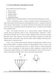
<strong>36</strong>.2 Otolith Distributed Parameter Model<br />
The otoliths are an overdamped second-order system whose structure is shown in Fig. <strong>36</strong>.1. In this model<br />
the otoconial layer is assumed to be a rigid and nondeformable, the gel layer is a deformable layer of<br />
isotropic viscoelastic material, and the fluid endolymph is assumed to be a newtonian fluid. A small<br />
element of the layered structure with surface area dA is cut from the surface, and a vertical view of this<br />
surface element, of width dx, is shown in Fig. <strong>36</strong>.2. To evaluate the forces that are present, free-body<br />
diagrams are constructed of each elemental layer of the small differential strip. See the nomenclature<br />
table for a description of all variables used in the following formulas, and for derivation details see Grant<br />
and colleagues [1984, 1991].<br />
FIGURE <strong>36</strong>.1 Schematic of the otolith organ: (a) Top view showing the peripheral region with differential area dA<br />
where the model is developed. (b) Cross-section showing the layered structure where dx is the width of the differential<br />
area dA shown in the top view at the left.<br />
© 2000 by CRC Press LLC