Bronchiectasis
Bronchiectasis
Bronchiectasis

You also want an ePaper? Increase the reach of your titles
YUMPU automatically turns print PDFs into web optimized ePapers that Google loves.
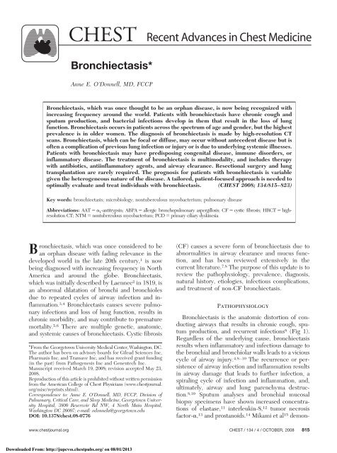
CHEST Recent Advances in Chest Medicine<br />
<strong>Bronchiectasis</strong>*<br />
Anne E. O’Donnell, MD, FCCP<br />
<strong>Bronchiectasis</strong>, which was once thought to be an orphan disease, is now being recognized with<br />
increasing frequency around the world. Patients with bronchiectasis have chronic cough and<br />
sputum production, and bacterial infections develop in them that result in the loss of lung<br />
function. <strong>Bronchiectasis</strong> occurs in patients across the spectrum of age and gender, but the highest<br />
prevalence is in older women. The diagnosis of bronchiectasis is made by high-resolution CT<br />
scans. <strong>Bronchiectasis</strong>, which can be focal or diffuse, may occur without antecedent disease but is<br />
often a complication of previous lung infection or injury or is due to underlying systemic illnesses.<br />
Patients with bronchiectasis may have predisposing congenital disease, immune disorders, or<br />
inflammatory disease. The treatment of bronchiectasis is multimodality, and includes therapy<br />
with antibiotics, antiinflammatory agents, and airway clearance. Resectional surgery and lung<br />
transplantation are rarely required. The prognosis for patients with bronchiectasis is variable<br />
given the heterogeneous nature of the disease. A tailored, patient-focused approach is needed to<br />
optimally evaluate and treat individuals with bronchiectasis. (CHEST 2008; 134:815–823)<br />
Key words: bronchiectasis; microbiology; nontuberculous mycobacterium; pulmonary disease<br />
Abbreviations: AAT 1-antitrypsin; ABPA allergic bronchopulmonary aspergillosis; CF cystic fibrosis; HRCT highresolution<br />
CT; NTM nontuberculous mycobacterium; PCD primary ciliary dyskinesia<br />
<strong>Bronchiectasis</strong>, which was once considered to be<br />
an orphan disease with fading relevance in the<br />
developed world in the late 20th century, 1 is now<br />
being diagnosed with increasing frequency in North<br />
America and around the globe. <strong>Bronchiectasis</strong>,<br />
which was initially described by Laennec2 in 1819, is<br />
an abnormal dilatation of bronchi and bronchioles<br />
due to repeated cycles of airway infection and inflammation.<br />
3,4 <strong>Bronchiectasis</strong> causes severe pulmonary<br />
infections and loss of lung function, results in<br />
chronic morbidity, and may contribute to premature<br />
mortality. 5,6 There are multiple genetic, anatomic,<br />
and systemic causes of bronchiectasis. Cystic fibrosis<br />
*From the Georgetown University Medical Center, Washington, DC.<br />
The author has been on advisory boards for Gilead Sciences Inc,<br />
Pharmaxis Inc, and Transave Inc, and has received grant funding<br />
(in the past) from Pathogenesis Inc and Genentech Inc.<br />
Manuscript received March 19, 2008; revision accepted May 23,<br />
2008.<br />
Reproduction of this article is prohibited without written permission<br />
from the American College of Chest Physicians (www.chestjournal.<br />
org/misc/reprints.shtml).<br />
Correspondence to: Anne E. O’Donnell, MD, FCCP, Division of<br />
Pulmonary, Critical Care, and Sleep Medicine, Georgetown University<br />
Hospital, 3800 Reservoir Rd NW, 4 North Main Hospital,<br />
Washington DC 20007; e-mail: odonnela@georgetown.edu<br />
DOI: 10.1378/chest.08-0776<br />
(CF) causes a severe form of bronchiectasis due to<br />
abnormalities in airway clearance and mucus function,<br />
and has been reviewed extensively in the<br />
current literature. 7,8 The purpose of this update is to<br />
review the pathophysiology, prevalence, diagnosis,<br />
natural history, etiologies, infectious complications,<br />
and treatment of non-CF bronchiectasis.<br />
Pathophysiology<br />
<strong>Bronchiectasis</strong> is the anatomic distortion of conducting<br />
airways that results in chronic cough, sputum<br />
production, and recurrent infections 8 (Fig 1).<br />
Regardless of the underlying cause, bronchiectasis<br />
results when inflammatory and infectious damage to<br />
the bronchial and bronchiolar walls leads to a vicious<br />
cycle of airway injury. 4,8–10 The recurrence or persistence<br />
of airway infection and inflammation results<br />
in airway damage that leads to further infection, a<br />
spiraling cycle of infection and inflammation, and,<br />
ultimately, airway and lung parenchyma destruction.<br />
8,10 Sputum analyses and bronchial mucosal<br />
biopsy specimens have shown increased concentrations<br />
of elastase, 11 interleukin-8, 12 tumor necrosis<br />
factor-, 13 and prostanoids. 14 Mikami et al 15 demon-<br />
www.chestjournal.org CHEST / 134 /4/OCTOBER, 2008 815<br />
Downloaded From: http://jupcvss.chestpubs.org/ on 08/01/2013
strated increased chemotactic activity of sputum in<br />
samples obtained from 19 patients with bronchiectasis<br />
and bronchitis. Systemic markers of inflammation<br />
are also elevated in stable patients with<br />
bronchiectasis. 16 Hence, anatomic factors, chronic<br />
infection and inflammation, and host defense all<br />
play important, yet poorly understood roles in the<br />
development of bronchiectasis. 17<br />
Prevalence<br />
Based on a review of an insurance claim database,<br />
it is estimated that at least 110,000 persons in the<br />
United States are currently being treated for non-CF<br />
bronchiectasis. 9,18 Weycker et al 18 reported a prevalence<br />
in the United States of 4.2 per 100,000 persons<br />
aged 18 to 34 years and 272 per 100,000 persons<br />
among those 75 years of age. <strong>Bronchiectasis</strong> is<br />
being recognized with increasing frequency because<br />
of the widespread use of high-resolution chest CT<br />
(HRCT) scanning. 19 In addition, there are increasing<br />
numbers of patients in nontuberculous mycobacteria<br />
(NTM) lung infections and bronchiectasis have been<br />
diagnosed. 20 Outside of North America, bronchiectasis<br />
is a common clinical problem, but the worldwide<br />
prevalence is also unknown. Tsang and Tipoe 21<br />
reported a prevalence rate of 1 per 6,000 persons in<br />
Auckland, New Zealand, children and a hospital<br />
admission rate of 16.4 per 100,000 population in<br />
Hong Kong. Globally, certain demographic groups<br />
have been recognized as having an increased risk for<br />
the development of bronchiectasis, including individuals<br />
with poor access to health care or high rates<br />
of pulmonary infection in childhood. 4,22<br />
Figure 1. This gross lung specimen from a patient with CF<br />
demonstrates the pathology of bronchiectasis, as follows: dilated<br />
peripheral airways filled with purulent mucus and increased<br />
vascularity that may result in hemoptysis. Image courtesy of<br />
Gwen Huitt, MD, National Jewish Medical and Research Center<br />
(Denver, CO).<br />
Diagnosis<br />
<strong>Bronchiectasis</strong> should be suspected in patients<br />
who present with chronic cough productive of mucopurulent<br />
sputum. Occasionally, a dry nonproductive<br />
cough is the manifesting symptom. Other symptoms<br />
of bronchiectasis include dyspnea, hemoptysis,<br />
and nonspecific constitutional complaints like fatigue<br />
and weight loss. <strong>Bronchiectasis</strong> is more common in<br />
women than in men and, though seen across the age<br />
spectrum, is more frequently encountered in middleaged<br />
and elderly persons. 18 Physical findings in<br />
bronchiectasis patients are nonspecific but may include<br />
crackles and wheezes on lung examination and<br />
clubbing of the digits. Pulmonary function testing<br />
results generally show airflow obstruction ranging<br />
from modest to severe. The diagnosis of bronchiectasis<br />
is confirmed by HRCT scan. Initially described<br />
by Naidich and colleagues 23 in 1982, CT scanning<br />
has replaced contrast bronchography as the current<br />
“gold standard” for the radiologic diagnosis of bronchiectasis.<br />
Plain chest radiography and conventional<br />
CT scanning are insufficiently sensitive for diagnosing<br />
bronchiectasis, but HRCT scanning is able to<br />
detect the airway abnormalities of bronchiectasis.<br />
These include bronchial dilatation (an internal bronchial<br />
diameter greater than the diameter of the<br />
accompanying bronchial artery [ie, the “signet ring”<br />
formation]) and a lack of bronchial tapering on<br />
sequential slices. 24,25 Traditional radiographic descriptions<br />
of bronchiectasis include cylindrical, varicose,<br />
and cystic/saccular (Fig 2); many patients have<br />
elements of all three findings, and cystic bronchiectasis<br />
is associated with a higher likelihood of Pseudomonas<br />
aeruginosa infection and poorer prognosis. 26<br />
The extent of disease seen on HRCT scans has been<br />
correlated with functional change and clinical outcomes.<br />
27,28 Helical, 16 multidetector, HRCT scanning<br />
with narrower collimation and faster acquisition<br />
times compared to conventional HRCT scanning may<br />
eventually supplant HRCT scanning as the optimal<br />
imaging technique for detecting bronchiectasis. 29<br />
Natural History<br />
The clinical course of non-CF bronchiectasis is<br />
variable. Some patients have few to no symptoms,<br />
and others have daily symptoms and progressive loss<br />
of lung function. One large clinical trial 30 showed a<br />
frequency of 1.5 exacerbations per year in patients<br />
from North America, the United Kingdom, and<br />
Ireland who were receiving “usual” care for their<br />
bronchiectasis. In the past few years, two studies 5,31<br />
have demonstrated a decline of approximately 50<br />
mL/yr in FEV 1 in patients with non-CF bronchiectasis.<br />
Factors associated with an accelerated rate of<br />
816 Recent Advances in Chest Medicine<br />
Downloaded From: http://jupcvss.chestpubs.org/ on 08/01/2013
decline of lung function include chronic colonization<br />
by Pseudomonas aeruginosa, a history of severe<br />
exacerbations, and evidence of systemic inflammation.<br />
31 Alzeer et al 32 showed that non-CF bronchiectasis<br />
was associated with cardiac abnormalities,<br />
including right ventricular and left ventricular systolic<br />
and diastolic dysfunction. Mortality due to<br />
bronchiectasis is highest in patients with chronic<br />
hypoxemia, hypercapnia, and radiographic extent of<br />
disease. 33 If admitted to an ICU for respiratory<br />
failure, bronchiectasis patients have a poor prognosis<br />
with a 60% 4-year survival rate found in one cohort. 6<br />
Etiologies<br />
Establishing the cause of bronchiectasis may be<br />
difficult. Even with exhaustive clinical, laboratory,<br />
and pathologic testing, up to 50 to 80% of cases of<br />
bronchiectasis may still be idiopathic. 21,34–36 Several<br />
cohorts of US and UK bronchiectasis patients have<br />
been characterized (Table 1). Nicotra et al 34 reviewed<br />
123 patients with bronchiectasis who were<br />
seen at the University of Texas Health Center at<br />
Tyler and found that they were predominantly white,<br />
female nonsmokers in the sixth decade of life. Most<br />
patients had symptoms for several years prior to<br />
diagnosis, and 30% of patients had no history of lung<br />
injury. Another 35% of patients did have a history of<br />
pneumonia predating by years the onset of bronchiectasis.<br />
Childhood infections, including pertussis,<br />
were thought to have caused 11% of the bronchiectasis<br />
cases, and 10% of cases were related to prior<br />
granulomatous disease. Pasteur et al 35 undertook an<br />
evaluation of 150 bronchiectatic adults in England;<br />
patients were assessed for underlying genetic, immune<br />
deficiency, or immune-mediated disease in<br />
addition to a review of their history. Most patients in<br />
that series 35 were also found to have idiopathic<br />
bronchiectasis, though previously unsuspected ciliary<br />
dysfunction, CF, and allergic bronchopulmonary<br />
aspergillosis (ABPA) were diagnosed. Chronic aspiration<br />
was found to be the etiology in 4% of patients<br />
in that cohort. Recently, another group of 165<br />
bronchiectasis patients who were seen at the Royal<br />
Brompton Hospital in London were characterized<br />
with similar findings. 36<br />
Hence, the etiologies of bronchiectasis can be<br />
categorized as idiopathic, postinfectious, or due to an<br />
underlying anatomic or systemic disease. The large<br />
Figure 2. Top, A: the HRCT scan slice shows cylindrical<br />
bronchiectasis. Center, B: the HRCT scan slice shows varicose<br />
bronchiectasis with a loss of normal bronchial tapering. Bottom,<br />
C: the HRCT scan slice shows cystic/saccular bronchiectasis<br />
changes.<br />
www.chestjournal.org CHEST / 134 /4/OCTOBER, 2008 817<br />
Downloaded From: http://jupcvss.chestpubs.org/ on 08/01/2013
Etiology<br />
cohort of patients with idiopathic bronchiectasis<br />
represents a poorly understood subtype who may<br />
have unrecognized or currently undetectable immunologic<br />
dysfunction or autoimmune abnormalities. 21<br />
Congenital causes of bronchiectasis include CF,<br />
primary ciliary dyskinesia (PCD) and 1-antitrypsin<br />
(AAT) deficiency. Though CF usually presents in<br />
infancy or childhood, adult presentation is not unusual<br />
and should be considered, particularly if there<br />
is a family history of suppurative lung disease. Extrapulmonary<br />
abnormalities such as sinusitis, pancreatic<br />
insufficiency, or infertility may or may not be<br />
present in the adult patient with CF. Sweat chloride<br />
testing is needed to make the diagnosis, and genetic<br />
testing is used to confirm the presence of a mutation<br />
on the CF transmembrane conductance regulator. 7<br />
When 100 Australian bronchiectasis patients were<br />
genetically screened, CF transmembrane conductance<br />
regulator gene mutations were found in 4 of<br />
them, all heterozygotes. 37 PCD is a rare genetic disorder<br />
also causing bronchiectasis, rhinosinusitis, ear infections,<br />
and infertility. About one half of patients with<br />
PCD have situs inversus totalis or heterotaxy, and a<br />
smaller percentage have pectus excavatum. 38 Kennedy<br />
et al 38 have reported on a cohort of 29 adult patients<br />
with PCD; all of them had bronchiectasis. With<br />
regard to AAT deficiency, emphysema is the most<br />
commonly associated pulmonary abnormality. However,<br />
Parr et al 39 have demonstrated that 27% of 74<br />
AAT-deficient patients had HRCT scan evidence of<br />
bronchiectasis.<br />
Immune deficiencies such as primary hypogammaglobulinemia<br />
can contribute to the onset of<br />
bronchiectasis. 40 Ig G subclass deficiencies have<br />
been implicated in bronchiectasis, but the evidence<br />
is mixed, and antibody production deficiency<br />
may need to be present in addition to<br />
decreased levels. 41,42 Rarely, defects of neutrophil<br />
adhesion, respiratory burst, and chemotaxis lead to<br />
bronchiectasis. 35 HIV/AIDS has been associated<br />
with bronchiectasis. 43<br />
Table 1—Etiologies of <strong>Bronchiectasis</strong>*<br />
Nicotra et al 34 /1995<br />
(n 123)<br />
Study/Year, %<br />
Pasteur et al 35 /2000<br />
(n 150)<br />
Shoemark et al 36 /2007<br />
(n 165)<br />
Postinfectious 35 29 32<br />
Idiopathic 30 53 26<br />
Genetic disease (CF, PCD, and AAT deficiency) 4 4.5 11<br />
Aspiration/GERD Not specified 4 1<br />
Immune deficiency Not specified 8 7<br />
Rheumatoid arthritis Not specified 3 2<br />
Ulcerative colitis Not specified 1 3<br />
ABPA<br />
*GERD gastroesophageal reflux disease.<br />
Not specified 7 8<br />
Immune-related diseases such as ABPA, collagen<br />
vascular diseases, and inflammatory bowel diseases<br />
all may contribute to the development of bronchiectasis.<br />
In ABPA patients, the bronchiectasis is typically<br />
central with a “finger-in-glove” distribution and<br />
results in increased cough and sputum production.<br />
<strong>Bronchiectasis</strong> is a complication of several collagen<br />
vascular diseases, notably rheumatoid arthritis and<br />
Sjögren syndrome. 19,44 The pathophysiology of bronchiectasis<br />
in patients with autoimmune diseases is<br />
unknown; there has been speculation about the role<br />
of chronic inflammation, aspiration, or a genetic link<br />
(not yet identified) between the articular and lung<br />
manifestations. 19 <strong>Bronchiectasis</strong> is also an underrecognized<br />
comorbidity in patients with inflammatory<br />
bowel disease. The prevalence is unknown, but many<br />
patients with ulcerative colitis and Crohn disease<br />
have chronic respiratory symptoms due to bronchiectasis.<br />
45 To date, the search for a common pathophysiology<br />
to account for the bowel and lung disease<br />
manifestations has been unsuccessful.<br />
<strong>Bronchiectasis</strong> is being noted with increasing frequency<br />
in patients with COPD and also rarely in<br />
asthma patients. Patel et al 46 found HRCT scan<br />
evidence of bronchiectasis in 50% of a cohort of<br />
stable COPD patients who had a mean FEV 1 of 0.96<br />
L. They found that COPD patients with bronchiectasis<br />
had more severe COPD exacerbations, lower<br />
airway bacterial colonization, and increased levels of<br />
sputum inflammatory markers. A 3% incidence of<br />
bronchiectasis was recently reported 47 in a small<br />
group of patients with asthma, mainly those with<br />
severe persistent asthma. Airway abnormalities such<br />
as endobronchial tumors and foreign bodies can<br />
result in bronchiectasis distal to the obstructing<br />
lesion. Chronic gastric aspiration also may lead to the<br />
development of bronchiectasis. 35 Investigators 48<br />
have attempted to assess whether Helicobacter pylori<br />
might have a pathogenic role in the development<br />
of bronchiectasis, but bronchial biopsy findings were<br />
not confirmative.<br />
818 Recent Advances in Chest Medicine<br />
Downloaded From: http://jupcvss.chestpubs.org/ on 08/01/2013
NTM infections are associated with and may cause<br />
nodular bronchiectasis. 20 The association of right<br />
middle lobe and lingular predominant bronchiectasis<br />
with NTM lung infection was first reported 49 in 1989<br />
and is being increasingly recognized around the<br />
globe. Whether NTM actually causes the bronchiectatic<br />
destruction is still uncertain. Fujita et al 50<br />
demonstrated the presence of Mycobacterium avium<br />
intracellulare complex organisms in bronchiectatic<br />
areas of lung tissue that had been removed from nine<br />
patients who underwent surgery for bronchiectasis,<br />
suggesting that there might be a causative relationship.<br />
Microbiology<br />
Nonenteric Gram-negative bacteria commonly infect<br />
areas of bronchiectasis, though Staphylococcus<br />
aureus and NTM are also commonly encountered as<br />
well (see Table 2 for a summary of the bacteriology<br />
of bronchiectasis). About one third of patients with<br />
bronchiectasis are chronically colonized with P<br />
aeruginosa. Patients with P aeruginosa experience an<br />
accelerated decline in lung function and more frequent<br />
exacerbations. 31 Patients with no pathogens<br />
isolated from their sputum had the mildest disease. 51<br />
The presence of S aureus raises the suspicion for the<br />
presence of CF. 52<br />
Treatment<br />
The goals of bronchiectasis treatment are to reduce<br />
the number of exacerbations and to improve<br />
quality of life (Table 3). If an underlying systemic<br />
etiology is identified and is treatable, then it should<br />
be addressed. For example, Ig replacement for documented<br />
deficiency or steroid therapy for ABPA are<br />
indicated, although it is unclear whether those interventions<br />
alter the natural history of bronchiectasis.<br />
Table 2—Bacteriology of <strong>Bronchiectasis</strong><br />
Organisms<br />
Nicotra<br />
et al 34 /1995<br />
(n 123)<br />
Study/Year, %<br />
Pasteur<br />
et al 35 /2000<br />
(n 150)<br />
King<br />
et al 51 /2007<br />
(n 89)<br />
H influenzae 30 35 47<br />
P aeruginosa<br />
(including mucoid)<br />
31 31 12<br />
Moraxella catarrhalis 2.4 20 8<br />
Streptococcus<br />
pneumoniae<br />
10.6 13 7<br />
S aureus 7.3 14 4<br />
No organism Not specified 23 21<br />
Mycobacterium 17 0 2<br />
Table 3—Potential Therapies for <strong>Bronchiectasis</strong><br />
Treat underlying condition, if possible<br />
Antimicrobial therapy<br />
Pathogen specific<br />
Antiinflammatory therapy<br />
Inhaled steroids<br />
Macrolides<br />
Mobilization of secretions<br />
Pharmacologic<br />
Mechanical<br />
Surgery<br />
Localized or refractory disease<br />
Transplantation<br />
End-stage disease<br />
Antimicrobial therapies should be aimed at identified<br />
pathogens; hence, sputum cultures need to<br />
obtained frequently and antibiotic sensitivity patterns<br />
as well as antibiotic usage need to be monitored.<br />
The reduction of airway inflammation and the<br />
mobilization of airway secretions may be important<br />
components of therapy. Occasionally, surgical resection<br />
is advisable. Transplantation has been performed for<br />
the treatment of end-stage disease. Because of the<br />
heterogeneity of patients with bronchiectasis and the<br />
small number of therapeutic trials, care of the patient<br />
must be individualized.<br />
Antimicrobial Therapy<br />
The role of the use of maintenance antibiotic<br />
therapy is uncertain in patients with non-CF bronchiectasis.<br />
Rotating oral antibiotic strategies have<br />
been commonly used but without evidence from<br />
controlled trials. A retrospective report 53 of 26 patients<br />
with bronchiectasis who were treated with<br />
cycles of alternating antibiotics, including a quinolone,<br />
showed radiographic stability of disease in 77%<br />
of patients; the length of therapy was from 6 to 84<br />
months. An older study 54 of 10 patients who had<br />
been treated with 90 days of oral ciprofloxin<br />
therapy showed decreased numbers of exacerbations,<br />
but resistance developed in 2 patients. Maintenance<br />
therapy with inhaled tobramycin has shown<br />
a microbiological benefit in two studies, 55,56 but<br />
those studies were not powered to detect a clinical<br />
benefit. In the first study, 55 74 patients were randomized<br />
to participate in a 4-week trial of inhaled<br />
tobramycin, 300 mg twice a day, vs taste-masked<br />
placebo, and the treated patients showed decreased<br />
P aeruginosa density in their sputum 2 weeks after<br />
completing therapy. The second study 56 was an<br />
open-label trial of 41 patients who were treated for<br />
three cycles of 2 weeks on/2 weeks off with inhaled<br />
www.chestjournal.org CHEST / 134 /4/OCTOBER, 2008 819<br />
Downloaded From: http://jupcvss.chestpubs.org/ on 08/01/2013
tobramycin, 300 mg twice a day, and again a microbiological<br />
benefit was demonstrated in addition to<br />
improvement in pulmonary symptom scores. A subset<br />
of treated patients in both studies had significant<br />
drug-related pulmonary adverse effects. Other inhaled<br />
antibiotic trials have included a short course of<br />
inhaled gentamicin in a pilot cohort of 28 patients 57<br />
and an open label trial 58 of colistin in 18 patients.<br />
The number of patients in these trials was insufficient<br />
to draw firm conclusions, though they suggested<br />
improvement in levels of inflammatory markers<br />
and pulmonary function. Based on these studies,<br />
there may be a benefit to a maintenance antibiotic<br />
regimen in patients who frequently experience exacerbations,<br />
but the evidence base is relatively weak<br />
and there is concern for the development of antimicrobial<br />
resistance. 59<br />
When bronchiectasis patients experience an exacerbation,<br />
antibiotic therapy should be tailored to<br />
their sputum microbiology results. Mild-to-moderate<br />
exacerbations may be treated with therapy with oral<br />
antibiotics for 2 to 3 weeks, though the optimal<br />
duration of therapy is unknown. Tsang et al 60 compared<br />
therapy with oral levofloxacin to that with IV<br />
ceftazidime in 35 consecutive patients with bronchiectasis<br />
exacerbations, most of whom had P aeruginosa<br />
or Haemophilus influenzae found in their sputum.<br />
There was no difference in clinical outcomes<br />
between the two treatment groups. One trial 61 explored<br />
the addition of inhaled tobramycin to therapy<br />
with oral ciprofloxacin for the treatment of exacerbations<br />
due to ciprofloxacin-sensitive P aeruginosa<br />
infections. This strategy improved the microbiological<br />
outcome but did not confer an additional clinical<br />
benefit over therapy with the oral agent alone, and<br />
treatment-emergent wheezing was noted due to<br />
inhalation of the drug. Severe exacerbations, particularly<br />
in patients who are infected with organisms<br />
that are resistant to therapy with oral quinolones,<br />
may require IV antibiotic therapy in the home or<br />
hospital setting. In light of the paucity of clinical<br />
trials, treatment of exacerbations must be individualized<br />
to the patient and to the infecting organism.<br />
Reduction of Airway Inflammation<br />
Therapy with inhaled corticosteroids and oral<br />
macrolides may reduce airway inflammation in patients<br />
with bronchiectasis. Tsang et al 62 demonstrated<br />
that therapy with inhaled fluticasone reduced<br />
sputum levels of inflammatory markers, and they<br />
have subsequently published a 12-month clinical<br />
trial 63 that showed clinical improvement in patients<br />
who had been treated with 500 g of inhaled<br />
fluticasone twice per day compared to placebo. Of<br />
note, therapy with systemic steroids has never been<br />
studied in a controlled fashion in patients with<br />
non-CF bronchiectasis.<br />
Macrolide antibiotics are thought to have an antiinflammatory<br />
effect in airways diseases such as panbronchiolitis<br />
or bronchiolitis obliterans, despite their<br />
lack of antimicrobial activity against many of the<br />
infecting pathogens. Oral macrolide therapy has<br />
been shown to reduce the 24-h sputum volume and<br />
improve lung function in a pilot study, 64 which<br />
compared erythromycin, 500 mg twice per day,<br />
compared to placebo. A small open label trial 65 of<br />
azithromycin, 500 mg twice per week for 6 months,<br />
also suggested a clinical benefit as the patients had a<br />
decreased number of exacerbations. Although these<br />
pilot studies are provocative, the results must be<br />
viewed with caution because of the small numbers of<br />
patients who have been studied. The risk of increasing<br />
the infectious burden with inhaled steroids or<br />
improperly treating unrecognized mycobacterial infections<br />
with single-agent macrolide therapy must be<br />
weighed against any possible benefit.<br />
Mobilization of Airway Secretions<br />
Pharmacologic agents and the mechanical mobilization<br />
of secretions have been evaluated to a limited<br />
degree in patients with non-CF bronchiectasis.<br />
Short-acting or long-acting bronchodilator adrenergic<br />
and anticholinergic agents are commonly prescribed,<br />
but there have been no randomized controlled<br />
trials to support their use. 66,67 The mucolytic<br />
agent, recombinant human DNase I, had adverse<br />
effects when studied in patients with non-CF bronchiectasis<br />
and hence should not be used as a maintenance<br />
medication. 30 Inhaled mannitol may have a<br />
benefit, but clinical trials have not yet been published<br />
in this patient population. 68 Preliminary results<br />
for nebulized hypertonic saline solution (7%)<br />
have shown promise in the treatment of patients with<br />
both CF and non-CF bronchiectasis, but long-term<br />
prospective trials are needed. 69,70 The use of mechanical<br />
aids, including chest physical therapy with<br />
postural drainage, active cycle of breathing, oscillatory<br />
positive expiratory pressure devices, and highfrequency<br />
assisted airway clearance, also constitute<br />
potential adjunct therapies for patients with bronchiectasis.<br />
71–73 Though these modalities are considered<br />
to be standard therapy for patients with CF bronchiectasis,<br />
their utility is less well proven in patients<br />
with non-CF bronchiectasis. Finally, pulmonary rehabilitation<br />
has been shown to be effective in patients<br />
with bronchiectasis. 74<br />
820 Recent Advances in Chest Medicine<br />
Downloaded From: http://jupcvss.chestpubs.org/ on 08/01/2013
Surgery<br />
Resectional surgery for the treatment of bronchiectasis<br />
may be considered in those patients with focal<br />
disease, in those who do not respond to conventional<br />
management, and in those with uncontrolled hemoptysis<br />
despite the use of interventional radiology<br />
techniques. The complete resection of bronchiectasis<br />
was reported 75 in 118 of 143 young patients with<br />
bronchiectasis (mean age, 23.4 years) with a 23%<br />
morbidity rate and a 1.3% mortality rate. Successful<br />
localized resection has been reported 76 in four hypogammaglobulinemic<br />
patients. End-stage bronchiectasis<br />
has been successfully treated with lung transplantation.<br />
Beirne et al 77 reported a 1-year survival<br />
rate of 68% in bronchiectasis patients who underwent<br />
single-lung and double-lung transplants.<br />
Approach to the Patient With<br />
<strong>Bronchiectasis</strong><br />
Patients in whom bronchiectasis has been diagnosed<br />
should be evaluated for potential underlying<br />
causes. Patients with focal disease may require bronchoscopy<br />
to evaluate for a localized airway obstruction<br />
as the cause of the bronchiectasis. 78 Rarely,<br />
acute pneumonia can result in “pseudobronchiectasis,”<br />
so patients need to undergo an HRCT scan<br />
when they are clinically stable. 79 Patients with diffuse<br />
bronchiectasis should be assessed for underlying<br />
systemic abnormalities including congenital disorders<br />
and immune-mediated dysfunction. <strong>Bronchiectasis</strong><br />
should be suspected in patients with rheumatologic<br />
disorders and inflammatory bowel disease<br />
when they complain of chronic cough. All patients<br />
with bronchiectasis should have a microbiological<br />
examination of their sputum for routine bacterial and<br />
NTM organisms. An individualized plan of therapy<br />
should be devised for all patients with bronchiectasis.<br />
There may be a role for antimicrobial, antiinflammatory,<br />
and airway clearance therapies for<br />
patients with bronchiectasis. Surgery and lung transplantation<br />
are rarely required.<br />
Because there are so few randomized controlled<br />
trials of therapies for non-CF bronchiectasis and<br />
there are no US Food and Drug Administrationapproved<br />
therapies for non-CF bronchiectasis, patients<br />
must be evaluated and treated on an individual<br />
basis. Patients with mild-to-moderate bronchiectasis<br />
who have infrequent exacerbations may need no<br />
maintenance therapy. Patients with more severe<br />
disease may benefit from one or more of the therapeutic<br />
options summarized above.<br />
Conclusions<br />
<strong>Bronchiectasis</strong>, which was once thought to be<br />
decreasing in prevalence, is now resurging in the<br />
developed world and continues to be a common<br />
respiratory disease in areas of the world in which<br />
people have less access to health care. Clinicians<br />
need to be vigilant for patients with bronchiectasis,<br />
so that a tailored clinical evaluation can be performed<br />
to detect underlying causes and an appropriate<br />
multimodality treatment plan can be initiated.<br />
References<br />
1 Barker AF, Bardana EJ. <strong>Bronchiectasis</strong>: update of an orphan<br />
disease. Am Rev Respir Dis 1988; 137:969–978<br />
2 Laennec RTH. A treatise on the disease of the chest. New<br />
York, NY: Library of the New York Academy of Medicine,<br />
Hafner Publishing, 1962; 78<br />
3 Reid LM. Reduction in bronchial subdivision in bronchiectasis.<br />
Thorax 1950; 5:233–247<br />
4 Barker AF. <strong>Bronchiectasis</strong>. N Engl J Med 2002; 346:1383–<br />
1393<br />
5 King PT, Holdsworth SR, Freezer NJ, et al. Outcome in adult<br />
bronchiectasis. COPD 2005; 2:27–34<br />
6 Alzeer AH, Masood M, Basha SJ, et al. Survival of bronchiectatic<br />
patients with respiratory failure in ICU. BMC Pulm<br />
Med 2007; 7:17<br />
7 Strausbaugh SD, Davis PB. Cystic fibrosis: a review of<br />
epidemiology and pathobiology. Clin Chest Med 2007; 28:<br />
279–288<br />
8 Morrissey BM. Pathogenesis of bronchiectasis. Clin Chest<br />
Med 2007; 28:289–296<br />
9 Prasad M, Tino G. <strong>Bronchiectasis</strong>: part 1. Presentation and<br />
diagnosis. J Respir Dis 2007; 28:545–554<br />
10 Cole PJ. Inflammation: a two-edged sword; the model of<br />
bronchiectasis. Eur J Respir Dis Suppl 1986; 147:6–15<br />
11 Tsang KW, Chan K, Ho P, et al. Sputum elastase in steadystate<br />
bronchiectasis. Chest 2000; 117:420–426<br />
12 Richman-Eisenstat JB, Jorens PG, Hebert CA, et al. Interleukin-8:<br />
an important chemoattractant in sputum of patients with<br />
bronchiectasis. Am J Physiol 1993; 264:L413–L418<br />
13 Shum DK, Chan SC, Ip MS. Neutrophil-mediated degradation<br />
of lung proteoglycans: stimulation by tumor necrosis<br />
factor-alpha in sputum of patients with bronchiectasis. Am J<br />
Respir Crit Care Med 2000; 162:1925–1931<br />
14 Tamaoki J, Chiyorani A, Kobayashi K, et al. Effect of<br />
indomethacin on bronchorrhea in patients with chronic bronchitis,<br />
diffuse panbronchiolitis or bronchiectasis. Am Rev<br />
Respir Dis 1992; 145:548–552<br />
15 Mikami M, Llewellyn-Jones CG, Bayley D, et al. The chemotactic<br />
activity of sputum from patients with bronchiectasis.<br />
Am J Respir Crit Care Med 1998; 157:723–728<br />
16 Wilson CB, Jones PW, O’Leary CJ, et al. Systemic markers of<br />
inflammation in stable bronchiectasis. Eur Respir J 1998;<br />
12:820–824<br />
17 Fuschillo S, De Felice A, Baizano G. Mucosal inflammation<br />
in idiopathic bronchiectasis: cellular and molecular mechanisms.<br />
Eur Respir J 2008; 31:396–406<br />
18 Weycker D, Edelsberg J, Oster G, et al. Prevalence and<br />
economic burden of bronchiectasis. Clin Pulm Med 2005;<br />
12:205–209<br />
19 Cohen M, Sahn S. <strong>Bronchiectasis</strong> in systemic disease. Chest<br />
1999; 116:1063–1074<br />
www.chestjournal.org CHEST / 134 /4/OCTOBER, 2008 821<br />
Downloaded From: http://jupcvss.chestpubs.org/ on 08/01/2013
20 Glassroth J. Pulmonary disease due to nontuberculous mycobacterium.<br />
Chest 2008; 133:243–251<br />
21 Tsang KW, Tipoe GL. <strong>Bronchiectasis</strong>: not an orphan disease<br />
in the East. Int J Tuberc Lung Dis 2004; 8:691–702<br />
22 Singleton R, Morris A, Redding G, et al. <strong>Bronchiectasis</strong> in<br />
Alaska native children: causes and clinical courses. Pediatr<br />
Pulmonol 2000; 29:182–187<br />
23 Naidich DP, McCauley DI, Khouri NF, et al. Computed<br />
tomography of bronchiectasis. J Comput Assist Tomogr 1982;<br />
6:437–444<br />
24 McGuiness G, Naidich D. CT of airways disease and bronchiectasis.<br />
Radiol Clin North Am 2002; 40:1–19<br />
25 Pasteur M, Helliwell S, Houghton S, et al. An investigation into<br />
causative factors in patients with bronchiectasis. Am J Respir<br />
Crit Care Med 2000; 162:1277–1284<br />
26 Lynch DA, Newell J, Hale V, et al. Correlation of CT findings<br />
with clinical evaluations in 261 patients with symptomatic<br />
bronchiectasis. AJR Am J Roentgenol 1999; 173:53–58<br />
27 Sheehan RE, Wells AU, Copley SJ, et al. A comparison of<br />
serial computed tomography and functional change in bronchiectasis.<br />
Eur Respir J 2002; 20:581–587<br />
28 Eshed I, Minski I, Katz R, et al. <strong>Bronchiectasis</strong>: correlation of<br />
high resolution CT findings with health-related quality of life.<br />
Clin Radiol 2007; 62:152–159<br />
29 Dodd JD, Souza CA, Muller NL. Conventional high-resolution<br />
CT versus helical high-resolution MDCT in the detection of<br />
bronchiectasis. AJR Am J Roentgenol 2006; 187:414–420<br />
30 O’Donnell AE, Barker AF, Ilowite JS, et al. Treatment of<br />
idiopathic bronchiectasis with aerosolized recombinant human<br />
DNase I. Chest 1998; 113:1329–1334<br />
31 Martinez-Garcia MA, Soler-Cataluna JJ, Perpina-Tordera M,<br />
et al. Factors associated with lung function decline in adult<br />
patients with stable non-cystic fibrosis bronchiectasis. Chest<br />
2007; 132:1565–1572<br />
32 Alzeer AH, Al-Mobeirek AF, Al-Otair HA, et al. Right and<br />
left ventricular function and pulmonary artery pressure in<br />
patients with bronchiectasis. Chest 2008; 133:468–473<br />
33 Onen ZP, Gulbay BE, Sen E, et al. Analysis of the factors<br />
related to mortality in patients with bronchiectasis. Respir<br />
Med 2007; 101:1390–1397<br />
34 Nicotra MB, Rivera M, Dale AM, et al. Clinical, pathophysiologic,<br />
and microbiologic characterization of bronchiectasis<br />
in an aging cohort. Chest 1995; 108:955–961<br />
35 Pasteur MC, Helliwell SM, Houghton SJ, et al. An investigation<br />
into causative factors in patients with bronchiectasis.<br />
Am J Respir Crit Care Med 2000; 162:1277–1284<br />
36 Shoemark A, Ozerovitch L, Wilson R. Aetiology in adult<br />
patients with bronchiectasis. Respir Med 2007; 101:1163–<br />
1170<br />
37 King PT, Freezer NJ, Holmes PW, et al. Role of CFTR<br />
mutations in adult bronchiectasis. Thorax 2004; 59:357–358<br />
38 Kennedy MP, Noone PG, Leight MW, et al. High-resolution CT<br />
of patients with primary ciliary dyskinesia. AJR Am J Roentgenol<br />
2007; 188:1232–1238<br />
39 Parr DG, Guest PG, Reynolds JH, et al. Prevalence and impact<br />
of bronchiectasis in 1-antitrypsin deficiency. Am J Respir Crit<br />
Care Med 2007; 176:1215–1221<br />
40 Bondioni MP, Duse M, Plebani A, et al. Pulmonary and<br />
sinusal changes in 45 patients with primary immunodeficiencies:<br />
computed tomography evaluation. J Comput Assist<br />
Tomogr 2007; 31:620–628<br />
41 Vendrell M, de Gracia J, Rodrigo MJ, et al. Antibody<br />
production deficiency with normal Ig G levels in bronchiectasis<br />
of unknown etiology. Chest 2005; 127:197–204<br />
42 De Gracia J, Rodrigo MJ, Morell F, et al. IgG subclass<br />
deficiencies associated with bronchiectasis. Am J Respir Crit<br />
Care Med 1996; 153:65–655<br />
43 McGuiness G, Naidich D, Garay S, et al. AIDS associated<br />
bronchiectasis: CT features. J Comput Assist Tomogr 1993;<br />
17:260–266<br />
44 McMahon MJ, Swinson DR, Shettar S, et al. <strong>Bronchiectasis</strong><br />
and rheumatoid arthritis: a clinical study. Ann Rheum Dis<br />
1993; 52:776–779<br />
45 Black H, Mendoza M, Murin S. Thoracic manifestations of<br />
inflammatory bowel disease. Chest 2007; 131:524–532<br />
46 Patel IS, Vlahos I, Wilkinson T, et al. <strong>Bronchiectasis</strong>, exacerbation<br />
indices and inflammation in chronic obstructive pulmonary<br />
disease. Am J Respir Crit Care Med 2004; 170:400–<br />
407<br />
47 Oguzulgen IK, Kervan F, Ozis T, et al. The impact of<br />
bronchiectasis in the clinical presentation of asthma. South<br />
Med J 2007; 100:468–471<br />
48 Angrill J, Sanchez N, Agusti C, et al. Does Helicobacter pylori<br />
have a pathogenic role in bronchiectasis? Respir Med 2006;<br />
100:1202–1207<br />
49 Prince DS, Peterson DD, Steiner RM, et al. Infection with<br />
Mycobacterium avium complex in patients without predisposing<br />
conditions. N Engl J Med 1989; 321:863–868<br />
50 Fujita J, Ohtsuki Y, Shigeto E, et al. Pathologic findings of<br />
bronchiectasis caused by Mycobacterium avium intracellulare<br />
complex. Respiratory Med 2003; 97:933–938<br />
51 King PT, Holdsworth SR, Freezer NJ, et al. Microbiologic<br />
follow-up study in adult bronchiectasis. Respir Med 2007;<br />
101:1633–1638<br />
52 Shah PL. Mawdsley S, Nash K, et al. Determinants of chronic<br />
infection with Staphylococcus aureus in patients with bronchiectasis.<br />
Eur Respir J 1999; 14:1340–1344<br />
53 Biewend ML, Waller EA, Aduen JF, et al. Radiographic<br />
evolution and antimicrobial resistance patterns in patients<br />
with bronchiectasis treated with cyclic antibiotics [abstract].<br />
Proc Am Thorac Soc 2005; 2:A176<br />
54 Rayner CF, Tillotson G, Cole PJ, et al. Efficacy and safety of<br />
long-term ciprofloxacin in the management of severe bronchiectasis.<br />
J Antimicrob Chemother 1994; 34:149–156<br />
55 Barker AF, Couch L, Fiel SB, et al. Tobramycin solution for<br />
inhalation reduces sputum Pseudomonas aeruginosa density<br />
in bronchiectasis. Am J Respir Crit Care Med 2000; 162:481–<br />
485<br />
56 Scheinberg P, Shore E. A pilot study of the safety and efficacy<br />
of tobramycin solution for inhalation in patients with severe<br />
bronchiectasis. Chest 2005; 127:1420–1426<br />
57 Lin HC, Cheng HF, Wang CH, et al. Inhaled gentamicin<br />
reduces airway neutrophil activity and mucus secretion in<br />
bronchiectasis. Am J Respir Crit Care Med 1997; 155:2024–<br />
2029<br />
58 Steinfort DP, Steinfort C. Effect of long-term nebulized<br />
colistin on lung function and quality of life in patients with<br />
chronic bronchial sepsis. Intern Med J 2007; 37:495–498<br />
59 Evans DJ, Greenstone M. Long-term antibiotics in the<br />
management of non-CF bronchiectasis: do they improve<br />
outcome? Respir Med 2003; 97:851–858<br />
60 Tsang KW, Chan W-M, Ho P-L, et al. A comparative study on<br />
the efficacy of levofloxacin and ceftazidime in acute exacerbations<br />
of bronchiectasis. Eur Respir J 1999; 14:1206–1209<br />
61 Bilton D, Henig N, Morrissey B, et al. Addition of inhaled<br />
tobramycin to ciprofloxacin for acute exacerbations of<br />
Pseudomonas aeruginosa infection in adult bronchiectasis.<br />
Chest 2006; 130:1503–1510<br />
62 Tsang KW, Ho PL, Lam WK, et al. Inhaled fluticasone<br />
reduces sputum inflammatory indices in severe bronchiectasis.<br />
Am J Respir Crit Care Med 1998; 158:723–727<br />
63 Tsang KW, Tan KC, Ho PL, et al. Inhaled fluticasone in<br />
bronchiectasis: a 12 month study. Thorax 2005; 60:239–243<br />
64 Tsang KW, Ho PI, Chan KN, et al. A pilot study of low-dose<br />
822 Recent Advances in Chest Medicine<br />
Downloaded From: http://jupcvss.chestpubs.org/ on 08/01/2013
erythromycin in bronchiectasis. Eur Respir J 1999; 13:361–<br />
364<br />
65 Cymbala AA, Edmonds LC, Bauer MA, et al. The disease<br />
modifying effects of twice-weekly oral azithromycin in patients<br />
with bronchiectasis. Treat Respir Med 2005; 4:117–122<br />
66 Franco F, Sheikh A, Greenstone M. Short acting 2-agonists<br />
for bronchiectasis. Cochrane Database Syst Rev (database<br />
online). Issue 3, 2003<br />
67 Restrepo RD. Inhaled adrenergics and anticholinergics in<br />
obstructive lung disease: do they enhance mucociliary clearance?<br />
Respir Care 2007; 52:1159–1173<br />
68 Daviskas E, Anderson SD, Eberl S, et al. Effect of increasing<br />
doses of mannitol on mucus clearance in patients with<br />
bronchiectasis. Eur Respir J 2008; 31:765–772<br />
69 Elkins MR, Robinson R, Rose BR, et al. A controlled trial of<br />
long-term inhaled hypertonic saline in patients with cystic<br />
fibrosis. N Engl J Med 2006; 354:229–240<br />
70 Kellett F, Redfern J, Niven RM. Evaluation of nebulised<br />
hypertonic saline (7%) as an adjunct to physiotherapy in<br />
patients with stable bronchiectasis. Respir Med 2005; 99:<br />
27–31<br />
71 Eaton T, Young P, Zeng I, et al. A randomized evaluation of<br />
the acute efficacy, acceptability and tolerability of flutter and<br />
active cycle of breathing with and without postural drainage<br />
in non-cystic fibrosis bronchiectasis. Chron Respir Dis 2007;<br />
4:23–30<br />
72 Myers TR. Positive expiratory pressure and oscillatory positive<br />
expiratory pressure therapies. Respir Care 2007; 52:<br />
1308–1326<br />
73 Chatburn RL. High-frequency assisted airway clearance.<br />
Respir Care 2007; 52:1224–1235<br />
74 Newall C, Stockley RA, Hill SL. Exercise training and<br />
inspiratory muscle training in patients with bronchiectasis.<br />
Thorax 2005; 60:943–948<br />
75 Eren S, Esme H, Avci A. Risk factors affecting outcome and<br />
morbidity in the surgical management of bronchiectasis.<br />
J Thorac Cardiovasc Surg 2007; 134:392–398<br />
76 Cohen AJ, Roifman C, Brendan J, et al. Localised pulmonary<br />
resection for bronchiectasis in hypogammaglobulinaemic patients.<br />
Thorax 1994; 49:509–510<br />
77 Beirne PA, Banner NR, Khaghani A, et al. Lung transplantation<br />
for non-cystic fibrosis bronchiectasis: analysis of a 13 year<br />
experience. J Heart Lung Transplant 2005; 24:1530–1535<br />
78 Bertolani MF, Marotti F, Bergamini BM, et al. Extraction of<br />
a rubber bullet from a bronchus after 1 year: complete<br />
resolution of chronic pulmonary damage. Chest 1999; 115:<br />
1210–1213<br />
79 Agarwal R. <strong>Bronchiectasis</strong> in acute pneumonia: pseudobronchiectasis.<br />
Chest 2007; 132:2054–2055<br />
www.chestjournal.org CHEST / 134 /4/OCTOBER, 2008 823<br />
Downloaded From: http://jupcvss.chestpubs.org/ on 08/01/2013