
Create successful ePaper yourself
Turn your PDF publications into a flip-book with our unique Google optimized e-Paper software.
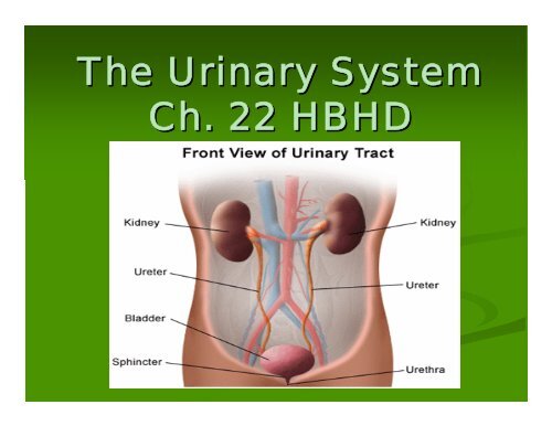
<strong>The</strong> <strong>Urinary</strong> <strong>System</strong><br />
<strong>Ch</strong>. <strong>22</strong> <strong>HBHD</strong>
<strong>Urinary</strong> <strong>System</strong> =<br />
Excretory <strong>System</strong><br />
We are focusing on the urinary system,<br />
but the following systems work<br />
interdependently to maintain homeostasis<br />
<strong>Urinary</strong> <strong>System</strong>: <strong>System</strong>:<br />
excretes water, salts, &<br />
waste products = urine<br />
Digestive <strong>System</strong>: <strong>System</strong>:<br />
excretes water, some<br />
salts, bile, & residue of digestion = feces<br />
Respiratory <strong>System</strong>: <strong>System</strong>:<br />
excretes CO2 & water<br />
(vapor)<br />
Integumentary <strong>System</strong>: <strong>System</strong>:<br />
water, salts &<br />
nitrogenous wastes = perspiration
Structures of the <strong>Urinary</strong><br />
2 Kidneys<br />
<strong>System</strong><br />
Extract waste from blood,<br />
balance body fluids, form<br />
urine<br />
2 Ureters<br />
Transports urine from<br />
kidneys to bladder<br />
<strong>Urinary</strong> Bladder<br />
Receives & stores urine<br />
Urethra<br />
Urethra<br />
Urine elimination
Lie against the<br />
<strong>The</strong> Kidneys<br />
muscles of the back<br />
in the upper<br />
abdomen. <strong>The</strong>y are<br />
protected by the lower<br />
ribs & costal<br />
cartilages<br />
Located in the<br />
retroperitoneal space
Structure:<br />
<strong>The</strong> Kidneys<br />
“Bean Bean” shaped<br />
Measurements:<br />
Measurements:<br />
Length: 4” 4<br />
Width: 2” 2<br />
Thickness: 1”<br />
1
External:<br />
Capsule<br />
Kidney Structure<br />
Capsule: : covers &<br />
protects the kidney; also<br />
has a protective fatty<br />
layer called an adipose<br />
capsule<br />
Hilus: : notch where the<br />
renal artery, renal vein,<br />
and ureters connect with<br />
the kidney<br />
Hilus
Internal<br />
Kidney Structure<br />
Internal<br />
Cortex: Cortex:<br />
outer portion of the<br />
kidney<br />
Medulla: Medulla:<br />
interior portion;<br />
contains tubes that collect urine<br />
(urine formed here)<br />
Renal Pyramids: Pyramids:<br />
cone-shaped<br />
cone shaped<br />
structures – point toward the<br />
renal pelvis<br />
Renal Pelvis: Pelvis:<br />
“funnel funnel-shaped shaped”<br />
forms the upper end of the<br />
ureter<br />
Calyces (calyx): (calyx) : cuplike<br />
extensions that collect urine
Kidney Blood Supply<br />
Renal Artery: supplies<br />
the kidney with blood<br />
Branch of the abdominal<br />
aorta<br />
Branching of vessels to<br />
supply entire kidney<br />
Eventually reaches<br />
nephrons<br />
Nephron: functional unit<br />
of the kidneys<br />
Renal Vein: blood<br />
leaves the kidneys<br />
Carries blood to inferior<br />
vena cava
<strong>The</strong> Microscopic Kidney<br />
Nephron:<br />
Structure<br />
<strong>The</strong> basic unit of the<br />
kidney<br />
Most of the work done<br />
here<br />
Important Facts:<br />
<strong>The</strong>re are approx. 1 million<br />
nephrons in each kidney<br />
If all uncoiled and placed<br />
end-to end to-end end ≈ 75 miles
Structures of a nephron<br />
Bowman’s s capsule:<br />
Bowman<br />
A bulb at the end of the coiled tube of the nephron<br />
Surrounds a cluster a capillaries called the<br />
glomerulus<br />
Glomerulus:<br />
Glomerulus:<br />
A cluster of capillaries inside the Bowman’s Bowman s<br />
capsule<br />
Where water and impurities leave the blood<br />
Afferent arteriole = carries blood TO the glomerulus<br />
Efferent arteriole = carries blood AWAY FROM the<br />
glomerulus
Structures of a nephron<br />
Proximal convoluted tubule:<br />
<strong>The</strong> coiled portion leading from Bowman<br />
<strong>The</strong> coiled portion leading from Bowman’s s capsule<br />
Loop of Henle:<br />
<strong>The</strong> bottom portion of the nephron between the Proximal & Distal<br />
Convoluted Tubules<br />
Distal Convoluted Tubule:<br />
<strong>The</strong> coiled portion farthest from Bowman<br />
<strong>The</strong> coiled portion farthest from Bowman’s s capsule<br />
Excess water is reabsorbed & urine is concentrated<br />
Amount of water reabsorbed is controlled by ADH (antidiuretic<br />
hormone –produced produced by pituitary)<br />
Empties into a collecting duct, which then continues through the<br />
medulla toward the renal pelvis<br />
Juxtaglomerular apparatus:<br />
“near near the glomerulus”<br />
glomerulus<br />
Helps regulate kidney functions by releasing hormones
Kidney Functions<br />
Excretion of unwanted substances<br />
excess salts, toxins, & waste products<br />
from cells<br />
Maintain water balance<br />
pH balance (acids vs. bases)<br />
Production of Hormones<br />
Renin – helps regulate BP<br />
Erthropoietin – stimulates red bone<br />
marrow to produce RBCs in an effort to<br />
prevent anemia
Urine Formation<br />
Glomerular filtration allows all<br />
diffusible material to pass from<br />
the blood to the nephron.<br />
Tubular reabsorption moves<br />
useful materials back to the blood<br />
Tubular secretion moves<br />
additional substances from the<br />
blood to the nephron<br />
Countercurrent mechanism<br />
concentrates the urine and<br />
reduces the volume excreted.<br />
Hormone involved is ADH<br />
(antidiuretic hormone)
<strong>The</strong> Ureters<br />
Long, Slender muscular tubes<br />
that extend from the kidneys to<br />
the bladder<br />
Length is dependent on individual<br />
structure, but it ranges from 10- 10<br />
13” 13<br />
~1” ~1 of the lower ureter enters the<br />
bladder by passing obliquely<br />
through the bladder wall.<br />
Causes a full bladder to compress the<br />
ureters and prevent backflow of urine<br />
Urine is moved from the kidney to<br />
the bladder by gravity &<br />
peristalsis
<strong>The</strong> Bladder<br />
Temporary storage pouch for urine<br />
Moderately full bladder holds about 1 pint<br />
Contains rugae which allow the bladder<br />
to stretch<br />
Empty bladder is firm due to thick<br />
muscular wall<br />
As the bladder fills the muscle wall<br />
becomes thinner and the organ may<br />
increase in size<br />
increase in size<br />
Can stretch 2-5” 2<br />
Has an internal sphincter (which<br />
contracts to prevent urine from escaping<br />
from the bladder involuntarily<br />
Voluntary Control – extrenal sphincter<br />
around the urethra, located below<br />
internal sphincter<br />
Urination: Urination:<br />
(micturition) – the<br />
process of expelling urine from the<br />
bladder
<strong>The</strong> Urethra<br />
<strong>The</strong> tube that extends from the bladder to the<br />
outside of the body<br />
Means by which the bladder is emptied<br />
Male & Female differ<br />
Male: part of both urinary & reproductive systems<br />
Much longer than females<br />
~8 inches : passes through the prostate<br />
Female: ~ 1.5 inches long<br />
Separate from reproductive system<br />
Opens in front of the vagina
Disorders<br />
Can be acute or chronic<br />
Acute: : typically result from infection.<br />
Imflammation of nephrons. Run course in a<br />
few weeks and heel completely<br />
Acute<br />
<strong>Ch</strong>ronic: : arise slowly and can result in<br />
eventual loss of kidney function<br />
<strong>Ch</strong>ronic
Kidney Disorders<br />
Acute Acute glomerulonephritis:<br />
glomerulonephritis<br />
Most common disease of the kidneys.<br />
Usually occurs in children about 1-4 1 4 weeks after a strep<br />
infection<br />
Antibodies injure the glomeruli so that blood and protein<br />
filter into urine<br />
Usually treatable, but can be chronic<br />
Pylonephritis:<br />
Pylonephritis:<br />
Inflammation of renal pelvis and kidney tissue<br />
May be acute or chronic<br />
Cause: passed on by a urinary tract infection<br />
Treatment: antibiotics, fever control, rest, and adequate fluids
Kidney Disorders<br />
Hydronephrosis:<br />
Distention of the renal pelvis and calyces with<br />
accumulated fluid caused by obstruction of urine<br />
flow.<br />
Pregnant women, enlarged prostate, kidney<br />
stones, tumor<br />
Treatment: prompt removal of the obstruction<br />
Polycystic Kidney:<br />
Many fluid-containing fluid containing sacs develop in the active<br />
tissue and gradually destroy functioning parts<br />
Runs in the family<br />
Treatment: transplant or dialysis
Tumors:<br />
Tumors:<br />
Kidney Disorders<br />
Typically slow growing<br />
Signs:<br />
Signs:<br />
Blood in urine<br />
Dull pain in kidney region<br />
Treatment:<br />
Treatment:<br />
Surgical removal of the kidney<br />
<strong>Ch</strong>emo and radiation are not effective
Renal Failure<br />
Acute renal failure:<br />
Sudden, serious decrease in kidney function – may be<br />
fatal<br />
Occurs as a complication of other serious illnesses<br />
<strong>Ch</strong>ronic renal failure:<br />
Due to gradual loss of nephrons resulting in loss of<br />
normal kidney function<br />
Signs & Symptoms: Dehydration, edema, electrolyte<br />
imbalance, hypertension, anemia, uremia<br />
Causes: uremia: accumulation of nitrogenous waste<br />
in blood or renal insufficiency
Kidney Disorders<br />
Kidney Stones:<br />
Kidney Disorders<br />
Calculi: Made of Calcium salts<br />
or uric acid. Precipitate out of the<br />
urine instead of staying in<br />
solution<br />
Calculi:<br />
Formation site: renal pelvis or<br />
bladder<br />
Causes: dehydration, stagnation of<br />
urine, urinary tract infection<br />
Size: variable<br />
Many are impassable so surgery is<br />
required.<br />
Lithotriptor: “stone stone-cracker cracker”
Finish reading pages 408-411, 408 411, beginning with<br />
treatment of kidney failure<br />
Classwork pg 450-451 450 451 # 16-23 16 23