Classical lissencephaly syndromes: doesthe face reflect the brain?
Classical lissencephaly syndromes: doesthe face reflect the brain?
Classical lissencephaly syndromes: doesthe face reflect the brain?

Create successful ePaper yourself
Turn your PDF publications into a flip-book with our unique Google optimized e-Paper software.
920<br />
90Med Genet 1998;35:920-923<br />
Children's Hospital of<br />
Eastern Ontario and<br />
University of Ottawa,<br />
Ottawa, Ontario,<br />
Canada<br />
J E Allanson<br />
Center for Medical<br />
Genetics, University of<br />
Chicago, Chicago, IL,<br />
USA<br />
D H Ledbetter<br />
University of<br />
Minnesota,<br />
Minneapolis, MN, USA<br />
W B Dobyns<br />
Correspondence to:<br />
Dr Allanson, Department of<br />
Genetics, Children's Hospital<br />
of Eastern Ontario, 401<br />
Smyth Road, Ottawa, ON<br />
K1H 8L1, Canada.<br />
Received 30 January 1998<br />
Revised version accepted for<br />
publication 20 April 1998<br />
<strong>Classical</strong> <strong>lissencephaly</strong> <strong>syndromes</strong>: does <strong>the</strong> <strong>face</strong><br />
<strong>reflect</strong> <strong>the</strong> <strong>brain</strong>?<br />
J E Allanson, D H Ledbetter, W B Dobyns<br />
Abstract<br />
Both Miller-Dieker syndrome and isolated<br />
<strong>lissencephaly</strong> sequence are associated<br />
with classical <strong>lissencephaly</strong>. Both<br />
have been shown to be associated with<br />
deletions and mutations in LIS1 on 17p.<br />
Traditionally, <strong>the</strong> two disorders have been<br />
distinguished by <strong>the</strong> presence of a characteristic<br />
facial appearance in Miller-Dieker<br />
syndrome. The forehead is tall and prominent<br />
and may have vertical furrowing.<br />
There is narrowing at <strong>the</strong> temples. Eyes<br />
are widely spaced with upward slanting<br />
fissures. The nose is very short with<br />
anteverted nares. The upper lip is long,<br />
wide, and thick. The ears may have minor<br />
flattening of <strong>the</strong> helices. By contrast, <strong>the</strong>se<br />
features are not seen in isolated <strong>lissencephaly</strong><br />
sequence.<br />
We have measured five children with<br />
Miller-Dieker syndrome (MDS) and 25<br />
children and adolescents with isolated <strong>lissencephaly</strong><br />
sequence (ILS). Z score<br />
(standard deviation score) pattern profiles<br />
have been formulated and compared.<br />
Patients with ILS at all ages show reduced<br />
head circumference, a round head, and a<br />
wide and flat <strong>face</strong> with a broad nose and<br />
widely spaced eyes. The most unexpected<br />
finding is <strong>the</strong> similarity of pattern profiles<br />
ofILS and MDS in <strong>the</strong> age group 6 months<br />
to 4 years. Correlation coefficient is 0.812<br />
(p
The <strong>face</strong> in <strong>lissencephaly</strong> <strong>syndromes</strong><br />
Figure 2 The <strong>face</strong> in isolated <strong>lissencephaly</strong> syndrome.<br />
smaller submicroscopic deletions are observed<br />
in about 40% of children with ILS.7 Recently,<br />
several subjects with ILS have been found to<br />
have point mutations within <strong>the</strong> LIS1 gene.8<br />
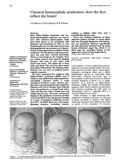
In MDS (fig 1), <strong>lissencephaly</strong> is associated<br />
with a particular facial appearance, consisting<br />
of a tall, prominent forehead, which may show<br />
vertical furrowing in up to one quarter of<br />
affected patients; bitemporal narrowing; widely<br />
spaced eyes with upward slanting palpebral fissures;<br />
a markedly short nose with a low bridge,<br />
anteverted nares, and flared, thin alae; a long<br />
and thick upper lip with rounded philtral pillars<br />
eu eu<br />
zyt<br />
go o go<br />
pr<br />
and thin, inverted, and downward facing<br />
vermilion border; flat mid<strong>face</strong>; small chin; and<br />
flattened ear helices.9 In contrast, no typical<br />
gestalt has been defined in ILS (fig 2).<br />
Material and methods<br />
We have evaluated five subjects with MDS<br />
(aged 3 months to 6 years) and 25 with ILS<br />
(aged 1 to 15 years) by clinical examination. In<br />
addition, a series of craniofacial measurements<br />
was obtained on each subject following <strong>the</strong><br />
method published by Farkas.'" Measurements<br />
were recorded to <strong>the</strong> nearest 0.5 millimetre<br />
using GPM sliding and spreading, blunt ended<br />
calipers, and a paper metric tape measure.<br />
These dimensions were chosen to represent<br />
craniofacial widths, lengths, depths, and circumferences<br />
plus details of ear, eye, nose, and<br />
mouth structure (fig 3). For each dimension,<br />
age and sex matched normal standards were<br />
available. The population norms were derived<br />
from measurements of <strong>the</strong> head and <strong>face</strong> in<br />
2326 healthy North American white children<br />
and young adults."<br />
Measurements were taken by one of <strong>the</strong><br />
authors (JEA). The raw data were compared to<br />
normal standards and converted to Z scores to<br />
control for age and sex differences. Pattern<br />
profiles were compiled for three groups of subjects:<br />
ages 6 months to 4 years, 4 to 9 years, and<br />
16 and over. Correlation coefficients and variability<br />
indices were generated by <strong>the</strong> Statistical<br />
Package for Social Scientists (SPSS), using <strong>the</strong><br />
methods published by Garn et al." 1'3<br />
Results<br />
The first pattern profile divides <strong>the</strong> 25 subjects<br />
with isolated <strong>lissencephaly</strong> sequence into three<br />
age groups: 6 months to 4 years, 4 to 9 years,<br />
and 9 to 16 years (fig 4). There is clear similarity<br />
of patterns, which is validated by <strong>the</strong> high<br />
Figure 3 Craniofacial measurements used in this study. Figure adaptedfrom "Anthropometric facial proportions in<br />
medicine", by Farkas LG and Munro IR, Charles C Thomas, Springfield, Illinois, 1987, with permission.<br />
921
922A22anson,<br />
Ledbetter, Dobyns<br />
Table I Isolated <strong>lissencephaly</strong> syndrome: age group<br />
correlation coefficients<br />
1 2 3<br />
1 1.000 0.7327** 0.5723*<br />
2 0.7327** 1.000 0.8935**<br />
3 0.5723* 0.8935** 1.000<br />
* p
The <strong>face</strong> in <strong>lissencephaly</strong> <strong>syndromes</strong><br />
When <strong>the</strong> cohort with ILS is divided by age,<br />
it is clear that certain craniofacial dimensions<br />
exhibit accelerated growth rates with time.<br />
Maxillary and mandibular depths and circumferences,<br />
in particular, are closer to <strong>the</strong> mean<br />
and may exceed <strong>the</strong> mean with increasing age.<br />
Total facial height becomes more average while<br />
minimal frontal distance exceeds average. Subjectively,<br />
<strong>the</strong>se age related changes are seen as a<br />
transition from <strong>the</strong> round <strong>face</strong> of a young child<br />
to a longer, more oval <strong>face</strong>, with heavy broad<br />
brows, at an older age.<br />
Molecular differences between MDS and<br />
ILS are beginning to emerge which may eventually<br />
explain <strong>the</strong> craniofacial and cerebral<br />
similarities and dissimilarities. Both MDS and<br />
ILS are associated with deletions within<br />
chromosome band 17p 1 3.3.? Visible deletions<br />
or o<strong>the</strong>r structural rearrangements resulting in<br />
deletion are observed in about two-thirds of<br />
children with MDS, while large submicroscopic<br />
deletions are found in almost all <strong>the</strong><br />
remainder.6 In contrast, smaller submicroscopic<br />
deletions are observed in about 40% of<br />
children with ILS.7 The <strong>lissencephaly</strong> minimal<br />
critical region spans approximately 100 to 150<br />
kb and coincides with <strong>the</strong> LIS 1 gene, which is<br />
orientated 5' to 3' from telomere to centromere.<br />
ILS translocation breakpoints and partial deletions<br />
of LIS 1 in ILS and MDS deletion<br />
patients strongly suggest that LIS 1 is <strong>the</strong><br />
<strong>lissencephaly</strong> causative gene. Recently, LIS 1<br />
point mutations have been identified in subjects<br />
with less severe <strong>lissencephaly</strong> and intellectual<br />
handicap.8 Subjective evaluation of <strong>the</strong>se<br />
subjects is completely unremarkable. Objective<br />
phenotypes have yet to be established.<br />
Complete deletions of LIS 1 occur in both<br />
ILS and MDS. Deletions extending several<br />
hundred kilobases centromeric to <strong>the</strong> LIS 1<br />
gene have been documented in two ILS<br />
923<br />
patients but not in any with MDS. Distal<br />
breakpoints of MDS patients are more telomeric<br />
than those of ILS patients. These observations<br />
are consistent with <strong>the</strong> concept of an<br />
additional gene or genes telomeric to LIS1<br />
contributing to <strong>the</strong> facial phenotype of MDS<br />
and do not support <strong>the</strong> hypo<strong>the</strong>sis that LIS 1 is<br />
wholly responsible for <strong>the</strong> facial dysmorphism<br />
seen in MDS.<br />
We are very grateful to <strong>the</strong> families who took part in <strong>the</strong> study<br />
and <strong>the</strong> help of <strong>the</strong> Lissencephaly Network.<br />
1 Dobyns WB, Truwit CL. Lissencephaly and o<strong>the</strong>r malformations<br />
of cortical development: 1995 update. Neuropediatrics<br />
1995;26:132-47.<br />
2 Dobyns WB, Elias ER, Newlin AC, Pagon RA, Ledbetter<br />
DH. Causal heterogeneity in isolated <strong>lissencephaly</strong>. Neurology<br />
1992;42:1375-88.<br />
3 Ledbetter SA, Kuwano A, Dobyns WB, Ledbetter DH.<br />
Microdeletions of chromosome 17p13 as a cause of<br />
isolated <strong>lissencephaly</strong>. Am J7 Hum Genet 1992;50: 182-9.<br />
4 Chong SS, Pack SD, Roschke AV, et al. A revision of <strong>the</strong> <strong>lissencephaly</strong><br />
and Miller-Dieker syndrome critical regions in<br />
chromosome 17pl3.3. Hum Mol Genet 1997;6:147-55.<br />
5 Dobyns WB, Andermann E, Andermann F, et al. X-linked<br />
malformations of neuronal migration. Neurology 1996;47:<br />
331-9.<br />
6 Dobyns WB, Reiner 0, Carrozzo R, Ledbetter DH.<br />
Lissencephaly: a human <strong>brain</strong> malformation associated<br />
with deletion of <strong>the</strong> IJS1 gene located at chromosome<br />
l7pl3.3. JAMA 1993;270:2838-42.<br />
7 Pilz DT, Macha ME, Precht KS, Dobyns WE, Smith ACM,<br />
Ledbetter DH. FISH analysis in 100 patients with isolated<br />
<strong>lissencephaly</strong> sequence (ILS): LIS probes significantly<br />
increase deletion detection rate. Am I Hum Genet 1997;61:<br />
A32.<br />
8 LoNigro C, Chong SS, Smith ACM, Dobyns WB, Ledbetter<br />
DH. Point mutations and an intragenic deletion in<br />
LIS1, <strong>the</strong> <strong>lissencephaly</strong> causative gene, in isolated <strong>lissencephaly</strong><br />
sequence and Miller-Dieker syndrome. Hum Mol<br />
Genet 1997;6: 157-64.<br />
9 Jones KL, Gilbert EF, Kaveggia EG, Opitz JM. The Miller-<br />
Dieker syndrome. Pediatrics 1980;66:277-81.<br />
10 Farkas LG. Anthropometry of <strong>the</strong> head and <strong>face</strong> in medicine.<br />
New York: Elsevier, 1981.<br />
11 Farkas LG. Anthropometry of <strong>the</strong> head and <strong>face</strong>. 2nd ed. New<br />
York: Raven Press, 1994.<br />
12 Gam SM, Smith BH, Lavelle M. Applications of pattern<br />
profile analysis to malformations of <strong>the</strong> head and <strong>face</strong>.<br />
Radiology 1984;150:683-90.<br />
13 Garn SM, Smith BH, Lavelle M. Quantification of<br />
dysmorphogenesis: pattern variability index, Oz. Am J<br />
Radiol 1985;144:365-9.