Molecular Characterization of a 130-kb Terminal ... - ResearchGate
Molecular Characterization of a 130-kb Terminal ... - ResearchGate
Molecular Characterization of a 130-kb Terminal ... - ResearchGate

Create successful ePaper yourself
Turn your PDF publications into a flip-book with our unique Google optimized e-Paper software.
Am. J. Hum. Genet. 60:113-120, 1997<br />
<strong>Molecular</strong> <strong>Characterization</strong> <strong>of</strong> a <strong>130</strong>-<strong>kb</strong> <strong>Terminal</strong> Microdeletion at<br />
22q in a Child with Mild Mental Retardation<br />
Andrew C. C. Wong,' Yi Ning,2 Jonathan Flint,3 Kevin Clark,3 Jan P. Dumanski,4<br />
David H. Ledbetter, * and Heather E. McDermid1<br />
'Department <strong>of</strong> Biological Sciences, University <strong>of</strong> Alberta, Edmonton; 2Diagnostic Development Branch, National Center for Human Genome<br />
Research, National Institutes <strong>of</strong> Health, Bethesda; 3Institute <strong>of</strong> <strong>Molecular</strong> Medicine, John Radcliffe Hospital, Oxford; and 4Department <strong>of</strong><br />
<strong>Molecular</strong> Medicine, Clinical Genetics Unit, Karolinska Institute, Stockholm<br />
Summary<br />
We have analyzed a recently described 22q13.3 microdeletion<br />
in a child with some overlapping features <strong>of</strong> the<br />
cytologically visible 22q13.3 deletion syndrome. Patient<br />
NT, who shows mild mental retardation and delay <strong>of</strong><br />
expressive speech, was previously found to have a paternal<br />
microdeletion in the subtelomeric region <strong>of</strong> 22q. In<br />
order to characterize this abnormality further, we have<br />
constructed a cosmid/P1 contig covering the terminal<br />
150 <strong>kb</strong> <strong>of</strong> 22q, which encompasses the <strong>130</strong>-<strong>kb</strong> microdeletion.<br />
The microdeletion breakpoint is within the<br />
VNTR locus D22S163. The cloning <strong>of</strong> the breakpoint<br />
sequence revealed that the broken chromosome end was<br />
healed by the addition <strong>of</strong> telomeric repeats, indicating<br />
that the microdeletion is terminal. This is the first cloned<br />
terminal deletion breakpoint on a human chromosome<br />
other than 16p. The cosmid/P1 contig was mapped by<br />
pulsed-field gel electrophoresis analysis to within 120<br />
<strong>kb</strong> <strong>of</strong> the arylsulfatase A gene, which places the contig<br />
in relation to genetic and physical maps <strong>of</strong> the chromosome.<br />
The acrosin gene maps within the microdeletion,<br />
-70 <strong>kb</strong> from the telomere. With the distal end <strong>of</strong> chromosome<br />
22q cloned, it is now possible to isolate genes<br />
that may be involved in the overlapping phenotype <strong>of</strong><br />
this microdeletion and 22q13.3 deletion syndrome.<br />
Introduction<br />
Cytogenetically visible deletions <strong>of</strong> 22q13.3 are associated<br />
with generalized developmental delay, normal or<br />
accelerated growth, hypotonia, severe delays in expressive<br />
speech, and mildly dysmorphic facial features<br />
Received July 15, 1996; accepted for publication October 16, 1996.<br />
Address for correspondence and reprints: Dr. Heather E. McDermid,<br />
Department <strong>of</strong> Biological Science, CW 405 Biological Sciences<br />
Centre, University <strong>of</strong> Alberta, Edmonton, AB, T6G 2E9 Canada. Email:<br />
hmcdermi@gpu.srv.ualberta.ca<br />
*Present affiliation: Center for Medical Genetics, The University <strong>of</strong><br />
Chicago, Chicago.<br />
© 1997 by The American Society <strong>of</strong> Human Genetics. All rights reserved.<br />
0002-9297/97/6001-0017$02.00<br />
(Nesslinger et al. 1994). <strong>Molecular</strong> studies <strong>of</strong> seven cases<br />
revealed no correlation between the severity <strong>of</strong> the phenotype<br />
and the proximal extent <strong>of</strong> deletion. The smallest<br />
region <strong>of</strong> overlap between the 22q13.3 deletion patients<br />
(critical region) extends from below locus D22S97 proximally<br />
to below arylsulfatase A (ARSA). Since ARSA is<br />
the most distally mapped locus on chromosome 22q<br />
(Dumanski et al. 1991; Collins et al. 1995), it has not<br />
been possible to assess the extent <strong>of</strong> the deletions below<br />
this locus.<br />
The critical region <strong>of</strong> the 22q13.3 deletion syndrome<br />
covers a genetic distance <strong>of</strong> 25.5 cM (Nesslinger et al.<br />
1994), which represents >a5 Mb as measured by pulsedfield<br />
gel electrophoresis (PFGE) (H. E. McDermid, unpublished<br />
data). This large size therefore makes impractical<br />
a search for genes that may be involved in the<br />
production <strong>of</strong> the phenotype <strong>of</strong> this syndrome. However,<br />
microdeletions within a critical region have been<br />
used in many deletion syndromes to narrow the focus<br />
<strong>of</strong> gene searches (Greger et al. 1993; Huber et al. 1994;<br />
Levy et al. 1995). Such a microdeletion <strong>of</strong> 22q13.3 has<br />
recently been reported. In a study <strong>of</strong> 99 mentally retarded<br />
individuals screened for subtelomeric chromosomal<br />
rearrangements, Flint et al. (1995) described a<br />
12-year-old boy (NT) with delayed expressive speech,<br />
mild mental retardation (IQ = 64), normal facial features,<br />
and a negative family history for mental retardation.<br />
Although his high-resolution karyotype was normal,<br />
the paternal allele <strong>of</strong> the minisatellite probe<br />
D22S163 (MS607) was found to be deleted. ARSA and<br />
other 22q13.3 probes were present in two copies, indicating<br />
that NT carried a microdeletion.<br />
We now describe the molecular characterization <strong>of</strong><br />
this microdeletion, which spans the terminal <strong>130</strong> <strong>kb</strong> <strong>of</strong><br />
22q. A cosmid/P1 contig covering the deleted region was<br />
constructed, allowing the localization <strong>of</strong> the deletion<br />
breakpoint and the mapping <strong>of</strong> the acrosin gene (ACR)<br />
to the region. Analysis <strong>of</strong> the deletion breakpoint revealed<br />
that it is actually within locus D22S163, which<br />
therefore is only partially deleted. The breakpoint was<br />
healed by the addition <strong>of</strong> telomeric sequences. With the<br />
distal end <strong>of</strong> chromosome 22q cloned, it becomes feasible<br />
to isolate the genes that may be involved in the<br />
113
114<br />
overlapping phenotype <strong>of</strong> this microdeletion and<br />
22q13.3 deletion syndrome.<br />
Material and Methods<br />
Cell Lines<br />
The establishment <strong>of</strong> Epstein Barr virus (EBV)-transformed<br />
lymphoblastoid cell lines for NT and his parents<br />
has been described elsewhere (Flint et al. 1995). The<br />
normal control lymphoblastoid cell line (GM03657)<br />
was obtained from the Coriell Human Genetic Mutant<br />
Cell Repository. The fibroblasts from FB, who has the<br />
22q13.3 deletion syndrome (Nesslinger et al. 1994),<br />
were used for comparison <strong>of</strong> the copy number <strong>of</strong> probes<br />
in NT's microdeletion region.<br />
Probes<br />
The following probes were used in this study: CP8, a<br />
partial cDNA <strong>of</strong> ARSA (Herzog et al. 1990); pHU4A,<br />
a cDNA <strong>of</strong> ACR (Baba et al. 1989); D22S163 (probe<br />
MS607), a minisatellite probe (Armour et al. 1990); and<br />
D21S15 and D21S110, two reference probes on chromosome<br />
21 (Stewart et al. 1985; Spinner et al. 1989).<br />
Three probes were produced from cosmid N66C4: a<br />
5.0-<strong>kb</strong> HindIII fragment (H5.0), a 1.2-<strong>kb</strong> XhoI fragment<br />
(Xhl.2), and a 7.0-<strong>kb</strong> HindIIIIEcoRI fragment (HE7.0).<br />
DNA Studies<br />
DNA extraction, digestion, electrophoresis, transfer,<br />
and hybridization were done as described by Nesslinger<br />
et al. (1994). The copy number <strong>of</strong> a probe was preferably<br />
determined by RFLP analysis <strong>of</strong> NT and his parents.<br />
For noninformative probes, quantitative analysis <strong>of</strong><br />
Southern blots was performed, comparing NT to a normal<br />
individual and a 22q13.3 deletion patient (FB). For<br />
each probe, five replicates were analyzed as described<br />
by Nesslinger et al. 1994, by using control probes<br />
D21S15 or D21S110 for comparison.<br />
PFGE<br />
PFGE was performed using a contour-clamped heterogeneous<br />
electrical field-DRII apparatus (BioRad).<br />
Cells from three normal individuals were embedded in<br />
agarose and processed as described by McDermid et<br />
al. (1993). DNA was digested with NotI or NruI and<br />
separated by PFGE at 150 V with a 120-s switch time<br />
for 23 h followed by 180-s switch time for 23 h. The<br />
resulting blot was probed with HE7.0 (fig. 1). The blot<br />
was then stripped <strong>of</strong> signal and reprobed with ARSA.<br />
Construction <strong>of</strong> a Cosmid Contig Spanning the NT<br />
Microdeletion Region<br />
Two cosmid contigs that together span the NT microdeletion<br />
region were constructed independently by<br />
screening two different cosmid libraries (fig. 1). The construction<br />
<strong>of</strong> the "B" contig from the chromosome 22-<br />
A [<br />
B [<br />
1 50<strong>kb</strong> 1 00<br />
I~<br />
_<br />
CEN<br />
_1&. ,.lllllr<br />
H5.0 Xhl.2<br />
8163 HE7.0<br />
L uLUJU<br />
Am. J. Hum. Genet. 60:113-120, 1997<br />
ACR<br />
!-<br />
50 0<br />
11111 1111111 1f (TTAQ00)n<br />
- N65E7<br />
- N66C4<br />
N55A3<br />
- 01220<br />
N94H12<br />
- N1G3<br />
126<br />
- C202<br />
_ 02421<br />
M221<br />
P103<br />
4439(P1)<br />
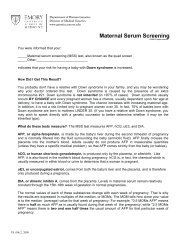
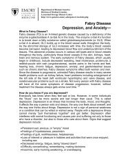
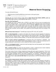
Figure 1 Cosmid/P1 contig, spanning the NT microdeletion.<br />
The upper bar (with the arrow) shows the chromosome, with the<br />
deleted region represented by an open bar. Vertical lines on the chromosome<br />
represent BamHI sites. A, Cosmid contig, constructed by<br />
screening the chromosome 22-specific library LL22NCO3. B, Cosmid<br />
contig, constructed by screening the chromosome 22-specific library<br />
ICRFc1O6 (modified from Ning et al. 1996). In contig B, the full name<br />
<strong>of</strong> each cosmid is "ICRFc1O6," followed by the designations shown<br />
in the figure. Locus D22S163 is abbreviated as "S163." The BamHI<br />
site marked with an asterisk (*) was found only in 44B9.<br />
specific library ICRFc1O6 is described elsewhere (Ning<br />
et al. 1996). It started from a P1 clone containing the<br />
22q telomere and extended proximally. The prefix<br />
ICRFc1O6 has been left <strong>of</strong>f each cosmid name, for brevity.<br />
The "A" contig was begun from overlapping cosmid<br />
clones N85E7 and N66C4, which were isolated from the<br />
chromosome 22-specific library LL22NC03 (De Jong<br />
et al. 1989) by probing with D22S163. Cosmid end<br />
fragments were isolated by detecting SmaI-digested vector-insert<br />
junction fragments with a vector probe on<br />
Southern blots. These vector-insert junction fragments<br />
were then used to identify XhoI and XhoIIHindIII fragments<br />
flanking but not including vector sequences. End<br />
fragments were tested for copy number in NT by RFLP<br />
or densitometric analysis before being used to isolate<br />
overlapping cosmid clones within the microdeletion region.<br />
A BamHI restriction map <strong>of</strong> both contigs was constructed<br />
by using BamHI fragments as probes against<br />
HindIII and/or SmaI cosmid digests electrophoresed and<br />
Southern blotted. This allowed unambiguous assignment<br />
<strong>of</strong> contiguous BamHI fragments.<br />
FISH<br />
Chromosome preparations were made from lymphoblast<br />
cultures by conventional methods. Probe labeling,<br />
suppression <strong>of</strong> repetitive sequences, denaturation, hybridization,<br />
and fluorescence detection were performed
Wong et al.: <strong>Characterization</strong> <strong>of</strong> 22q <strong>Terminal</strong> Microdeletion<br />
as described by Kuwano et al. (1991). DNA from cosmid<br />
P103 was labeled with digoxigenin-1ldUTP by nicktranslation.<br />
A chromosome 22 alpha-satellite probe generated<br />
by using a mouse-human hybrid containing a<br />
single human chromosome 22 (Weier et al. 1991) was<br />
labeled with biotin-16-dUTP and used as a chromosome<br />
22 control. Rhodamine antidigoxigenin and fluorescein<br />
isothiocyanate (FITC) -avidin were used to detect digoxigenin<br />
and biotin-labeled probes, respectively. Slides<br />
were counterstained with 4,6-diamidino-2-phenylindole<br />
(DAPI). Analysis was performed using a Zeiss Axiophot<br />
microscope equipped with filters to detect rhodamine,<br />
FITC, and DAPI separately, as well as a triple band pass<br />
filter set to detect signals simultaneously. Images were<br />
collected and merged using a cooled CCD camera (KAF<br />
1400, Photometrics) and IP Lab Spectrum s<strong>of</strong>tware.<br />
BAL31 Analysis<br />
Agarose-embedded DNA from NT was used to avoid<br />
DNA shearing. Plugs <strong>of</strong> 50 gl were added to the BAL31<br />
digest reaction mix and adjusted to a final volume <strong>of</strong> 1<br />
ml. DNA was digested with 20 U BAL31 (New England<br />
Biolab) at 30'C for 0, 0.5, 1, and 1.5 h. DNA plugs were<br />
then equilibrated in ice-cold 100 mM EGTA (ethylene<br />
glycol-bis[3-Aminoethyl ether] N, N, N', N'-tetraacetic<br />
acid) to inactivate BAL31, followed by an ice-cold TE<br />
(10 mM Tris pH8.0, 1 mM EDTA) wash before digestion<br />
with EcoRV. DNA plugs were then loaded in a<br />
0.6% agarose gel and separated by standard electrophoresis,<br />
Southern blotted, and probed with D22S163.<br />
Cloning and Sequencing <strong>of</strong> PCR Products<br />
The sequence <strong>of</strong> D22S 163 locus (Armour and Jeffreys<br />
1991) was used to design a primer to amplify the region<br />
adjacent to the deletion breakpoint. A 25-nt forward<br />
primer (5' GTGACTTGACTTCTCTGAACCTTGG 3')<br />
was used in conjunction with a reverse primer containing<br />
a telomeric sequence (5' TATGGATCCCTA-<br />
ACCCTAACCCTAACCC 3'). Amplification conditions<br />
were 30 cycles <strong>of</strong> 94°C (1 min), 60°C (1 min), and<br />
72°C (3 min) in 100-gl reaction with Taq polymerase<br />
purchased from Boehringer Mannheim and the manufacturer's<br />
buffer. PCR products were cloned into M13<br />
and sequenced with forward and reverse primers on an<br />
ABI 373 sequencing machine. Six independent clones<br />
were sequenced in both orientations. Normal sequence<br />
was obtained by sonicating the cosmid<br />
N66C4, size separating the fragments, and subcloning<br />
a 2-<strong>kb</strong> fraction into M13. The M13 library was plated<br />
out, and plaques were transferred to Hybond N (Amersham<br />
International) nylon membrane. Filters were<br />
then hybridized, with the forward primer used for<br />
PCR and positive plaques sequenced on the ABI sequencer.<br />
Results<br />
PFGE Localizes the NT Microdeletion Region to within<br />
120 <strong>kb</strong> <strong>of</strong> ARSA<br />
Flint et al. (1995) detected a paternal deletion <strong>of</strong> locus<br />
D22S163 (MS607) in NT but not <strong>of</strong> ARSA or other<br />
22q13.3 probes. D22S163 had previously been mapped<br />
to 22q by somatic cell hybrid analysis and linked to<br />
CRI-L1272 (D22S18) by genetic analysis (Armour et al.<br />
1990). However, the exact location <strong>of</strong> D22S163 was<br />
unknown. In order to determine the location <strong>of</strong><br />
D22S163 relative to ARSA, the most distal probe in<br />
chromosome 22 genetic and physical maps (Dumanski<br />
et al. 1991; Collins et al. 1995), we used PFGE <strong>of</strong> DNA<br />
from normal individuals. The resulting blots were<br />
probed with HE7.0, a fragment from a cosmid (N66C4)<br />
containing D22S163 (fig. 2A). The resulting NotI and<br />
NruI banding patterns were identical to the same blot<br />
stripped and probed with ARSA (fig. 2B). The smallest<br />
NotI band (120 <strong>kb</strong>, arrow) gives a maximal distance<br />
between the two probes. Densitometric analysis (Flint<br />
et al. 1995) and FISH (Ning et al. 1996) results both<br />
indicate that ARSA is not deleted in NT. Therefore, the<br />
NT microdeletion is within 120 <strong>kb</strong> <strong>of</strong> ARSA, although<br />
these results did not indicate whether it lays proximally<br />
or distally.<br />
Production <strong>of</strong> a Cosmid Contig from D22S163/P1 to<br />
the 22q Telomere<br />
To characterize the NT microdeletion fully, we constructed<br />
contigs that span the deleted region. One cosmid<br />
walk started from a P1 clone containing the 22q<br />
telomere and walked proximally (fig. 1, contig B) (Ning<br />
et al. 1996). A second cosmid walk (fig. 1, contig A)<br />
started from the cosmid clones N85E7 and N66C4,<br />
Not1 190<strong>kb</strong>.<br />
Notl 120<strong>kb</strong>.<br />
A HE7.0 B ARSA<br />
r-Noti-m P-Nrul-r Noti-, -N<br />
115<br />
-Nrul 270<strong>kb</strong><br />
-Nrul 210<strong>kb</strong><br />
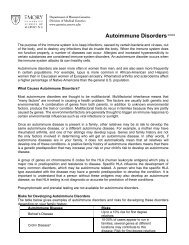
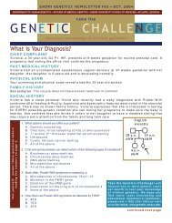
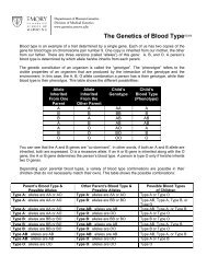
Figure 2 Localization <strong>of</strong> the NT microdeletion to within 120<br />
<strong>kb</strong> <strong>of</strong> ARSA. DNA from three normal individuals was digested with<br />
NotI or NruI and was then separated by PFGE under conditions stated<br />
in Material and Methods. The resulting blot was probed with the<br />
HE7.0 fragment (fig. 2) from N66C4 (A). The blot was then stripped<br />
<strong>of</strong> signal and reprobed with ARSA (B). The arrows indicate the smallest<br />
Nod band (120 <strong>kb</strong>) containing both probes. Bands were sized by<br />
comparison to lambda concatamers.
116<br />
which were isolated using a probe from D22S163. End<br />
fragments <strong>of</strong> these cosmids were tested by densitometric<br />
or RFLP analysis, to assess copy number in NT. Densitometric<br />
analysis showed that the proximal end <strong>of</strong> N85E7<br />
was not deleted in NT (data not shown). However, the<br />
distal end fragment <strong>of</strong> N66C4 (Xhl.2, fig. 1) showed a<br />
paternal deletion by RFLP analysis (fig. 3). The cosmid<br />
walk was therefore extended from the distal end <strong>of</strong><br />
N66C4 for three rounds <strong>of</strong> end-fragment walking. The<br />
two contigs overlap by about three cosmid lengths<br />
('100 <strong>kb</strong>) and span '150 <strong>kb</strong> between the proximal<br />
end <strong>of</strong> N85E7 and the 22q telomere. Since ARSA was<br />
not present by hybridization within the combined contig,<br />
D22S163 maps distal to ARSA. This also places<br />
ARSA at a maximum distance <strong>of</strong> 250 <strong>kb</strong> (120 <strong>kb</strong> + <strong>130</strong><br />
<strong>kb</strong>) from the 22q telomere.<br />
FISH Indicates That the NT Microdeletion Is <strong>Terminal</strong><br />
When the cosmid walk reached the N94H12/N1G3<br />
region, densitometric analysis could no longer be used<br />
to determine the copy number in NT; presumably this<br />
is because <strong>of</strong> the presence <strong>of</strong> the subtelomeric repeats<br />
shared with other chromosomes (Ning et al. 1996). As<br />
a result, FISH was used to test for deletion in NT. The<br />
most distal cosmid <strong>of</strong> the contig (P103) was labeled and<br />
hybridized to NT metaphase spreads, in combination<br />
with the chromosome 22 alpha-satellite probe to mark<br />
the centromere <strong>of</strong> both chromosome 22s. P103 hybridized<br />
to many chromosome ends, as well as 2q13, the<br />
fusion site <strong>of</strong> two ancestral chromosomes (Ijdo et al.<br />
1991). Only one chromosome 22 hybridized to P103<br />
(fig. 4). This result indicates that the entire cosmid contig<br />
is deleted in NT distal to D22S163.<br />
Localization <strong>of</strong> the Breakpoint <strong>of</strong> the NT Microdeletion<br />
Since the proximal end <strong>of</strong> N85E7 was not deleted,<br />
while the distal end <strong>of</strong> N66C4 showed a paternal dele-<br />
L0j<br />
-2.7 <strong>kb</strong><br />
-1.3 <strong>kb</strong><br />
-0.9 <strong>kb</strong><br />
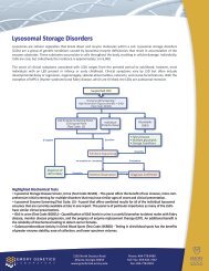
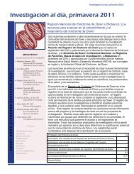
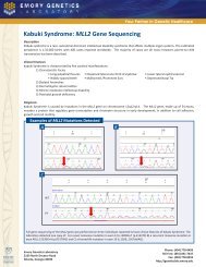
Figure 3 RFLP analysis, showing that Xhl.2 is deleted in NT.<br />
The autoradiograph shows the NT family genomic DNA digested with<br />
TaqI and probed with Xhl.2. Each individual shows a constant 2.7<strong>kb</strong><br />
band. The father is homozygous for the 0.9-<strong>kb</strong> allele, and the<br />
mother has both the 1.3-<strong>kb</strong> and 0.9-<strong>kb</strong> allele. NT inherited the 1.3<strong>kb</strong><br />
allele from the mother but no allele from the father.<br />
Am. J. Hum. Genet. 60:113-120, 1997<br />
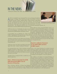
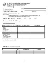
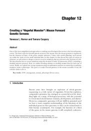
Figure 4 FISH, showing that the most distal cosmid in the contig<br />
is deleted in NT. A chromosome 22-specific alpha-satellite probe,<br />
appearing as green (FITC) centromeric signals, was used to identify<br />
the two chromosome 22 homologues. Cosmid P103, appearing as red<br />
signal (rhodamine), was observed on one homologue and deleted on<br />
the other homologue (arrows). Since cosmid P103 contains subtelomeric<br />
repetitive sequences, the red signals are also present on some<br />
telomeres <strong>of</strong> other chromosomes as well as chromosome 2q13 (arrow<br />
head).<br />
tion by RFLP analysis, these results indicated that the<br />
breakpoint <strong>of</strong> the NT microdeletion is within the N66C4<br />
and N85E7 region. An additional probe (H5.0), which<br />
is 6.5 <strong>kb</strong> proximal to D22S163 (fig. 1), showed no deletion<br />
by densitometric analysis (data not shown), further<br />
narrowing down the breakpoint. In order to look for<br />
DNA rearrangement fragments indicative <strong>of</strong> the<br />
breakpoint, genomic DNAs from a normal control and<br />
the NT family were digested with HindIII, SmaI, EcoRV,<br />
and NgoMI, electrophoresed, blotted, and hybridized to<br />
the D22S163 probe. Novel smeared bands in the 9-11<strong>kb</strong><br />
range were present in HindIII and SmaI digestions<br />
<strong>of</strong> NT DNA (fig. 5). This smear is characteristic <strong>of</strong><br />
breakpoint-junction fragments fused with telomeric sequences<br />
that are heterogeneous in length (Wilkie et al.<br />
1990). D22S163 also detected the presence <strong>of</strong> a large<br />
rearrangement band in NT with EcoRV and NgoMI<br />
digests (fig. 5). The presence <strong>of</strong> these wide rearrangement<br />
bands suggested that NT has terminal deletion and<br />
that the breakpoint is within D22S163. One discrepancy<br />
between this study and that <strong>of</strong> Flint et al. (1995) is<br />
that we detected HindIII-, SmaI-, EcoRV-, and NgoMIrearrangement<br />
fragments with the D22S163 probe,<br />
while Flint et al. (1995) detected a paternal deletion with<br />
the same probe, using Sau3AI. We hypothesized that<br />
under normal electrophoretic conditions, the smaller<br />
Sau3AI-smeared rearrangement band could be diffused
Wong et al.: <strong>Characterization</strong> <strong>of</strong> 22q <strong>Terminal</strong> Microdeletion<br />
23.1 <strong>kb</strong>-<br />
9.4<strong>kb</strong>-<br />
6.5<strong>kb</strong>-<br />
rHindlill 1-SmaI rEcoRVn rNgoMI, A<br />
O0 O0 OL-j<br />
Figure 5 Rearrangement bands, detected by the D22S163<br />
probe. The autoradiograph shows the NT family genomic DNA digested<br />
with HindIII, SmaI, EcoRV, and NgoMI, followed by hybridization<br />
to the D22S163 probe. Novel smeared bands in the 9-11-<strong>kb</strong><br />
range were present in HindIII and SmaI digest <strong>of</strong> NT DNA (arrows).<br />
SmaI also identifies a polymorphic locus with a 9.4-<strong>kb</strong> allele in NT<br />
and his mother and 8.0/9.0-<strong>kb</strong> alleles in the father, which confirms<br />
the paternal deletion <strong>of</strong> NT. D22S163 also detected the presence <strong>of</strong><br />
large rearrangement bands in NT with EcoRV and NgoMI (arrows).<br />
Slight differences in the apparent size <strong>of</strong> the large bands common to<br />
all individuals are due to electrophoresis artifact.<br />
to the point <strong>of</strong> being not visible with a standard exposure<br />
(fig. 6A). We therefore repeated the Sau3AI digestions<br />
<strong>of</strong> the NT family DNAs and electrophoresed the<br />
fragments over a short distance to maximize the chance<br />
<strong>of</strong> seeing a smeared rearrangement band. Under these<br />
conditions, a novel smeared band was seen in NT (fig.<br />
6B), indicating that D22S163 contains the breakpoint<br />
rather than being deleted. This also confirms the explanation<br />
given by Flint et al. (1995) as to why cosmids<br />
containing D22S163 did not show a deletion for NT<br />
when FISH was used, since only part <strong>of</strong> the cosmid is<br />
deleted.<br />
BAL31 Analysis<br />
If NT is a terminal deletion, then the rearrangement<br />
fragment should be sensitive to BAL31 exonuclease digestion.<br />
When agarose-embedded NT DNA was subjected<br />
to BAL31 digest from 0 to 1.5 h, followed by<br />
EcoRV digestion, the D22S163 probe detected a reduction<br />
in the size <strong>of</strong> the rearrangement band (fig. 7). The<br />
larger, normal band detected by D22S163 served as a<br />
control to show that the genomic DNA was intact. Since<br />
D22S163 is normally located <strong>130</strong> <strong>kb</strong> from the telomere,<br />
it is not sensitive to BAL31 digestion on a normal chromosome.<br />
PCR Amplification <strong>of</strong> the Breakpoint Junction<br />
Sequence<br />
To determine the sequence at the breakpoint <strong>of</strong> the<br />
NT microdeletion, DNA was amplified by PCR by using<br />
a D22S163 locus-specific primer and a primer that contains<br />
three telomeric repeats. PCR products were then<br />
cloned, and the breakpoint was sequenced (fig. 8A) and<br />
compared with a corresponding normal chromosome 22<br />
Z0<br />
-5.6<strong>kb</strong><br />
_4.6<strong>kb</strong><br />
.3.5<strong>kb</strong><br />
-3.4<strong>kb</strong><br />
B<br />
LI-<br />
117<br />
-5.8<strong>kb</strong><br />
-4.6<strong>kb</strong><br />
.3.5<strong>kb</strong><br />
3.4<strong>kb</strong><br />
Figure 6 Sau3AI-digested NT family genomic DNA, hybridized<br />
to the D22S163 probe under two different electrophoretic conditions.<br />
In A, using conditions reported in Flint et al. (1995), NT appears to<br />
inherit one 5.8-<strong>kb</strong> allele from his mother but no allele from his father,<br />
suggesting a paternal deletion <strong>of</strong> D22S163 in NT. In B, electrophoresing<br />
the DNA over a much shorter distance and exposing the autoradiograph<br />
for a longer time, D22S163 detected a smeared rearrangement<br />
band in NT, indicating the microdeletion breakpoint is close to, or<br />
within, D22S163.<br />
sequence (fig. 8B). The breakpoint was located within<br />
the D22S163 locus and can be identified by the presence<br />
<strong>of</strong> telomeric repeats (ttaggg, in fig. 8A) substituted for<br />
the normal sequence. This result agrees with the localization<br />
<strong>of</strong> the breakpoint by Southern blot and BAL31<br />
analyses. D22S163 consists <strong>of</strong> two distinct minisatellite<br />
arrays, namely, MS607A and MS607B (Armour and<br />
Jeffreys 1991). The flanking sequence surrounding the<br />
MS607A minisatellite array has been determined for<br />
500 bp proximally (European <strong>Molecular</strong> Biology Library<br />
[EMBLI accession number X58043) and 866 bp<br />
distally (EMBL accession number X58044). The location<br />
<strong>of</strong> the breakpoint is 26 bp distal to the 3' end <strong>of</strong><br />
the 866-bp flanking sequence. Therefore, the breakpoint<br />
is 892 bp (866 bp + 26 bp) distal to the MS607A. The<br />
distance between minisatellite MS607A and MS607B is<br />
0 0.5 1 1.5<br />
-_ 23.1 <strong>kb</strong><br />
---12.2<strong>kb</strong><br />
Figure 7 BAL31 sensitivity <strong>of</strong> the rearrangement band in NT.<br />
Genomic DNA from NT was digested with BAL31 before digestion<br />
with EcoRV. The number at the top <strong>of</strong> each lane indicates the incubation<br />
time <strong>of</strong> BAL31 (in hours). DNA was then separated by standard<br />
electrophoresis in a 0.6% agarose gel. The resulting blot probed with<br />
D22S163 shows a decrease in size <strong>of</strong> the rearrangement band after<br />
BAL31 digestion. The positions <strong>of</strong> DNA size markers from lambda<br />
phage digested with HindIII and the 1-<strong>kb</strong> ladder (BRL) are shown on<br />
the right.
118<br />
A5'AGGGGGTGGAGAGGGGGGTGGTGGAGagggttagggttagggtta3'<br />
B5'AGGGGGTGGAGAGGGGGGTGGTGGAGGGGGGTGGTGGCACAGGGG3'<br />
Figure 8 Sequence at the NT microdeletion breakpoint, compared<br />
to that <strong>of</strong> a normal chromosome. A, Breakpoint sequence <strong>of</strong><br />
NT. The 5' end <strong>of</strong> the sequence starts from the first base after the<br />
866-bp 3' flanking sequence <strong>of</strong> the MS607A minisatellite at the<br />
D22S163 locus. The breakpoint is within D22S163, identified by the<br />
presence <strong>of</strong> telomeric repeats (ttaggg, in small letters) substituted for<br />
the normal sequence. B, Sequence <strong>of</strong> a normal individual.<br />
not known, but an upper size is estimated to be 1 <strong>kb</strong><br />
(J. A. L. Armour, personal communication). If the NT<br />
deletion breakpoint is 891 bp distal to MS607A, it is<br />
either at the 5' end or within the MS607B minisatellite.<br />
Localization <strong>of</strong> ACR to the NT Microdeletion<br />
PFGE studies <strong>of</strong> the region indicated that ARSA,<br />
D22S163, and the ACR locus all mapped to a 190-<strong>kb</strong><br />
NotI fragment (data not shown). When the ACR cDNA<br />
probe was hybridized to a Southern blot containing<br />
BamHI digests <strong>of</strong> all the cosmid DNAs, fragments <strong>of</strong><br />
4.6, 6.9, and 17 <strong>kb</strong> in cosmids N94H12, 126, and C202<br />
were positive. The BamHI fragment pattern matched<br />
the ACR restriction map reported by Vazquez-Levin et<br />
al. (1992). Therefore, the ACR locus maps to the NT<br />
microdeletion, 60 <strong>kb</strong> distal to D22S163 and -70 <strong>kb</strong><br />
from the telomere (fig. 1). Densitometric analysis confirmed<br />
that ACR is deleted in NT (fig. 9), as well as the<br />
in 22q13.3 deletion patient FB.<br />
Discussion<br />
We have shown that NT carries a terminal microdeletion<br />
<strong>of</strong> the last <strong>130</strong> <strong>kb</strong> <strong>of</strong> chromosome 22q. Cloning<br />
<strong>of</strong> the breakpoint sequence and BAL31 analysis <strong>of</strong> rearrangement<br />
fragments suggest that the deletion<br />
breakpoint was healed by the addition <strong>of</strong> telomeric<br />
repeats. This explains the characteristic smeared rearrangement<br />
bands seen with HindIII, SmaI, and Sau3AI.<br />
The healing <strong>of</strong> broken human chromosomes by the<br />
addition <strong>of</strong> telomeric repeats has been described in<br />
individuals with x-thalassemia as a result <strong>of</strong> terminal<br />
16pl3.3 deletions (Wilkie et al. 1990; Lamb et al.<br />
1993; Flint et al. 1994). However, the NT microdeletion<br />
is the first case <strong>of</strong> a cloned terminal deletion<br />
breakpoint on a human chromosome other than 16p.<br />
The cosmid/P1 contig spanning the NT microdeletion<br />
serves to anchor the physical map <strong>of</strong> chromosome 22.<br />
PFGE analysis indicates that this contig lies 6 120 <strong>kb</strong><br />
from ARSA, previously the most distally mapped locus<br />
on chromosome 22 (Dumanski et al. 1991; Collins et<br />
al. 1995). The order <strong>of</strong> known loci at the end <strong>of</strong> 22q is<br />
therefore ARSA-D22S163-ACR-telomere. This confirms<br />
the subtelomeric location <strong>of</strong> the VNTR at D22S1 63 used<br />
for detecting cryptic 22q terminal rearrangements (Flint<br />
et al. 1995).<br />
Am. J. Hum. Genet. 60:113-120, 1997<br />
A patient with the 22q13.3 deletion syndrome (FB)<br />
was also shown to have a deletion <strong>of</strong> ACR (fig. 9). This<br />
suggests that FB also has a terminal deletion. Subsequent<br />
analyses <strong>of</strong> other 22q13.3 deletion syndrome patients<br />
have shown deletions <strong>of</strong> D22S163 in all cases tested<br />
(H. E. McDermid, unpublished data). Thus, the NT microdeletion<br />
overlaps and most likely falls entirely within<br />
the critical region for this syndrome. Although it is possible<br />
that the 22q13.3 microdeletion is not related to the<br />
phenotype <strong>of</strong> NT, this region may contain genes critical<br />
to the phenotypic features <strong>of</strong> NT that overlap with the<br />
22q13.3 deletion syndrome: specifically, delayed speech<br />
and mental retardation.<br />
The only gene mapped to the microdeletion to date<br />
is ACR, a serine protease present in the acrosome <strong>of</strong><br />
the sperm head (Klemn et al. 1991). Protease inhibition<br />
studies suggest that ACR plays a key role in the acrosome-reacted<br />
binding <strong>of</strong> sperm to the zona pellucida and<br />
the penetration <strong>of</strong> sperm into the oocyte (Liu and Baker<br />
1993; Takano et al. 1993). It is highly unlikely that<br />
the deletion <strong>of</strong> ACR is related to any present visible<br />
phenotype <strong>of</strong> NT. However, the reduction <strong>of</strong> ACR activity<br />
in sperm has been associated with male infertility<br />
(Mohsenian et al. 1982; Koukoulis et al. 1989), suggesting<br />
that the deletion <strong>of</strong> ACR may affect the fertility<br />
<strong>of</strong> NT and male 22q13.3 deletion patients.<br />
We are currently searching for other genes deleted in<br />
NT. Subtelomeric regions <strong>of</strong> chromosomes are believed<br />
to be gene-rich (Saccone et al. 1992) and may have an<br />
average <strong>of</strong> one gene every 23.4 <strong>kb</strong> (Fields et al. 1993).<br />
We have cloned <strong>130</strong> <strong>kb</strong> from the 22q telomere; however,<br />
not all <strong>of</strong> this region will likely contain genes. FISH<br />
results indicate that cosmid P103 (fig. 1) cross-hybridizes<br />
with 2q13 and many chromosome ends and therefore<br />
contains subtelomeric repeats (fig. 4; Ning et al.<br />
co<br />
E<br />
0<br />
z NT FB<br />
-ACR (6.4<strong>kb</strong>)<br />
- D21Si15 (2.0<strong>kb</strong>)<br />
Figure 9 Densitometric analysis <strong>of</strong> ACR. The copy number <strong>of</strong><br />
ACR was compared by densitometric analysis <strong>of</strong> TaqI-digested DNA<br />
from a normal individual, NT, and FB. The ACR gene probe was<br />
compared to a reference probe from chromosome 21 (D21S15). Both<br />
NT and FB indicate the presence <strong>of</strong> only one copy <strong>of</strong> ACR.
Wong et al.: <strong>Characterization</strong> <strong>of</strong> 22q <strong>Terminal</strong> Microdeletion 119<br />
1996). Dosage analysis using the end fragment <strong>of</strong><br />
N94H12 as a probe was unreliable, presumably because<br />
<strong>of</strong> the presence <strong>of</strong> subtelomeric repeats shared with other<br />
chromosomes. Although the NT microdeletion encompasses<br />
the terminal <strong>130</strong> <strong>kb</strong> <strong>of</strong> 22q, we estimate that at<br />
least the distal 60 <strong>kb</strong> (from the telomere to the end<br />
<strong>of</strong> N94H12) is probably rich in subtelomeric repeats.<br />
Therefore, only the proximal 70 <strong>kb</strong> may contain genes.<br />
Since the ACR locus has already been mapped to this<br />
region, we expect there are at least one to two genes in<br />
the microdeletion region yet to be mapped.<br />
Acknowledgments<br />
The authors wish to thank Richard Wintle, for providing<br />
his BAL31 digestion protocol; Nikolaus Blin, for the ARSA<br />
CP8 clone; Tadashi Baba, for the ACR cDNA clone; Marcia<br />
Budarf, for providing somatic cell hybrid information about<br />
the location <strong>of</strong> ACR; and John Armour, for sharing the information<br />
about the sequence within the D22S163 locus. This<br />
research was funded by the Children's Health Foundation <strong>of</strong><br />
Northern Alberta and the Canadian Genome Analysis and<br />
Technology Program.<br />
References<br />
Armour JAL, Jefferys AC (1991) STS for minisatellite MS607<br />
(D22S163) Nucleic Acids Res 19:3158<br />
Armour JAL, Povey S, Jeremiah S, Jeffreys AJ (1990) Systematic<br />
cloning <strong>of</strong> human minisatellites from ordered array<br />
charomid libraries. Genomics 8:501-512<br />
Baba T. Watanabe K, Kashiwabara S-I, Arai Y (1989) Primary<br />
structure <strong>of</strong> human proacrosin deduced from its cDNA<br />
sequence. FEBS Lett 244:296-300<br />
Collins JE, Cole CG, Smink LJ, Garrett CL, Leversha MA,<br />
Soderlund CA, Maslen GL, et al (1995) A high-density YAC<br />
contig map <strong>of</strong> human chromosome 22. Nature Suppl 377:<br />
267-379<br />
De Jong PJ, Yokobata K, Chen C, Lohman F, Pederson L,<br />
McNinch J, van Dilla M (1989) Human chromosome-specific<br />
partial digest libraries in K and cosmid vectors (A2333).<br />
Cytogenet Cell Genet 51:985<br />
Dumanski JP, Carlborn E, Collins VP, Nordenskjold M,<br />
Emanuel BS, Budarf ML, McDermid HE, et al (1991) A<br />
map <strong>of</strong> 22 loci on human chromosome 22. Genomics 11:<br />
709-719<br />
Fields C, Adams MD, White 0, Venter JC (1994) How many<br />
genes in human genome? Nat Genet 7:345-346<br />
Flint J, Craddock CF, Villegas A, Bentley DP, Williams HJ,<br />
Galanello R, Cao A, et al (1994) Healing <strong>of</strong> broken human<br />
chromosomes by the addition <strong>of</strong> telomeric repeats. Am J<br />
Hum Genet 55:505-512<br />
Flint J, Wilkie AOM, Buckle VJ, Winter RM, Holland AJ,<br />
McDermid HE (1995) The detection <strong>of</strong> subtelomeric chromosomal<br />
rearrangements in idiopathic mental retardation.<br />
Nat Genet 9:132-140<br />
Greger V, Woolf E, Lalande M (1993) Cloning <strong>of</strong> the<br />
breakpoints <strong>of</strong> a submicroscopic deletion in an Angleman<br />
syndrome patient. Hum Mol Genet 2:921-924<br />
Herzog R, Holzmann K, Blin N (1990) A TaqI RFLP for the<br />
human arylsulfatase A gene. Nucleic Acids Res 18:6746<br />
Huber I, Bitner-Glindzicz M, de Kok YJM, van der Maarel<br />
SM, Ishikawa-Brush Y, Monaco AP, Robinson D, et al<br />
(1994) X-linked mixed deafness (DFN3): cloning and characterization<br />
<strong>of</strong> the critical region allows the identification<br />
<strong>of</strong> novel microdeletions. Hum Mol Genet 3:1151-1154<br />
Ijdo JW, Baldini A, Ward DC, Reeders ST, Wells RA (1991)<br />
Origin <strong>of</strong> human chromosome 2: an ancestral telomeretelomere<br />
fusion. Proc Natl Acad Sci USA 88:9051-9055<br />
Klemn U, Muller-Esterl W, Engel W (1991) Acrosin, the peculiar<br />
sperm-specific serine protease. Hum Genet 87:635-641<br />
Koukoulis GN, Vantman D, Dennison L, Banks SM, Sherins<br />
RJ (1989) Low acrosin activity in a subgroup <strong>of</strong> men with<br />
idiopathic infertility does not correlate with sperm density,<br />
percent motility, curvilinear velocity, or linearity. Fertil<br />
Steril 52:120-127<br />
Kuwano A, Ledbetter SA, Dobyns WB, Emanuel BS, Ledbetter<br />
DH (1991) Detection <strong>of</strong> deletions and cryptic translocations<br />
in Miller-Dieker syndrome by in situ hybridization. Am J<br />
Hum Genet 49:707-714<br />
Lamb J, Harris PC, Wilkie AOM, Wood WG, Dauwerse JHG,<br />
Higgs DR (1993) De novo truncation <strong>of</strong> chromosome 16p<br />
and healing with (TTAGGG),, in the a-thalassemia/mental<br />
retardation syndrome (ATR-16). Am J Hum Genet 52:668-<br />
676<br />
Levy A, Demczuk S, Aurias A, Depetris D, Mattei M-G, Philip<br />
N (1995) Interstitial 22q11 microdeletion excluding the<br />
ADU breakpoint in a patient with DiGeorge syndrome.<br />
Hum Mol Genet 4:2417-2419<br />
Liu DY, Baker HWG (1993) Inhibition <strong>of</strong> acrosin activity with<br />
a trypsin inhibitor blocks human sperm penetration <strong>of</strong> the<br />
zona pellucida. Biol Reprod 48:340-348<br />
McDermid HE, Budarf ML, Emanuel BS (1993) Long-range<br />
restriction map <strong>of</strong> human chromosome 22qll-22ql2 between<br />
the lambda immunoglobulin locus and the Ewing sarcoma<br />
breakpoint. Genomics 18:308-318<br />
Mohsenian M, Syner FN, Moghissi KS (1982) A study <strong>of</strong><br />
sperm acrosin in patients with unexplained infertility. Fertil<br />
Steril 37:223-229<br />
Nesslinger NJ, Gorski JL, Kurczynski TW, Shapira SK, Siegel-<br />
Bartelt J, Dumanski JP, Cullen Jr RF, et al (1994) Clinical,<br />
cytogenetic, and molecular characterization <strong>of</strong> seven patients<br />
with deletions <strong>of</strong> chromosome 22q13.3. Am J Hum<br />
Genet 54:464-472<br />
Ning Y, Rosenberg M, Biesecker LG, Ledbetter DH (1996)<br />
Isolation <strong>of</strong> the human chromosome 22q telomere and its<br />
application to detection <strong>of</strong> cryptic chromosomal abnormalities.<br />
Hum Genet 97:765-769<br />
Saccone S. De Sario A, Della Valle G. Bernardi G (1992) The<br />
highest gene concentrations in the human genome are in<br />
telomeric bands <strong>of</strong> metaphase chromosomes. Proc Natl<br />
Acad Sci USA 89:4913-4917<br />
Spinner NB, Eunpu DL, Schmickel RD, Zackai EH, McEldrew<br />
D, Bunin GR, McDermid HE, et al (1989) The role <strong>of</strong> cytological<br />
NOR variants in the etiology <strong>of</strong> trisomy 21. Am J<br />
Hum Genet 44:631-638<br />
Stewart GD, Harris P, Galt J, Ferguson-Smith MA (1985)
120 Am. J. Hum. Genet. 60:113-120, 1997<br />
Cloned DNA probes regionally mapped to human chromosome<br />
21 and their use in determining the origin <strong>of</strong> nondisjunction.<br />
Nucleic Acids Res 13:4125-4132<br />
Takano H, Yanagimachi R, Urch UA (1993) Evidence that<br />
acrosin activity is important for the development <strong>of</strong> fusibility<br />
<strong>of</strong> mammalian spermatozoa with the oolemma: inhibitor<br />
studies using the golden hamster. Zygote 1:79-91<br />
Vazquez-Levin MH, ReventosJ, GordonJW (1992) <strong>Molecular</strong><br />
cloning, sequencing and restriction mapping <strong>of</strong> genomic se-<br />
quence encoding human proacrosin. Eur J Biochem 207:<br />
23-26<br />
Weier H-UG, Kleine H-D, Gray JW (1991) Labeling <strong>of</strong> the<br />
centromeric region on human chromosome 8 by in situ hybridization.<br />
Hum Genet 87:489-494<br />
Wilkie AOM, Lamb J. Harris PC, Finney RD, Higgs DR<br />
(1990) A truncated human chromosome 16 associated with<br />
a-thalassemia is stabilized by addition <strong>of</strong> telomeric repeat<br />
(TTAGGG)n. Nature 346:868-872