Lateral Calcaneal Artery Adipofascial Flap for Reconstruction of the ...
Lateral Calcaneal Artery Adipofascial Flap for Reconstruction of the ...
Lateral Calcaneal Artery Adipofascial Flap for Reconstruction of the ...

You also want an ePaper? Increase the reach of your titles
YUMPU automatically turns print PDFs into web optimized ePapers that Google loves.
METHODS<br />
Materials<br />
Between March 2003 and April 2007, five patients (3 males<br />
2<br />
Chung et al. <strong>Lateral</strong> <strong>Calcaneal</strong> <strong>Artery</strong> <strong>Adip<strong>of</strong>ascial</strong> <strong>Flap</strong><br />
Clinics in Orthopedic Surgery • Vol. 1, No. 1, 2009 • www.ecios.org<br />
and 2 females) with s<strong>of</strong>t-tissue defects over <strong>the</strong> posterior<br />
heel underwent a reconstruction using a lateral calcaneal<br />
artery adip<strong>of</strong>ascial flap and a full-thickness skin graft. The<br />
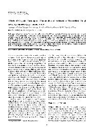
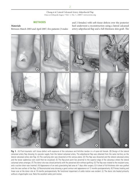
Fig. 1. (A) Post-traumatic s<strong>of</strong>t tissue defect with exposure <strong>of</strong> <strong>the</strong> calcaneus and Achilles tendon in a 4-year-old female. (B) Design <strong>of</strong> <strong>the</strong> lateral<br />
calcaneal artery flap showing its vascular supply from <strong>the</strong> lateral calcaneal artery. The adip<strong>of</strong>ascial flap was obtained from <strong>the</strong> same territory as <strong>the</strong><br />
lateral calcaneal artery skin flap. (C) The overlying skin was dissected at this venous plane. (D) The flap was dissected and <strong>the</strong> lateral calcaneal artery<br />
and <strong>the</strong> lesser saphenous vein could <strong>the</strong>n be visualized. (E) The flap pivot point lies proximal to <strong>the</strong> superior edge <strong>of</strong> <strong>the</strong> calcaneus where <strong>the</strong> lateral<br />
calcaneal artery emerges. (F) The donor site was closed primarily with <strong>the</strong> preserved skin without grafting. (G) The flap was rotated to <strong>the</strong> recipient area,<br />
and a suction drain was inserted. (H) Appearance <strong>of</strong> an early granulating bed area at 7 days after surgery. (I) A sheet <strong>of</strong> full-thickness skin was grafted<br />
onto <strong>the</strong> raw surface <strong>of</strong> <strong>the</strong> flap. (J) Reconstructed area showing a good contour and appearance at 12 months postoperatively. (K) Primary healing and<br />
linear scar at <strong>the</strong> donor site at 18 months postoperatively. No functional impairment <strong>of</strong> ankle motion was evident. (L) The donor site healed primarily<br />
without a hypertrophic scar. Note <strong>the</strong> excellent ankle joint motion.