Lateral Calcaneal Artery Adipofascial Flap for Reconstruction of the ...
Lateral Calcaneal Artery Adipofascial Flap for Reconstruction of the ...
Lateral Calcaneal Artery Adipofascial Flap for Reconstruction of the ...

Create successful ePaper yourself
Turn your PDF publications into a flip-book with our unique Google optimized e-Paper software.
Original Article Clinics in Orthopedic Surgery 2009;1:1-5 • doi:10.4055/cios.2009.1.1.1<br />
<strong>Lateral</strong> <strong>Calcaneal</strong> <strong>Artery</strong> <strong>Adip<strong>of</strong>ascial</strong> <strong>Flap</strong> <strong>for</strong><br />
<strong>Reconstruction</strong> <strong>of</strong> <strong>the</strong> Posterior Heel <strong>of</strong> <strong>the</strong> Foot<br />
Moon Sang Chung, MD, Goo Hyun Baek, MD, Hyun Sik Gong, MD, Seung Hwan Rhee, MD,<br />
Won Seok Oh, MD, Min Bum Kim, MD, Kyung Hag Lee, MD, Tae Woo Kim, MD, Young Ho Lee, MD<br />
Department <strong>of</strong> Orthopedic Surgery, Seoul National University College <strong>of</strong> Medicine, Seoul, Korea<br />
Background: S<strong>of</strong>t tissue defects <strong>of</strong> <strong>the</strong> posterior heel <strong>of</strong> <strong>the</strong> foot present difficult reconstructive problems. This paper reports <strong>the</strong><br />
authors’ early experience <strong>of</strong> five patients treated with a lateral calcaneal artery adip<strong>of</strong>ascial flap.<br />
Methods: Between 2003 and 2007, five patients (3 males and 2 females) with s<strong>of</strong>t-tissue defects over <strong>the</strong> posterior heel<br />
underwent a reconstruction using a lateral calcaneal artery adip<strong>of</strong>ascial flap and a full-thickness skin graft. The flap sizes ranged<br />
from 3.5 × 2.5 cm to 5.5 × 4.0 cm.<br />
Results: All five flaps survived completely with no subsequent breakdown <strong>of</strong> <strong>the</strong> grafted skin, even after regularly wearing normal<br />
shoes. The adip<strong>of</strong>ascial flap donor sites were closed primarily in all patients.<br />
Conclusions: <strong>Lateral</strong> calcaneal artery adip<strong>of</strong>ascial flaps should be included in <strong>the</strong> surgical armamentarium to cover difficult<br />
wounds <strong>of</strong> <strong>the</strong> posterior heel <strong>of</strong> <strong>the</strong> foot. These flaps do not require <strong>the</strong> sacrifice <strong>of</strong> a major artery to <strong>the</strong> leg or foot, <strong>the</strong>y are<br />
relatively thin with minimal morbidity at <strong>the</strong> donor site, and leave a simple linear scar over <strong>the</strong> lateral aspect <strong>of</strong> <strong>the</strong> foot.<br />
Keywords: Foot, Posterior heel, S<strong>of</strong>t tissue defect, <strong>Lateral</strong> calcaneal artery adip<strong>of</strong>ascial flap<br />
S<strong>of</strong>t tissue defects <strong>of</strong> <strong>the</strong> posterior heel <strong>of</strong> <strong>the</strong> foot present<br />
difficult reconstructive problems due to <strong>the</strong> bony prominence,<br />
limited availability <strong>of</strong> local tissue, requirement<br />
<strong>for</strong> specialized tissue, and <strong>the</strong> limitations imposed by<br />
donor site morbidity. Moreover, <strong>the</strong>re are many occasions<br />
where primary closure is impossible, even <strong>for</strong> small<br />
defects. 1) Moreover, skin grafts must heal, even in cases<br />
with exposed bone or tendons and when <strong>the</strong> recipient<br />
sites are stimulated continuously by footwear. In addition,<br />
local rotation, advancement, and transposition flaps are<br />
limited by <strong>the</strong> availability <strong>of</strong> mobile skin. Several types <strong>of</strong><br />
reverse flow island flaps have been developed in <strong>the</strong> <strong>for</strong>m<br />
<strong>of</strong> fasciocutaneous or cutaneous flaps but <strong>the</strong>y require<br />
Received April 10, 2008; Accepted June 3, 2008<br />
Correspondence to: Young Ho Lee, MD<br />
Department <strong>of</strong> Orthopedic Surgery, Seoul National University Hospital, 28<br />
Yeongeon-dong, Jongno-gu, Seoul 110-744, Korea<br />
Tel: +82-2-2072-2367, Fax: +82-2-764-2718<br />
E-mail: orthoyhl@snu.ac.kr<br />
Copyright © 2009 by The Korean Orthopaedic Association<br />
Clinics in Orthopedic Surgery • pISSN 2005-291X eISSN 2005-4408<br />
sacrifice <strong>of</strong> an important leg artery and create obvious<br />
contour de<strong>for</strong>mities at <strong>the</strong> donor site. The use <strong>of</strong> free<br />
flaps has improved <strong>the</strong> ability to cover s<strong>of</strong>t tissue defects.<br />
However, <strong>the</strong> flap bulk, <strong>the</strong> need <strong>for</strong> secondary procedures,<br />
and <strong>the</strong> risk <strong>of</strong> vascular failure are considerable obstacles.<br />
A lateral calcaneal artery skin flap is an axial<br />
pattern flap that includes <strong>the</strong> lateral calcaneal artery,<br />
lesser saphenous vein and <strong>the</strong> sural nerve. 2) Since its development<br />
in 1981, this flap has been demonstrated to<br />
be both an effective and reliable local flap <strong>for</strong> reconstructing<br />
s<strong>of</strong>t tissue defects about <strong>the</strong> posterior heel and<br />
both malle-oli. 3,4) Modifications <strong>of</strong> this flap include island<br />
arterial flaps, 3-6) distally based flaps 6) and free flaps, 7) all <strong>of</strong><br />
which have a wide variety <strong>of</strong> clinical applications. Lin et<br />
al. 8) modified this flap as an adip<strong>of</strong>ascial flap and used it to<br />
reconstruct s<strong>of</strong>t tissue defects <strong>of</strong> <strong>the</strong> posterior heel as well<br />
as <strong>the</strong> lateral malleolar and lateral supramalleolar areas.<br />
This study reports our early experience <strong>of</strong> five patients<br />
treated with this flap <strong>for</strong> a posterior heel reconstruction.
METHODS<br />
Materials<br />
Between March 2003 and April 2007, five patients (3 males<br />
2<br />
Chung et al. <strong>Lateral</strong> <strong>Calcaneal</strong> <strong>Artery</strong> <strong>Adip<strong>of</strong>ascial</strong> <strong>Flap</strong><br />
Clinics in Orthopedic Surgery • Vol. 1, No. 1, 2009 • www.ecios.org<br />
and 2 females) with s<strong>of</strong>t-tissue defects over <strong>the</strong> posterior<br />
heel underwent a reconstruction using a lateral calcaneal<br />
artery adip<strong>of</strong>ascial flap and a full-thickness skin graft. The<br />
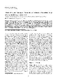
Fig. 1. (A) Post-traumatic s<strong>of</strong>t tissue defect with exposure <strong>of</strong> <strong>the</strong> calcaneus and Achilles tendon in a 4-year-old female. (B) Design <strong>of</strong> <strong>the</strong> lateral<br />
calcaneal artery flap showing its vascular supply from <strong>the</strong> lateral calcaneal artery. The adip<strong>of</strong>ascial flap was obtained from <strong>the</strong> same territory as <strong>the</strong><br />
lateral calcaneal artery skin flap. (C) The overlying skin was dissected at this venous plane. (D) The flap was dissected and <strong>the</strong> lateral calcaneal artery<br />
and <strong>the</strong> lesser saphenous vein could <strong>the</strong>n be visualized. (E) The flap pivot point lies proximal to <strong>the</strong> superior edge <strong>of</strong> <strong>the</strong> calcaneus where <strong>the</strong> lateral<br />
calcaneal artery emerges. (F) The donor site was closed primarily with <strong>the</strong> preserved skin without grafting. (G) The flap was rotated to <strong>the</strong> recipient area,<br />
and a suction drain was inserted. (H) Appearance <strong>of</strong> an early granulating bed area at 7 days after surgery. (I) A sheet <strong>of</strong> full-thickness skin was grafted<br />
onto <strong>the</strong> raw surface <strong>of</strong> <strong>the</strong> flap. (J) Reconstructed area showing a good contour and appearance at 12 months postoperatively. (K) Primary healing and<br />
linear scar at <strong>the</strong> donor site at 18 months postoperatively. No functional impairment <strong>of</strong> ankle motion was evident. (L) The donor site healed primarily<br />
without a hypertrophic scar. Note <strong>the</strong> excellent ankle joint motion.
s<strong>of</strong>t-tissue defects were caused by acute trauma in three<br />
patients and a chronic ulcer in two. The flaps ranged in<br />
size from 3.5 × 2.5 cm to 5.5 × 4.0 cm. The patients’ ages<br />
ranged from 4 to 72 years (mean, 37.2 years), and <strong>the</strong><br />
follow-up period ranged from 6 to 18 months (mean, 11<br />
months).<br />
Operative Procedure<br />
Be<strong>for</strong>e surgery, <strong>the</strong> peroneal and lateral calcaneal artery<br />
presence and patency were confirmed. The tissue used <strong>for</strong><br />
<strong>the</strong> adip<strong>of</strong>ascial flaps was in <strong>the</strong> same areas as <strong>the</strong> lateral<br />
calcaneal artery skin flap. 2) The flap sizes required to cover<br />
<strong>the</strong> heel defects were marked on <strong>the</strong> skin. Initially, a zigzag<br />
skin incision was marked in <strong>the</strong> central portion <strong>of</strong><br />
<strong>the</strong> proposed flap. Surgery was aided with a tourniquet<br />
and surgical loupes (× 2.5). The incision was deepened<br />
through <strong>the</strong> skin down to <strong>the</strong> subcutaneous tissue and<br />
superficial venous plexus. The overlying skin was dissected<br />
at this venous plane and <strong>the</strong> lesser saphenous vein and<br />
its contributors were identified and preserved on <strong>the</strong> flap<br />
surface <strong>of</strong> <strong>the</strong> flap.<br />
After <strong>the</strong> skin over <strong>the</strong> proposed flap had been<br />
dissected completely, <strong>the</strong> initial incision <strong>for</strong> elevation<br />
<strong>of</strong> <strong>the</strong> fascial side <strong>of</strong> <strong>the</strong> flap was made at <strong>the</strong> posterior<br />
aspect <strong>of</strong> <strong>the</strong> lateral malleolus and extended proximally<br />
along <strong>the</strong> fibula and lateral tendon <strong>of</strong> <strong>the</strong> peroneus<br />
longus muscle. Through traction and elevation <strong>of</strong> <strong>the</strong><br />
medial edge <strong>of</strong> <strong>the</strong> adip<strong>of</strong>ascial flap, <strong>the</strong> lateral calcaneal<br />
vessels and accompanying sural nerve were identified<br />
in <strong>the</strong> suprafascial layer. This neurovascular pedicle was<br />
protected and kept in <strong>the</strong> flap, and used as an anatomical<br />
landmark <strong>of</strong> <strong>the</strong> proper plane to undermine <strong>the</strong> fascia.<br />
The sural nerve was dissected by incising <strong>the</strong> fascia<br />
and preserved. However, its branches to <strong>the</strong> flap were<br />
separated interfascicularly and retained in <strong>the</strong> flap. The<br />
posterior edge <strong>of</strong> <strong>the</strong> flap was incised close to <strong>the</strong> Achilles<br />
tendon near <strong>the</strong> calcaneus and carried down to <strong>the</strong><br />
periosteum. Undermining <strong>of</strong> <strong>the</strong> fascial side <strong>of</strong> <strong>the</strong> flap<br />
was straight<strong>for</strong>ward when <strong>the</strong> dissection was per<strong>for</strong>med<br />
Table 1. Clinical Data <strong>of</strong> <strong>the</strong> Patients<br />
3<br />
Chung et al. <strong>Lateral</strong> <strong>Calcaneal</strong> <strong>Artery</strong> <strong>Adip<strong>of</strong>ascial</strong> <strong>Flap</strong><br />
Clinics in Orthopedic Surgery • Vol. 1, No. 1, 2009 • www.ecios.org<br />
immediately over <strong>the</strong> periosteum <strong>of</strong> <strong>the</strong> calcaneus and <strong>the</strong><br />
dissection could be accomplished through this site. 8)<br />
The pivot point <strong>of</strong> <strong>the</strong> flap lies proximal to <strong>the</strong><br />
superior edge <strong>of</strong> <strong>the</strong> calcaneus where <strong>the</strong> lateral calcaneal<br />
artery emerges. The proximal base <strong>of</strong> <strong>the</strong> flap was not<br />
narrowed and included <strong>the</strong> lateral calcaneal vessels, <strong>the</strong><br />
superficial venous system and fascicular branches <strong>of</strong> <strong>the</strong><br />
sural nerve. 8)<br />
After rotating <strong>the</strong> flap to <strong>the</strong> recipient area, <strong>the</strong> donor<br />
site was closed primarily with preserved skin, and<br />
multiple small silastic drains or a suction drain was inserted.<br />
A moist, loose 1% framycetin sulfate-impregnated<br />
sterile tulle gauze dressing with a wet dressing was simply<br />
placed over this flap until <strong>the</strong> skin graft was per<strong>for</strong>med.<br />
On postoperative days 5 to 7, <strong>the</strong> raw surface <strong>of</strong> <strong>the</strong> flap<br />
on <strong>the</strong> recipient site was covered with a full- thickness skin<br />
graft (Fig. 1).<br />
RESULTS<br />
Table 1 shows <strong>the</strong> patient data. All five flaps had good<br />
perfusion and survived completely. No venous congestion<br />
was noted but flap edema lasted <strong>for</strong> 3 days. The skin grafts<br />
on <strong>the</strong> adip<strong>of</strong>ascial flap had taken well in 4 patients. One<br />
case (case 3) suffered partial loss <strong>of</strong> <strong>the</strong> full-thickness skin<br />
graft on <strong>the</strong> flap that was associated with a thin hematoma<br />
beneath <strong>the</strong> skin graft. However, this healed spontaneously<br />
without <strong>the</strong> need <strong>for</strong> a secondary graft. In all patients, <strong>the</strong><br />
donor sites <strong>of</strong> <strong>the</strong> adip<strong>of</strong>ascial flap were closed primarily<br />
without any functional impairment and healed. One case<br />
(case 4) was complicated by a hematoma at <strong>the</strong> donor<br />
site, which was emptied by suction and compression.<br />
Marginal desquamation <strong>of</strong> <strong>the</strong> donor site wound occurred<br />
in one case (case 2) but it subsequently healed. All patients<br />
became ambulatory after wound healing, and ankle<br />
motion was not restricted. There was no subsequent<br />
breakdown <strong>of</strong> <strong>the</strong> grafted skin with <strong>the</strong> regular wearing <strong>of</strong><br />
shoes.<br />
Case Age (yr), Sex Cause <strong>of</strong> defect Site <strong>of</strong> defect Size <strong>of</strong> flap (cm) Duration <strong>of</strong> follow-up (mon) Complication<br />
1 4, F Acute trauma Posterior heel 4.5 × 3.0 18 None<br />
2 38, M Acute trauma Posterior heel 5.5 × 4.0 11 Marginal desquamation <strong>of</strong> <strong>the</strong> donor site<br />
3 65, F Chronic ulcer Posterior heel 5.5 × 3.5 14 Partial loss <strong>of</strong> <strong>the</strong> full-thickness skin graft<br />
4 7, M Acute trauma Posterior heel 3.5 × 2.5 8 Hematoma <strong>of</strong> <strong>the</strong> donor site<br />
5 72, M Chronic ulcer Posterior heel 5.0 × 4.0 6 None
DISCUSSION<br />
<strong>Adip<strong>of</strong>ascial</strong> flaps have inherent shortcomings that<br />
warrant consideration. 9) These include flap thinness,<br />
bleeding or hematoma, monitoring difficulties and skin<br />
graft associated problems. 10-12)<br />
The thinness <strong>of</strong> adip<strong>of</strong>ascial flaps makes <strong>the</strong>m<br />
vulnerable to pressure. Meland and Weimar 11) reported<br />
that <strong>the</strong> avoidance <strong>of</strong> external pressure is essential <strong>for</strong><br />
maintaining flap viability. They used a protective, clear<br />
plastic dome over <strong>the</strong> flap area, and no dressing material<br />
was placed in direct contact with <strong>the</strong> flaps. In order to<br />
avoid this problem, Walton et al. 12) dressed <strong>the</strong> flaps with a<br />
thin sheet <strong>of</strong> petrolatum-impregnated gauze but left <strong>the</strong>m<br />
o<strong>the</strong>rwise exposed. We also dressed <strong>the</strong> flaps using this<br />
method but used a 1% framycetin sulphate impregnated<br />
sterile tulle gauze instead.<br />
An axial pattern adip<strong>of</strong>ascial flap has a rich blood<br />
supply <strong>for</strong> <strong>the</strong> vessels to run and <strong>for</strong>m a redundant<br />
vascular network within <strong>the</strong> fascia. 10) There<strong>for</strong>e, <strong>the</strong><br />
potential difficulties associated with intraoperative<br />
and postoperative bleeding are a valid concern. 11)<br />
Intraoperative bleeding can be minimized by <strong>the</strong> careful<br />
use <strong>of</strong> bipolar cauterization. The problems associated a<br />
postoperative hematoma beneath flaps is best addressed<br />
using small-caliber suction drains, as recommended by<br />
Brent and Byrd, 13) ra<strong>the</strong>r by applying external pressure. 11)<br />
We currently use a number <strong>of</strong> small Silastic or suction<br />
drains.<br />
Ano<strong>the</strong>r problem associated with adip<strong>of</strong>ascial<br />
flaps is <strong>the</strong> tendency towards incomplete take-up <strong>of</strong> <strong>the</strong><br />
overlying skin graft. Meland and Weimar 11) reported<br />
that delaying skin grafting <strong>for</strong> 3 to 5 days allows an early<br />
granulating bed to develop on <strong>the</strong> surface <strong>of</strong> <strong>the</strong> adipose<br />
tissue, which eliminates <strong>the</strong> problem <strong>of</strong> immediate,<br />
postoperative oozing beneath <strong>the</strong> skin graft. In all our<br />
cases, <strong>the</strong> raw surface <strong>of</strong> <strong>the</strong> flap on <strong>the</strong> recipient site was<br />
covered with a full thickness skin graft 5 to 7 days after<br />
1. Lee ET. A new wound closure achieving and maintaining<br />
device using serial tightening <strong>of</strong> loop suture and its clinical<br />
applications in 15 consecutive patients <strong>for</strong> up to 102 days.<br />
Ann Plast Surg. 2004;53(5):436-41.<br />
2. Grabb WC, Argenta LC. The lateral calcaneal artery skin<br />
flap(<strong>the</strong> lateral calcaneal artery, lesser saphenous vein, and<br />
sural nerve skin flap). Plast Reconstr Surg. 1981;68(5):723-30.<br />
4<br />
Chung et al. <strong>Lateral</strong> <strong>Calcaneal</strong> <strong>Artery</strong> <strong>Adip<strong>of</strong>ascial</strong> <strong>Flap</strong><br />
Clinics in Orthopedic Surgery • Vol. 1, No. 1, 2009 • www.ecios.org<br />
REFERENCES<br />
surgery. The main shortcoming <strong>of</strong> this procedure is that<br />
an adip<strong>of</strong>ascial flap or fascial flap must act as an additional<br />
skin graft. Most surgeons use a split-thickness skin graft. 10-<br />
12,14,15) However, Lai et al. 16) used an adip<strong>of</strong>ascial turnover<br />
flap to reconstruct <strong>the</strong> dorsum <strong>of</strong> <strong>the</strong> foot, and Lin<br />
et al. 8,17) used a full-thickness skin graft obtained from<br />
<strong>the</strong> groin or inguinal areas as a distally based posterior<br />
tibial arterial adip<strong>of</strong>ascial flap and lateral calcaneal artery<br />
adip<strong>of</strong>ascial flap, which closed primarily. A full-thickness<br />
skin graft ra<strong>the</strong>r than a split-thickness graft minimizes <strong>the</strong><br />
breakdown <strong>of</strong> grafted skin. Full-thickness skin grafts are<br />
applied to raw adip<strong>of</strong>ascial flap surfaces to reconstruct <strong>the</strong><br />
foot and allow <strong>the</strong> regular wearing <strong>of</strong> shoes. In this study,<br />
a full-thickness skin graft was used in all <strong>of</strong> <strong>the</strong> five cases<br />
described.<br />
Overall, lateral calcaneal artery adip<strong>of</strong>ascial flaps<br />
should be included in <strong>the</strong> surgical armamentarium to cover<br />
difficult wounds <strong>of</strong> <strong>the</strong> posterior heel <strong>of</strong> <strong>the</strong> foot. They do<br />
not require sacrifice <strong>of</strong> a major artery to <strong>the</strong> leg or foot, are<br />
relatively thin with minimal morbidity at <strong>the</strong> donor sites,<br />
and leave only a simple linear scar over <strong>the</strong> lateral aspect<br />
<strong>of</strong> <strong>the</strong> foot. In addition, <strong>the</strong> flap dissecting technique is<br />
straight<strong>for</strong>ward, and has <strong>the</strong> advantages <strong>of</strong> better aes<strong>the</strong>tic<br />
results at <strong>the</strong> donor sites and sural nerve preservation<br />
compared with a lateral calcaneal artery skin flap.<br />
<strong>Lateral</strong> calcaneal artery adip<strong>of</strong>ascial flaps are limited<br />
in size but can fill defects <strong>of</strong> <strong>the</strong> posterior heel. Moreover,<br />
<strong>the</strong>ir s<strong>of</strong>tness and pliability make <strong>the</strong>m suitable <strong>for</strong> filling<br />
<strong>the</strong> subcutaneous spaces <strong>for</strong>med by debridement. In <strong>the</strong><br />
five patients described in this report, lateral calcaneal<br />
artery adip<strong>of</strong>ascial flaps were used to effectively cure<br />
intractable defects with minimal donor site morbidity.<br />
ACKNOWLEDGEMENTS<br />
This study was supported by Korean Human Technology<br />
Research Foundation (KOHTERF-2007-02).<br />
3. Holmes J, Rayner CR. <strong>Lateral</strong> calcaneal artery island flaps.<br />
Br J Plast Surg. 1984;37(3):402-5.<br />
4. Yanai A, Park S, Iwao T, Nakamura N. <strong>Reconstruction</strong> <strong>of</strong> a<br />
skin defect <strong>of</strong> <strong>the</strong> posterior heel by a lateral calcaneal flap.<br />
Plast Reconstr Surg. 1985;75(5):642-7.
5. Gang RK. <strong>Reconstruction</strong> <strong>of</strong> s<strong>of</strong>t-tissue defect <strong>of</strong> <strong>the</strong> posterior<br />
heel with a lateral calcaneal artery island flap. Plast<br />
Reconstr Surg. 1987;79(3):415-21.<br />
6. Ishikawa K, Isshiki N, Hoshino K, Mori C. Distally based<br />
lateral calcaneal flap. Ann Plast Surg. 1990;24(1):10-6.<br />
7. Ishikawa K, Kyutoku S, Takeuchi E. Free lateral calcaneal<br />
flap. Ann Plast Surg. 1993;30(2):167-70.<br />
8. Lin SD, Lai CS, Chiu YT, Lin TM. The lateral calcaneal<br />
artery adip<strong>of</strong>ascial flap. Br J Plast Surg. 1996;49(1):52-7.<br />
9. Lee YH, Rah SK, Choi SJ, Chung MS, Baek GH. Distally<br />
based lateral supramalleolar adip<strong>of</strong>ascial flap <strong>for</strong> reconstruction<br />
<strong>of</strong> <strong>the</strong> dorsum <strong>of</strong> <strong>the</strong> foot and ankle. Plast Reconstr<br />
Surg. 2004;114(6):1478-85.<br />
10. Jin YT, Cao HP, Chang TS. Clinical application <strong>of</strong> <strong>the</strong> free<br />
scapular fascial flap. Ann Plast Surg. 1989;23(2):170-7.<br />
11. Meland NB, Weimar R. Microsurgical reconstruction: experience<br />
with free fascia flaps. Ann Plast Surg. 1991;27(1):1-8.<br />
5<br />
Chung et al. <strong>Lateral</strong> <strong>Calcaneal</strong> <strong>Artery</strong> <strong>Adip<strong>of</strong>ascial</strong> <strong>Flap</strong><br />
Clinics in Orthopedic Surgery • Vol. 1, No. 1, 2009 • www.ecios.org<br />
12. Walton RL, Matory WE Jr, Petry JJ. The posterior calf fascial<br />
free flap. Plast Reconstr Surg. 1985;76(6):914-26.<br />
13. Brent B, Byrd HS. Secondary ear reconstruction with<br />
cartilage grafts covered by axial, random, and free flaps <strong>of</strong><br />
temporoparietal fascia. Plast Reconstr Surg. 1983;72(2):141-52.<br />
14. Gibstein LA, Abramson DL, Sampson CE, Pribaz JJ.<br />
Muscul<strong>of</strong>ascial flaps based on <strong>the</strong> dorsalis pedis vascular<br />
pedicle <strong>for</strong> coverage <strong>of</strong> <strong>the</strong> foot and ankle. Ann Plast Surg.<br />
1996;37(2):152-7.<br />
15. Ismail TI. The dorsalis pedis my<strong>of</strong>ascial flap. Plast Reconstr<br />
Surg. 1990;86(3):573-6.<br />
16. Lai CS, Lin SD, Yang CC, Chou CK. <strong>Adip<strong>of</strong>ascial</strong> turnover<br />
flap <strong>for</strong> reconstruction <strong>of</strong> <strong>the</strong> dorsum <strong>of</strong> <strong>the</strong> foot. Br J Plast<br />
Surg. 1991;44(3):170-4.<br />
17. Lin SD, Lai CS, Chou CK, Tsai CW. The distally based<br />
posterior tibial arterial adip<strong>of</strong>ascial flap. Br J Plast Surg.<br />
1992;45(4):284-7.