Strong Association of ARK5 with Tumor Invasion and Metastasis
Strong Association of ARK5 with Tumor Invasion and Metastasis
Strong Association of ARK5 with Tumor Invasion and Metastasis

You also want an ePaper? Increase the reach of your titles
YUMPU automatically turns print PDFs into web optimized ePapers that Google loves.
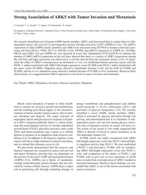
J. Exp. Clin. Cancer Res., 23, 2, 2004<br />
<strong>Strong</strong> <strong>Association</strong> <strong>of</strong> <strong>ARK5</strong> <strong>with</strong> <strong>Tumor</strong> <strong>Invasion</strong> <strong>and</strong> <strong>Metastasis</strong><br />
G. Kusakai 1,2* , A. Suzuki 1* , T. Ogura 1 , M. Kaminishi 2 , H. Esumi 1<br />
Investigative Treatment Division 1 , National Cancer Center Research Institute East, Chiba; Dept. <strong>of</strong> Gastrointestinal Surgery 2 , University<br />
<strong>of</strong> Tokyo, Tokyo; Japan<br />
We recently identified a novel human AMPK family member, <strong>ARK5</strong>, <strong>and</strong> discovered that is a major factor in Aktdependent<br />
cancer cell survival (1) <strong>and</strong> migration activity through activation <strong>of</strong> MT1-MMPs in vitro. The mRNA<br />
expression <strong>of</strong> other AMPK family members <strong>and</strong> <strong>ARK5</strong> was measured using RT-PCR in human colorectal carcinoma<br />
cell lines DLD-1, WiDr, HCT-15, SW620, LoVo, SW480, <strong>and</strong> mRNA expression <strong>of</strong> AMPK-α1, SNARK,<br />
MELK <strong>and</strong> <strong>ARK5</strong>, but not AMPK-α2, was detected in every line. Quantitative-PCR (Q-PCR) to estimate the<br />
amount <strong>of</strong> <strong>ARK5</strong> mRNA expression in the cell lines showed that there is a variety <strong>of</strong> <strong>ARK5</strong> expressions among<br />
the cell lines <strong>and</strong> high expression was observed in a cell line derived from the metastatic lesion, LoVo. To determine<br />
the effect <strong>of</strong> <strong>ARK5</strong> overexpression on metastasis in vivo, we established human pancreas cancer cell line<br />
PANC-1 stably transfected <strong>with</strong> <strong>ARK5</strong> full-length expression vector (P/ARK) <strong>and</strong> DLD-1 stably transfected <strong>with</strong><br />
the same vector (D/ARK). Migration assay showed a remarkable increase in the activity both in P/ARK <strong>and</strong><br />
D/ARK, <strong>and</strong> an in vivo metastasis assay showed a marked increase <strong>of</strong> P/ARK in liver metastasis. Based on these<br />
observations, it is suggested that <strong>ARK5</strong> expression is involved in cancer invasion <strong>and</strong> metastasis.<br />
Key Words: <strong>ARK5</strong>, <strong>Metastasis</strong>, <strong>Invasion</strong>, Glucose starvation, Migration<br />
Blood vessel formation <strong>of</strong> tumors is <strong>of</strong>ten insufficient<br />
to sustain its excessive growth <strong>and</strong> proliferation,<br />
<strong>and</strong> the resulting poor blood supply, in the microenvironment<br />
<strong>of</strong> tumor, usually exposes cancer cells to nutrient<br />
starvation <strong>and</strong> hypoxia. The major activator <strong>of</strong><br />
angiogenic factors <strong>and</strong> glycolysis in response to hypoxia<br />
is HIF-1 (hypoxia-inducible factor-1), which modulates<br />
the transcriptional activity <strong>of</strong> vascular endothelial<br />
growth factor (VEGF), glycolytic enzymes, nitric oxide<br />
(NO), <strong>and</strong> heme-oxygenase type I genes as a cellular<br />
defensive response or an adaptation under hypoxic conditions<br />
(2,3). In addition to the hypoxic response, some<br />
cancer cells have a high tolerance to nutrient starvation,<br />
<strong>and</strong> we termed this tolerance austerity (4).<br />
We previously demonstrated that the austerity <strong>and</strong><br />
hypoxic responses are essential for cancer cell survival<br />
<strong>and</strong> tumor formation, <strong>and</strong> that they are dependent on<br />
the activity <strong>of</strong> Akt <strong>and</strong> 5'-AMP activated protein kinase<br />
(AMPK) (5,6). AMPKs are a family <strong>of</strong> metabolitesensing<br />
serine/threonine protein kinases that are activated<br />
by various cellular stresses. AMPK plays a major<br />
role in protecting cells through mediating changes in<br />
energy metabolism <strong>and</strong> phosphorylates <strong>and</strong> inhibits<br />
acetyl-coenzyme A (CoA) carboxylase (ACC) <strong>and</strong><br />
activation <strong>of</strong> glucose transporters (7,8). We recently<br />
identified a novel AMPK family member, <strong>ARK5</strong>,<br />
which is activated by glucose starvation through Akt<br />
activity, <strong>and</strong> demonstrated that it is a mediator <strong>of</strong> Aktdependent<br />
cancer cell survival during glucose starvation<br />
<strong>and</strong> <strong>of</strong> migration activity in cancer cell lines (1).<br />
The results <strong>of</strong> our recent in vitro study suggested that<br />
<strong>ARK5</strong> is directly involved in cancer metastasis in an<br />
Akt-dependent manner (9).<br />
D/ARK which was stably transfected <strong>with</strong> <strong>ARK5</strong><br />
full-length expression vector into DLD-1 showed higher<br />
migration activity than DLD-1. We also established<br />
a PANC-1 cells derivative, P/ARK cells, by transfecting<br />
<strong>ARK5</strong> full-length expression vector into PANC-1<br />
cells, <strong>and</strong> comparison <strong>of</strong> the ability <strong>of</strong> PANC-1 <strong>and</strong><br />
P/ARK cells to give rise to liver metastasis in SCID<br />
mice revealed that overexpression <strong>of</strong> <strong>ARK5</strong> resulted in<br />
increased metastasis. Based on these findings, we concluded<br />
that <strong>ARK5</strong> is one <strong>of</strong> the factors associated <strong>with</strong><br />
cancer metastasis.<br />
263
G. Kusakai et al.<br />
Materials <strong>and</strong> Methods<br />
Homology Search for Subunit Structures <strong>of</strong> <strong>ARK5</strong><br />
<strong>and</strong> AMPK Catalytic Subunits. Based on their reported<br />
amino acid sequences, a BLAST search analysis was<br />
used to detect a putative consensus catalytic peptide<br />
sequence <strong>of</strong> the AMPK family members AMPK-α1,<br />
AMPK-2, MELK, SNARK, <strong>and</strong> <strong>ARK5</strong>. A homology<br />
search was performed by computer analysis using<br />
GENETYX 10.1 s<strong>of</strong>tware.<br />
Cell Lines <strong>and</strong> Culture. All human carcinoma cells<br />
were cultured in Dulbecco's modified Eagle Medium<br />
(DMEM: NISSUI, Japan) supplemented <strong>with</strong> 10%<br />
fetal bovine serum, 2 mM L-glutamine <strong>and</strong> 25 mM<br />
HEPES-NaOH (pH 7.3) at 37°C under an atmosphere<br />
<strong>of</strong> 5% CO 2 in air.<br />
Reverse Transcription-PCR (RT-PCR) <strong>and</strong> Quantitative-RT-PCR<br />
(Q-RT-PCR). Total RNA was isolated<br />
from cancer cells by the AGPC method using ISOGEN<br />
(NIPPONGENE, Japan), <strong>and</strong> 1 µg <strong>of</strong> total RNA was<br />
reverse transcripted into cDNA <strong>with</strong> oligo-dT primers<br />
<strong>and</strong> AMV-reverse transcriptase (TaKaRa, Japan)<br />
according to the manufacturer's instructions. The<br />
cDNA was subjected to 25 cycles <strong>of</strong> PCR amplification<br />
on a PCR Thermal Cycler 480 (TaKaRa, Japan), <strong>and</strong> the<br />
PCR primers used for <strong>ARK5</strong> detection were: forward,<br />
5'-ATGCTAAGTACCCTCTGAATG-3', <strong>and</strong> reverse,<br />
5'-GCAACAAGCAGTCAGTCGATC-3'. PCR products<br />
were fractionated by agarose electrophoresis,<br />
stained <strong>with</strong> ethidium bromide, <strong>and</strong> visualized under<br />
UV light. GAPDH served as a control for PCR.<br />
A Light-Cycler (Roche, Germany) was used to<br />
detect the number <strong>of</strong> <strong>ARK5</strong> <strong>and</strong> GAPDH gene copies.<br />
The amplification mixture contained cDNA derived<br />
from 100 ng <strong>of</strong> total RNA. The PCR primers used for<br />
GAPDH detection were: forward, 5'-AGGGCTG-<br />
GTTTTAACTCTGGT-3', <strong>and</strong> reverse, 5'-CCC-<br />
CACTTGATTTTGGAGGGA-3'. Q-PCR was performed<br />
under cycling conditions <strong>of</strong> 95°C for 10 min,<br />
followed by 40 cycles <strong>of</strong> denaturation at 95°C for 10<br />
sec, annealing at 60°C for 10 sec, extension at 72°C for<br />
20 sec.<br />
Transfection <strong>and</strong> Selection. For transfection, 1˘10 5<br />
cells were seeded into each well <strong>of</strong> a 6-well plate, <strong>and</strong><br />
<strong>ARK5</strong>-full length expression vector was transfected<br />
into DLD-1 <strong>and</strong> PANC-1 cells <strong>with</strong> TransFast transfection<br />
reagent (Promega Corp., USA) according to the<br />
manufacturer's instructions <strong>with</strong> some modifications.<br />
Stable transfectants were selected <strong>with</strong> G418.<br />
264<br />
Immunoblotting. Cells were washed <strong>with</strong> PBS containing<br />
1mM sodium ortho-vanadate, 10mM sodium<br />
fluoride <strong>and</strong> 25 mM sodium glycerophosphate. Cells<br />
were collected, <strong>and</strong> lyzed <strong>with</strong> a buffer containing 1%<br />
sodium dodecyl sulfate (SDS), 1 mM SOV4 <strong>and</strong> 10<br />
mM Tris-HC1 (pH 7.4). Proteins were separated on a<br />
10% SDS polyacrylamide gel, <strong>and</strong> transferred onto a<br />
polyvinylidene difluoride membrane. A polyclonal<br />
antibody to human SNARK was made by immunizing<br />
rabbits <strong>with</strong> a peptide based on the sequence <strong>of</strong><br />
SNARK (KKPRQRESGYYSSPEPS) <strong>and</strong> it was found<br />
to crossreact <strong>with</strong> <strong>ARK5</strong> (1). Membranes were incubated<br />
<strong>with</strong> the anti-SNARK antibody at dilution <strong>of</strong><br />
1:1000 <strong>and</strong> anti-rabbit antibody conjugated <strong>with</strong> horseradish<br />
peroxidase (HRP) was used as a second antibody.<br />
For visualisation, Western blotting detection<br />
reagent (ECL: Amersham Pharmacia Biotech, UK) <strong>and</strong><br />
Hyperfilm ECL (Amersham Pharmacia Biotech, UK)<br />
were used.<br />
Migration Assay. In vitro migration assay was performed<br />
<strong>with</strong> a matrigel-coated invasion chamber (Becton-DickinsoN,<br />
USA). DLD-1 cells (5˘10 4 /chamber)<br />
were seeded into each chamber, <strong>and</strong> the media in the<br />
inner <strong>and</strong> outer chamber were changed after 6 hrs. The<br />
cells that had invaded were counted under a phase-contrast<br />
microscope after 48 hrs.<br />
In vivo Liver <strong>Metastasis</strong> Assay in SCID Mice. SCID<br />
mice (CB-17/Icr Crj-scid, 5-week-old males) were<br />
anesthetized <strong>with</strong> pentobarbital-Na (Nembutal:<br />
ABBOTT, USA). Ventrotomy was performed, the<br />
spleen was exposed, <strong>and</strong> 200 µl <strong>of</strong> serum free DMEM<br />
containing 1˘10 6 PANC-1 or P/ARK cells was injected<br />
under the splenic capsule <strong>of</strong> ten mice each. The mice<br />
were sacrificed 5 weeks later, <strong>and</strong> liver metastasis was<br />
assessed by counting the number microscopically. The<br />
liver metastases were stained <strong>with</strong> hematoxylin <strong>and</strong><br />
eosin (H.E.) <strong>and</strong> confirmed pathologically.<br />
Results<br />
Subunit Structures <strong>of</strong> <strong>ARK5</strong> <strong>and</strong> AMPK Catalytic<br />
Subunits. The AMPK family consists <strong>of</strong> five members,<br />
AMPK-α1, AMPK-α2, MELK, SNARK, <strong>and</strong> <strong>ARK5</strong><br />
recently identified (Fig.1). As shown in Fig. 1, all<br />
AMPK members have a putative consensus catalytic<br />
domain structure. <strong>ARK5</strong> contained the putative consensus<br />
catalytic peptide sequence at amino acids 55 -<br />
305, <strong>and</strong> an Akt-phosphorylation site (RXRXXS) was<br />
found near the C-terminal (Fig.1). The C-terminal sub-
Fig. 1 - Peptide structure <strong>of</strong> the AMPK catalytic subunits <strong>of</strong><br />
AMPK-α1, AMPK-α2, MELK, SNARK, <strong>and</strong> <strong>ARK5</strong>.<br />
Analysis <strong>of</strong> the putative peptide catalytic domain <strong>and</strong> Akt<br />
phosphorylation motif was searched by GENETYX 10.1.1<br />
on a computer.<br />
unit-targeting site <strong>of</strong> AMPK-α1 <strong>and</strong> AMPK-α2 bound<br />
to β <strong>and</strong> γ subunits, <strong>and</strong> AMPK-α1 <strong>and</strong> AMPK-α2 possessed<br />
an autoregulatory domain, unlike the other three<br />
AMPK members (Fig.1). The overall homology <strong>of</strong><br />
<strong>ARK5</strong> calculated using GENETYX 10.1 was 47%<br />
compared <strong>with</strong> AMPK-α1 <strong>and</strong> 55% compared to<br />
SNARK.<br />
<strong>ARK5</strong> Expression in Cancer Cell Lines. We previously<br />
demonstrated that at least AMPK-α1 <strong>and</strong> 2 contribute<br />
to tumor progression <strong>and</strong> survival via austerity<br />
(4). RT-PCR to confirm mRNA expression <strong>of</strong> AMPK<br />
family members, in six human colorectal carcinoma<br />
cell lines, DLD-1, WiDr, HCT-15, SW620, LoVo, <strong>and</strong><br />
SW480 (Fig.2A), showed that all six cell lines<br />
expressed AMPK-α1, SNARK, <strong>and</strong> MELK mRNA,<br />
<strong>and</strong> that AMPK-α2 was strongly expressed in HCT-15,<br />
SW620, LoVo, <strong>and</strong> SW480, but not in DLD-1 or WiDr.<br />
The amount <strong>of</strong> <strong>ARK5</strong> mRNA expression was estimated<br />
by Q-PCR in the same human colorectal carcinoma<br />
cell lines, <strong>and</strong> the results showed that <strong>ARK5</strong> was highly<br />
expressed in LoVo <strong>and</strong> SW480 (Fig.2B).<br />
Correlation between <strong>ARK5</strong> Expression <strong>and</strong> Migration<br />
Activity. In our previous studies, <strong>ARK5</strong> expression<br />
is <strong>of</strong>ten associated <strong>with</strong> tumor cell survival <strong>and</strong> <strong>ARK5</strong><br />
is a direct downstream target <strong>of</strong> Akt1 (1). We asked if<br />
<strong>ARK5</strong> might be involved in tumor cell migration <strong>and</strong><br />
metastasis. In order to answer this question, an in vivo<br />
<strong>Strong</strong> <strong>Association</strong> <strong>of</strong> <strong>ARK5</strong> <strong>with</strong> <strong>Tumor</strong> <strong>Invasion</strong> <strong>and</strong> <strong>Metastasis</strong><br />
Fig. 2 - mRNA expression <strong>of</strong> other AMPK family members <strong>and</strong><br />
<strong>ARK5</strong> in human colorectal carcinoma cell lines. A. Expression<br />
<strong>of</strong> AMPK family member mRNA was estimated<br />
by RT-PCR in DLD-1, WiDr, HCT-15, SW620, LoVo,<br />
<strong>and</strong>SW480. B. <strong>ARK5</strong> mRNA expression in human colorectal<br />
carcinoma cell lines was measured by Q-PCR.<br />
*Significant at p < 0.05<br />
migration assay was performed using DLD-1 <strong>and</strong><br />
D/ARK cells. As shown in Fig. 3A, <strong>ARK5</strong> overexpression<br />
<strong>of</strong> D/ARK was confirmed by western blotting. In<br />
comparison <strong>with</strong> DLD-1, D/ARK showed an exceedingly<br />
higher activity <strong>with</strong> a significant difference<br />
(p
G. Kusakai et al.<br />
Fig. 3 - The correlation between <strong>ARK5</strong> overexpression <strong>and</strong> migration<br />
activity. A. Confirmation <strong>of</strong> <strong>ARK5</strong> expression in<br />
DLD-1 <strong>and</strong> D/ARK cells by using western blot. B. Migration<br />
activity induced by <strong>ARK5</strong> overexpression. **Significant<br />
at p < 0.01<br />
In vivo Liver <strong>Metastasis</strong> Assay in SCID Mice. To<br />
evaluate the effect <strong>of</strong> <strong>ARK5</strong> overexpression on in vivo<br />
metastasis, 1˘10 6 PANC-1 or P/ARK cells were transplanted<br />
under the splenic capsule <strong>of</strong> SCID mice. <strong>ARK5</strong><br />
overexpression <strong>of</strong> PANC-1 <strong>and</strong> P/ARK was examined<br />
by western blotting, <strong>and</strong> P/ARK cells were confirmed<br />
to overexpress <strong>ARK5</strong> protein (Fig.4A).<br />
Five weeks later, 40% <strong>of</strong> the mice transplanted <strong>with</strong><br />
P/ARK cells showed liver metastasis (Fig.4B,C). The<br />
number <strong>and</strong> size <strong>of</strong> tumor nodules <strong>of</strong> the liver metastases<br />
were counted (Table I). One <strong>of</strong> them also showed<br />
extensive peritoneal dissemination in the peritoneal<br />
wall, mesentery, omentum, <strong>and</strong> intra-abdominal lymph<br />
nodes. No liver metastasis or peritoneal dissemination<br />
was found in the SCID mice transplanted <strong>with</strong> PANC-<br />
1 cells (Fig.4C). In similar experiments in which the<br />
number <strong>of</strong> cells transplanted was doubled, the incidence<br />
<strong>of</strong> liver metastasis in the PANC-1 transplanted<br />
SCID mice increased by only 10% (data not shown).<br />
Discussion<br />
<strong>Tumor</strong>s are classified clinically into two types according<br />
to their vascularity: hypervascular <strong>and</strong> hypovascular.<br />
Since low circulating blood volume is caused<br />
by hypovascularity, <strong>and</strong> insufficient blood circulation<br />
can cause both hypoxia <strong>and</strong> nutrient starvation, hypovascular<br />
tumors are presumed to have acquired high<br />
tolerance to nutrient starvation. Indeed, we previously<br />
reported that cell lines isolated from pancreatic cancer,<br />
which is a representative hypovascular tumor showed<br />
extremely strong tolerance to nutrient starvation (4, 5),<br />
266<br />
Fig. 4 - in vivo liver metastasis assay after transplantation <strong>of</strong><br />
PANC-1 <strong>and</strong> P/ARK cells in SCID mice. A. Western blotting<br />
was performed to confirm <strong>ARK5</strong> expression in<br />
PANC-1 <strong>and</strong> P/ARK cells. B. Macroscopic view <strong>of</strong> liver<br />
metastasis after transplantation under the splenic capsule<br />
<strong>of</strong> SCID mice <strong>and</strong> between microscopic view after staining<br />
<strong>with</strong> hematoxylin <strong>and</strong> eosin. C. Comparison the incidence<br />
<strong>of</strong> liver metastasis in SCID mice after transplantation<br />
<strong>of</strong> PANC-1 <strong>and</strong> P/ARK cells.<br />
<strong>and</strong> we termed this tolerance austerity (4). The molecular<br />
mechanism <strong>of</strong> austerity has not been fully elucidated,<br />
but Akt <strong>and</strong> AMPK were found to be involved<br />
(1, 4, 5, 10, 11).<br />
Accumulating evidence has recently demonstrated<br />
that activation <strong>of</strong> AMPK during myocardial <strong>and</strong> skele-
Table I - Effect <strong>of</strong> <strong>ARK5</strong> overexpression on liver metastasis<br />
<strong>Tumor</strong> nodules <strong>of</strong> liver metastasis<br />
Mean number Mean size (mm)<br />
PANC-1 0 0<br />
P/ARK 5.3±10.9 (0-33)* 0.8±1.1 (0-3.0)*<br />
Mean±SD (range), *p
G. Kusakai et al.<br />
new biochemical target for cancer therapy. Cancer Res. 60:<br />
6201-6207, 2000.<br />
6. Chen Z.P., McConell G.K., Michell B.J., Snow R.J., Canny<br />
B.J., Kemp B.E.: AMPK signaling in contracting human<br />
skeletal muscle: acetyl-CoA carboxylase <strong>and</strong> NO synthase<br />
phosphorylation. Am. J. Physipl. Endocrinol. Metab. 279:<br />
1202-1206, 2000.<br />
7. Dale S., Wilson W.A., Edelman A.M., Hardie D.G.: Similar<br />
substrate recognition motifs for mammalian AMPK-activated<br />
protein kinase, higher plant HMG-CoA reductase kinase-A,<br />
yeast SNF1, <strong>and</strong> mammalian calmodulin-dependent protein<br />
kinase I. FEBS Lett. 361: 191-195, 1995.<br />
8. Gao G., Widmer J., Stapleton D., Teh T., Cox T., Kemp B. E.,<br />
Witters L.A.: Catalytic subunits <strong>of</strong> the porcine <strong>and</strong> rat 5'-<br />
AMP-activated protein kinase members <strong>of</strong> the SNF1 protein<br />
kinase family. Biochim. Biophys. Acta. 1266: 73-82, 1995.<br />
9. Suzuki A., Lu J., Kusakai G., Kishimoto A., Oguna T., Esumi<br />
H.: <strong>ARK5</strong> is a tumor invasion-associated factor downstream<br />
<strong>of</strong> AKT signalling. Mol. Cell. Biol. 24:3526-3535, 2004.<br />
10. Kato K., Ogra T., Kishimoto A., Minegishi Y., Nakajima N.,<br />
Esumi H.: Critical roles <strong>of</strong> AMP-activated kinase in constitutive<br />
tolerance cancer cells to nutrient deprivation <strong>and</strong> tumor<br />
formation. Oncogene 21: 6082-6090, 2002.<br />
11. Xi X., Han J., Zhang J.Z.: Stimulation <strong>of</strong> glucose transport by<br />
AMP-activated protein kinase via activation <strong>of</strong> p38 mitogenactivated<br />
protein kinase. J. Biol. Chem. 276: 41029-41034,<br />
2001.<br />
12. Samb<strong>and</strong>am N., Lopaschuk G.D.: AMP-activated protein kinase<br />
(AMPK) control <strong>of</strong> fatty acid glucose metabolism in the<br />
ischemic heart. Prog. Lipid Res. 42: 238-256, 2003.<br />
13. Hopkins T.A., Dyck J.R., Lopaschuk G.D.: AMP-activated<br />
protein kinase reglation <strong>of</strong> fatty acid oxidation in the ischemic<br />
heart. Biochem. Soc. Trans. 31: 207-212, 2003.<br />
268<br />
14. Marsin A.S., Bouzin C., Bertr<strong>and</strong> L., Hue L.: The stimulation<br />
<strong>of</strong> glycolysis by hypoxia in activated monocytes is mediated<br />
by AMP-activated protein kinase <strong>and</strong> inducible 6-phosph<strong>of</strong>ructo-2-kinase.<br />
J. Biol. Chem. 277: 30778-30783, 2002.<br />
15. Abbud W., Habinowski S., Zhang J.Z., Kendrew J., Elkairi<br />
F.S., Kemp B.E., Witters L.A., Ismail-Beigi F.: Stimulation <strong>of</strong><br />
AMP-activated protein kinase (AMPK) is associated <strong>with</strong> enhancement<br />
<strong>of</strong> Glut1-mediated glucose transport. Arch.<br />
Biochem. Biophys. 380: 347-352, 2000.<br />
16. Haas T.L., Davis S.J., Madri J.A.: Three-dimensional type I<br />
collagen lattices induce coordinate expression <strong>of</strong> matrix metalloproteases<br />
MT1-MMP <strong>and</strong> MMP-2 in microvascular endotherial<br />
cells. J. Biol. Chem. 273: 3604-3610, 1998.<br />
17. Lei T.C., Vieira W.D., Hearing V.J.: In vitro migration <strong>of</strong><br />
melanoblasts requires matrix metalloprotease-2: implications<br />
to vitiligo therapy by photochemotherapy. Pigment Cell Res.<br />
15: 426-432, 2002<br />
18. Tester A.M., Ruangpani N., Anderso R.L., Thompson E.W.:<br />
MMP-9 secretion <strong>and</strong> MMP-2 activation distinguish invasive<br />
<strong>and</strong> metastatic sublines <strong>of</strong> a mouse mammary carcinoma system<br />
showing epithelial-mesenchymal transition traits. Clin.<br />
Exp. <strong>Metastasis</strong> 18: 553-560, 2000.<br />
A selected article from JSGC<br />
Hiroyasu Esumi, M.D.<br />
Investigative Treatment Division,<br />
National Cancer Center Research Institute<br />
East, 6-5-1 Kashiwanoha, Kashiwa,<br />
Chiba 277-8577, Japan<br />
Tel.: +81-4-7134-6880; Fax: +81-4-7134-6859<br />
E-mail:hesumi@east.ncc.go.jp