The Retina Provides a Window to the Brain for Early ... - Cedars-Sinai
The Retina Provides a Window to the Brain for Early ... - Cedars-Sinai
The Retina Provides a Window to the Brain for Early ... - Cedars-Sinai

Create successful ePaper yourself
Turn your PDF publications into a flip-book with our unique Google optimized e-Paper software.
ADVANCES IN NEUROLOGY & NEUROSURGERY<br />
Th e <strong>Retina</strong> <strong>Provides</strong> a <strong>Window</strong> <strong>to</strong> <strong>the</strong><br />
<strong>Brain</strong> <strong>for</strong> <strong>Early</strong> Alzheimer’s Diagnosis<br />
Maya Koronyo-Hamaoui, PhD<br />
For physicians looking <strong>to</strong> diagnose Alzheimer’s<br />
disease (AD) at <strong>the</strong> earliest stages, <strong>the</strong> answer<br />
might be right in <strong>the</strong> eyes. An eye scan may allow<br />
early detection of AD, opening <strong>the</strong> possibility of<br />
earlier and more effective treatment intervention.<br />
A recent study at <strong>Cedars</strong>-<strong>Sinai</strong> provides <strong>the</strong> fi rst<br />
evidence of amyloid plaques (<strong>the</strong> hallmark pathological<br />
signs of AD) in <strong>the</strong> retinas of deceased<br />
patients, including retinas of those who were<br />
probably in early stages of <strong>the</strong> disease. Moreover,<br />
in live labora<strong>to</strong>ry mice genetically modifi ed <strong>to</strong><br />
model <strong>the</strong> human disease, similar retinal plaques<br />
were detected in vivo at unprecedentedly high<br />
resolution by a newly developed, noninvasive optical<br />
imaging approach. This work has generated<br />
considerable discussion among scientists, since it<br />
reveals that AD pathology is not restricted <strong>to</strong> <strong>the</strong><br />
brain as experts previously thought.<br />
<strong>The</strong> retina: a better target <strong>for</strong> live<br />
imaging of AD?<br />
AD is a devastating neurodegenerative condition<br />
and <strong>the</strong> leading cause of dementia among <strong>the</strong> elderly.<br />
It is diagnosed in patients at advanced stages<br />
of <strong>the</strong> disease, and defi nitive diagnosis is feasible<br />
only upon au<strong>to</strong>psy. His<strong>to</strong>logical examination of<br />
postmortem brain tissue reveals <strong>the</strong> presence of<br />
distinctive changes of AD: abnormal accumulation<br />
of neuro<strong>to</strong>xic amyloid-β (Aβ) peptide in its aggregated<br />
<strong>for</strong>m (Aβ plaques) and as soluble Aβ, as well<br />
as <strong>the</strong> presence of intracellular neurofi brillary tangles.<br />
A variety of new diagnostic approaches are<br />
currently under development. One such technique<br />
is <strong>the</strong> collection of cerebrospinal fl uid (CSF) by<br />
lumbar puncture (LP) <strong>for</strong> oligomeric Aβ analysis.<br />
Un<strong>for</strong>tunately, levels of CSF oligomeric Aβ have<br />
a considerable overlap in range between early<br />
AD patients and <strong>the</strong> non-AD elderly population.<br />
Fur<strong>the</strong>rmore, even an improved LP procedure <strong>for</strong><br />
CSF collection was reported <strong>to</strong> cause incidences<br />
of severe headache and is highly controversial in<br />
terms of safety and feasibility. Ano<strong>the</strong>r approach<br />
is <strong>the</strong> noninvasive imaging of amyloid burden in<br />
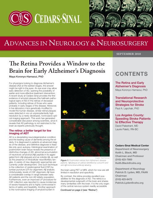
Images: Yosef Koronyo<br />
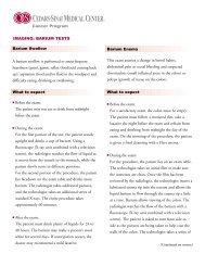
Figure 1: Postmortem retinas from Alzheimer’s disease<br />
patients. Aβ plaques (in yellow) identifi ed by curcumin and<br />
anti-Aβ monoclonal antibody labeling.<br />
<strong>the</strong> brain using PET or MRI, which <strong>for</strong> now are still<br />
limited in resolution and specifi city.<br />
By contrast, <strong>the</strong> retina provides excellent possibilities<br />
<strong>for</strong> <strong>the</strong> diagnosis of AD. <strong>The</strong> retina is a<br />
developmental outgrowth of <strong>the</strong> brain and shares<br />
many similarities with <strong>the</strong> brain. It is <strong>the</strong> only organ<br />
of <strong>the</strong> central nervous system readily accessible<br />
Continued on page 2 (see “<strong>Retina</strong>”)<br />
SEPTEMBER 2010<br />
CONTENTS<br />
<strong>The</strong> <strong>Retina</strong> and <strong>Early</strong><br />
Alzheimer’s Diagnosis<br />
Maya Koronyo-Hamaoui, PhD<br />
Translational Research<br />
and Neuroprotective<br />
Strategies <strong>for</strong> Stroke<br />
Paul A. Lapchak, PhD<br />
Los Angeles County:<br />
Speeding Stroke Patients<br />
<strong>to</strong> Effective <strong>The</strong>rapy<br />
David Palestrant, MD<br />
Laurie Paletz, RN-BC<br />
<strong>Cedars</strong>-<strong>Sinai</strong> Medical Center<br />
Department of Neurosurgery<br />
Keith L. Black, MD<br />
Chairman and Professor<br />
(310) 423-7900<br />
Keith.Black@cshs.org<br />
Department of Neurology<br />
Patrick D. Lyden, MD, FAAN<br />
Chairman<br />
(310) 423-6472<br />
Patrick.Lyden@cshs.org
<strong>Retina</strong>: continued from page 1<br />
<strong>for</strong> direct, noninvasive and repeated live<br />
optical imaging. Because existing noninvasive<br />
brain-imaging technologies can nei<strong>the</strong>r<br />
provide adequate identifi cation nor suffi cient<br />
detail regarding changes in Aβ plaques during<br />
early stages of AD, <strong>the</strong> <strong>Cedars</strong>-<strong>Sinai</strong><br />
research team considered <strong>the</strong> retina as a<br />
better target <strong>for</strong> direct, high resolution live<br />
imaging of AD.<br />
<strong>Retina</strong>l pathology in AD patients<br />
Studies in <strong>the</strong> 1980s and ’90s documented<br />
early and nonspecifi c visual dysfunctions in<br />
AD patients, as well as several retinal abnormalities.<br />
<strong>The</strong>se abnormalities were mostly<br />
related <strong>to</strong> reduction of nerve fi ber layer (NFL)<br />
thickness and loss of retinal ganglion cells in<br />
postmortem retinas of AD patients (1). Such<br />
changes are not specifi c <strong>to</strong> AD, however, as<br />
<strong>the</strong>y appear in o<strong>the</strong>r eye disorders and neurodegenerative<br />
conditions, including ocular<br />
hypertension, glaucoma, multiple sclerosis<br />
and Parkinson’s disease.<br />
Recent reports demonstrated accumulation<br />
of AD-specifi c Aβ deposits in retinal samples<br />
from genetically modifi ed mice modeling AD<br />
(2). <strong>Retina</strong>l plaques in mice were mainly detected<br />
in <strong>the</strong> NFL throughout <strong>the</strong> outer plexi<strong>for</strong>m<br />
layer, and <strong>the</strong>ir prevalence correlated<br />
with disease progression, retinal ganglion<br />
cell degeneration, microglial activation and<br />
functional impairment. Building on this ef<strong>for</strong>t,<br />
<strong>the</strong> <strong>Cedars</strong>-<strong>Sinai</strong> research team turned <strong>to</strong><br />
<strong>the</strong> examination of AD-specifi c deposits in<br />
postmortem retinas of human AD patients.<br />
<strong>The</strong> team was <strong>the</strong> fi rst <strong>to</strong> discover <strong>the</strong> hallmark<br />
Aβ plaque pathology in retinas from<br />
AD patients (Fig. 1), which did not appear in<br />
<strong>the</strong> retinas from non-AD controls of similar<br />
age. Fur<strong>the</strong>rmore, retinal plaque burden appears<br />
<strong>to</strong> correlate with clinical diagnosis and<br />
brain pathology. <strong>The</strong>se fi ndings emphasized<br />
<strong>the</strong> need <strong>to</strong> develop a method of visualizing<br />
retinal plaques, a specifi c diagnostic marker<br />
<strong>for</strong> AD, in live subjects.<br />
<strong>The</strong> role of curcumin labeling<br />
<strong>The</strong> <strong>Cedars</strong>-<strong>Sinai</strong> research team found that<br />
curcumin (diferuloylmethane), a natural and<br />
safe compound of <strong>the</strong> Indian spice turmeric,<br />
is a useful agent <strong>for</strong> imaging retinal plaques.<br />
Curcumin demonstrated an ability <strong>to</strong> cross<br />
<strong>the</strong> blood-brain barrier and binds tightly<br />
<strong>to</strong> brain plaques (3). Likewise, curcumin<br />
crosses <strong>the</strong> blood-retina barrier and binds <strong>to</strong><br />
retinal plaques. When injected <strong>to</strong> <strong>the</strong> peripheral<br />
blood of mice or given orally, curcumin<br />
enabled selective retinal plaque detection in<br />
vivo. By adapting an advanced microscope<br />
designed <strong>to</strong> examine live rodents’ retinas,<br />
<strong>the</strong> team was able <strong>to</strong> demonstrate noninvasive<br />
visualization of individual plaques<br />
at an unprecedentedly high resolution and<br />
sensitivity.<br />
Figure 2: Noninvasive high resolution imaging of Aß<br />
plaques (bright white spots) in <strong>the</strong> retina of live Alzheimer’s<br />
transgenic mouse. Images captured using<br />
Micron III retinal imaging microscope.<br />
<strong>Retina</strong>l Aβ plaques as an early<br />
biomarker<br />
In murine models of AD, <strong>the</strong> research team<br />
found that Aβ plaques emerged in <strong>the</strong> retina<br />
months prior <strong>to</strong> <strong>the</strong>ir appearance in <strong>the</strong><br />
brain and at a very early presymp<strong>to</strong>matic<br />
stage. In postmortem retinas from AD patients,<br />
plaques were found not only in <strong>the</strong><br />
retinas of individuals with defi nitive AD diagnosis,<br />
but also in <strong>the</strong> retinas of those who<br />
were suspected of having early-stage AD.<br />
<strong>Retina</strong>l plaque location and morphology<br />
were documented and compared with those<br />
found in <strong>the</strong> brain. <strong>The</strong> morphology of retinal<br />
plaques suggests that Aβ plaques may<br />
actually appear in <strong>the</strong> retina fi rst. Aβ plaques<br />
with early-stage morphology (condensed,<br />
hard cores containing aggregated Aβ and<br />
<strong>the</strong> absence of radiating fi bril arms, not often<br />
found in <strong>the</strong> brain) were frequently identifi<br />
ed in retinas from AD patients.<br />
<strong>The</strong> successful detection of retinal Aβ<br />
plaques in patients suspected of having AD<br />
is signifi cant <strong>for</strong> <strong>the</strong> future of AD diagnosis<br />
and treatment. O<strong>the</strong>r researchers have<br />
shown a signifi cantly high rate of trans<strong>for</strong>mation<br />
of patients with mild dementia and/<br />
or mild cognitive impairment <strong>to</strong> full AD (4).<br />
Moreover, our study identifi ed Aβ plaques in<br />
<strong>the</strong> retina of a cognitively normal individual<br />
who also had sparsely diffused plaques in<br />
<strong>the</strong> hippocampus, which was discovered<br />
upon brain au<strong>to</strong>psy. Fur<strong>the</strong>r investigation<br />
of retinal plaques in larger numbers of early<br />
patients is warranted <strong>to</strong> determine <strong>the</strong>ir time<br />
of appearance in <strong>the</strong> retina and <strong>to</strong> quantitatively<br />
correlate AD-specifi c pathology in <strong>the</strong><br />
brain <strong>to</strong> <strong>the</strong> retina.<br />
Immuno<strong>the</strong>rapy reduces<br />
retinal Aβ plaques<br />
Although AD currently has no effective cure,<br />
promising <strong>the</strong>rapies <strong>to</strong> slow progression<br />
are being explored. Accumulating research<br />
2 SEPTEMBER 2010 • CEDARS-SINAI ADVANCES IN NEUROLOGY AND NEUROSURGERY<br />
suggests that AD pathology starts very early<br />
during <strong>the</strong> prodromal phase and may occur<br />
decades be<strong>for</strong>e clinical manifestation of<br />
cognitive impairments. Detecting AD be<strong>for</strong>e<br />
symp<strong>to</strong>ms emerge is crucial <strong>for</strong> <strong>the</strong> testing<br />
of treatment interventions that might prevent<br />
or slow mental decline if initiated at a suffi<br />
ciently early stage of <strong>the</strong> disease.<br />
One such treatment in preclinical studies<br />
developed by our group is an immune-modulating<br />
<strong>the</strong>rapy found effective in arresting<br />
cognitive decline and brain pathology in AD<br />
mouse models. A vaccination administered<br />
<strong>to</strong> AD mice recruited monocytes from <strong>the</strong><br />
blood in<strong>to</strong> <strong>the</strong> brain, and <strong>the</strong>se monocytes<br />
were directly involved in Aβ plaque clearance<br />
and dampening of <strong>the</strong> local pro-infl amma<strong>to</strong>ry<br />
responses (5). As a result, a signifi -<br />
cant reduction in Aβ plaque burden in both<br />
retinas and brains was observed (6). <strong>The</strong>se<br />
fi ndings provide fur<strong>the</strong>r evidence <strong>for</strong> a correlation<br />
between retinal and brain plaques and<br />
suggest similar immune mechanisms<br />
of repair.<br />
Conclusion<br />
Future studies are needed <strong>to</strong> demonstrate<br />
<strong>the</strong> ability of optical imaging <strong>to</strong> detect Aβ<br />
plaques in <strong>the</strong> retinas of living AD patients,<br />
especially in early stages of disease. If<br />
this ability is proven, imaging of retinal Aβ<br />
plaques offers <strong>the</strong> possibility of early, noninvasive<br />
and defi nitive diagnosis of AD. It<br />
sets <strong>the</strong> stage <strong>for</strong> proof-of-concept studies<br />
<strong>to</strong> validate retinal plaques as an early biomarker<br />
<strong>for</strong> AD. Most importantly, it provides<br />
what could be a powerful new <strong>to</strong>ol <strong>to</strong> help<br />
evaluate early and more effective <strong>the</strong>rapeutic<br />
approaches <strong>for</strong> Alzheimer’s disease in human<br />
patients.<br />
References<br />
1. Hin<strong>to</strong>n et al, N Engl J Med 1986; Blanks et al,<br />
Neurobiol Aging 1996.<br />
2. Ning et al, Invest Ophthalmol Vis Sci 2008; Perez<br />
et al, Invest Ophthalmol Vis Sci 2009; Koronyo-<br />
Hamaoui et al, NeuroImage 2010.<br />
3. Yang et al, J Biol Chem 2005.<br />
4. Morris and Price, J Mol Neurosci 2001; Petersen<br />
et al, Arch Neurol 1999.<br />
5. Bu<strong>to</strong>vsky and Koronyo-Hamaoui et al, Proc<br />
Natl Acad Sci 2006 and Eur J Neurosci 2007;<br />
Koronyo-Hamaoui et al, J Neurochemistry 2009.<br />
6. Koronyo-Hamaoui et al, NeuroImage 2010.<br />
Dr. Koronyo-Hamaoui is<br />
an Assistant Professor<br />
of Neurosurgery and a<br />
neurosurgery research<br />
scientist at <strong>Cedars</strong>-<br />
<strong>Sinai</strong>.<br />
Maya.Koronyo@<br />
cshs.org
Translational Research and Neuroprotective<br />
Strategies <strong>to</strong> Treat Stroke<br />
Paul A. Lapchak, PhD, FAHA<br />
Alteplase (tissue plasminogen activa<strong>to</strong>r, or<br />
tPA) is currently <strong>the</strong> only FDA-approved<br />
treatment that can be given <strong>to</strong> acute ischemic<br />
stroke (AIS) patients if <strong>the</strong>y present<br />
within three hours of an ischemic stroke.<br />
After 14 years of alteplase clinical research,<br />
evidence from <strong>the</strong> European Cooperative<br />
Acute Stroke Study (ECASS) II now suggests<br />
that <strong>the</strong> <strong>the</strong>rapeutic treatment window<br />
can be expanded <strong>to</strong> 4.5 hours, although this<br />
has not been <strong>for</strong>mally approved by <strong>the</strong><br />
FDA (1).<br />
Translational research using an animal embolic<br />
stroke model with a clinically relevant<br />
endpoint leads <strong>the</strong> way <strong>for</strong> <strong>the</strong> testing of<br />
new neuroprotective strategies in AIS patients<br />
(2). More than 30 years of applied<br />
stroke research shows that <strong>the</strong>re are no<br />
easy fi xes <strong>for</strong> ischemic brain damage, but<br />
neuroprotection using pharmacological<br />
agents or devices shows promise in earlystage<br />
clinical trials.<br />
Near-infrared laser <strong>the</strong>rapy<br />
<strong>The</strong> strategy of using transcranial near-infrared<br />
laser <strong>the</strong>rapy (TLT) with a non-excision<br />
laser device <strong>to</strong> induce pho<strong>to</strong>biostimulation<br />
is based is upon <strong>the</strong> hypo<strong>the</strong>sis that TLT<br />
improves energy metabolism and potentially<br />
enhances cell viability. Moreover, in preclinical<br />
animal studies, TLT is safe, promotes<br />
behavioral recovery following embolic<br />
strokes when administered up <strong>to</strong> six hours<br />
after <strong>the</strong> event, and produces a durable<br />
effect (3). In <strong>the</strong> Neuro<strong>The</strong>ra Effectiveness<br />
and Safety Trial (NEST) 2, TLT was shown <strong>to</strong><br />
be effective in AIS patients with a National<br />
Institute of Health Stroke Scale score of<br />
less than 16 and a mean time-<strong>to</strong>-treatment<br />
of 14.6 hours, signifi cantly longer than <strong>the</strong><br />
<strong>the</strong>rapeutic window <strong>for</strong> alteplase (4, 5). <strong>The</strong><br />
fi nal, defi nitive NEST trial is pending FDA<br />
approval <strong>for</strong> initiation.<br />
Injectable and infusion Radicut ®<br />
Radicut is a multi-target free radical scavenger<br />
approved and marketed in Japan<br />
<strong>to</strong> treat AIS patients presenting within 24<br />
hours of <strong>the</strong> attack (6). Injectable Radicut<br />
ampoules (30 mg administered twice a day<br />
<strong>for</strong> 14 days) were fi rst approved on May 23,<br />
2001, and on January 19, 2010, <strong>the</strong> 30 mg<br />
Radicut Bag <strong>for</strong> IV infusion was approved<br />
<strong>for</strong> use by <strong>the</strong> Japanese Ministry of Health<br />
and Welfare. When administered six <strong>to</strong> 72<br />
hours following an ischemic stroke, <strong>the</strong> effi<br />
cacy of Radicut ranges from large signifi -<br />
cant clinical improvements <strong>to</strong> only modest<br />
improvements in clinical function measured<br />
using standard stroke scales. In a preclinical<br />
animal embolic stroke model, Radicut<br />
administered subcutaneously decreased<br />
behavioral defi cits when given up <strong>to</strong> three<br />
hours post-embolization. Moreover, <strong>the</strong><br />
study showed that Radicut could be administered<br />
in combination with standard dose<br />
intravenous alteplase <strong>the</strong>rapy (7).<br />
In a single randomized clinical trial and numerous<br />
retrospective studies of Radicut, all<br />
research shows Radicut has a signifi cant<br />
effect on clinical, functional or survival parameters<br />
of stroke patients, independent of<br />
<strong>the</strong> time-<strong>to</strong>-treatment and type of stroke.<br />
Based upon <strong>the</strong> available clinical data, Radicut<br />
is most effective when administered<br />
within 24 hours following a stroke. Radicut<br />
is currently being studied <strong>for</strong> safety and<br />
pharmacokinetics in AIS patients in a Phase<br />
IIa multi-center, randomized, double-blind,<br />
placebo-controlled clinical study.<br />
Statins <strong>for</strong> stroke prevention<br />
and recovery<br />
Ano<strong>the</strong>r novel method <strong>to</strong> promote neuroprotection<br />
is by using statins, such as 3-Hydroxy-3-methylglutaryl<br />
coenzyme A (HMG-<br />
CoA) reductase inhibi<strong>to</strong>rs, which were initially<br />
designed as cholesterol-lowering drugs<br />
<strong>for</strong> <strong>the</strong> primary and secondary prevention<br />
of coronary artery disease and AIS. More<br />
recently, <strong>the</strong> pleiotropic biological activities<br />
of statins have been recognized, and many<br />
preclinical stroke studies support <strong>the</strong> testing<br />
of statins in AIS patients. Recently, <strong>the</strong><br />
Stroke Prevention by Aggressive Reduction<br />
in Cholesterol Levels (SPARCL) trial studied<br />
<strong>the</strong> effects of a<strong>to</strong>rvastatin (80 mg per day)<br />
in patients with a his<strong>to</strong>ry of recent AIS or<br />
transient ischemic attack. <strong>The</strong> trial showed<br />
that a<strong>to</strong>rvastatin reduced <strong>the</strong> overall incidence<br />
of strokes and cardiovascular events,<br />
but caused an increase in hemorrhage (8),<br />
which was observed in patients with JNC 7<br />
Stage 2 hypertension.<br />
<strong>The</strong> Neuroprotection with Statin <strong>The</strong>rapy <strong>for</strong><br />
Acute Recovery Trial (NeuSTART) was designed<br />
<strong>to</strong> test <strong>the</strong> hypo<strong>the</strong>sis that high-dose<br />
statins (1 <strong>to</strong> 8 mg/kg/day) administered<br />
within 24 hours of an AIS can promote clinical<br />
recovery (9,10). <strong>The</strong> initial study showed<br />
that high-dose lovastatin (8 mg/kg/day)<br />
could be <strong>to</strong>lerated <strong>for</strong> three days without<br />
adverse effects. While a non-randomized<br />
trial is ongoing, a randomized, double-blind,<br />
placebo-controlled clinical study is required<br />
<strong>to</strong> determine statin effi cacy <strong>to</strong> treat AIS.<br />
With <strong>the</strong> recent failure of numerous neuroprotective<br />
and thrombolytic strategies due<br />
<strong>to</strong> inadequate preclinical testing or <strong>to</strong>xicity,<br />
an emphasis should be placed on effective<br />
translational research studies <strong>to</strong> ultimately<br />
provide <strong>the</strong> best treatment options <strong>to</strong> AIS<br />
patients.<br />
References<br />
1. Hacke W et al, N Engl J Med 2008 Sep<br />
25;359(13):1317-29.<br />
2. Lapchak PA, Translational Stroke Research<br />
2010;1(2):96-107.<br />
3. Lapchak PA et al, SPIE Proceedings<br />
2010;7552:75520R-R-13<br />
4. Lampl Y et al, Stroke 2007 Jun;38(6):1843-9.<br />
5. Zivin JA et al, Stroke 2009 Apr;40(4):1359-64.<br />
6. Lapchak PA, Expert Opinion on Pharmaco<strong>the</strong>rapy<br />
2010;11(10):1753-63.<br />
7. Lapchak PA, Zivin JA, Exp Neurol 2009<br />
Jan;215(1):95-100.<br />
8. Amarenco P et al, N Engl J Med 2006 Aug<br />
10;355(6):549-59.<br />
9. Elkind MS et al, Int J Stroke 2008<br />
Aug;3(3):210-8.<br />
10. Elkind MS et al, Cerebrovasc Dis 2009;<br />
28(3):266-75.<br />
Dr. Lapchak is Direc<strong>to</strong>r<br />
of Translational<br />
Research at <strong>Cedars</strong>-<br />
<strong>Sinai</strong>.<br />
Paul.Lapchak@<br />
cshs.org<br />
CEDARS-SINAI ADVANCES IN NEUROLOGY AND NEUROSURGERY • SEPTEMBER 2010 3
Los Angeles County: Speeding Stroke Patients <strong>to</strong><br />
Eff ective Th erapeutic Intervention<br />
David Palestrant, MD; Laurie Paletz, RN-BC<br />
When local and regional government entities<br />
adopt <strong>the</strong> concept of ambulance-directed<br />
stroke transport, area residents receive<br />
previously unprecedented access <strong>to</strong> early<br />
intervention as well as comprehensive care<br />
<strong>for</strong> especially complicated cases. Although<br />
a few networks have existed <strong>for</strong> more than a<br />
decade, <strong>the</strong> majority began <strong>to</strong> gel when <strong>the</strong><br />
Joint Commission started certifying Primary<br />
Stroke Centers a few years ago. Ef<strong>for</strong>ts by<br />
<strong>the</strong> <strong>Brain</strong> Attack Coalition, <strong>the</strong> American<br />
Stroke Association and o<strong>the</strong>r organizations<br />
encouraging aggressive intervention played<br />
a signifi cant role.<br />
Los Angeles County’s Emergency Medical<br />
Services (EMS) Agency instituted its stroke<br />
response plan last November. Patients suspected<br />
of suffering acute strokes are now<br />
transported directly <strong>to</strong> Approved Stroke<br />
Centers that are committed <strong>to</strong> being prepared<br />
24 hours a day—because while time<br />
<strong>to</strong> treatment is important, <strong>the</strong> type of treatment<br />
is critical. As many as one-third of all<br />
patients with stroke come <strong>to</strong> an emergency<br />
department within <strong>the</strong> appropriate time<br />
window <strong>to</strong> receive intravenous thrombolytic<br />
<strong>the</strong>rapy. A much smaller percentage, however,<br />
actually receive this life- and abilitysparing<br />
intervention.<br />
<strong>Cedars</strong>-<strong>Sinai</strong> was among <strong>the</strong> fi rst nine sites<br />
identifi ed in Los Angeles County as Approved<br />
Stroke Centers, and we have seen<br />
an increase in <strong>the</strong> number of EMS-transported<br />
patients. We currently evaluate 300<br />
<strong>to</strong> 400 patients per year <strong>for</strong> acute stroke,<br />
treating two <strong>to</strong> seven patients a month.<br />
Many patients now receive treatment that<br />
would not have been possible be<strong>for</strong>e <strong>the</strong><br />
county’s emergency stroke system was<br />
activated.<br />
<strong>The</strong> recent experience of a healthy, active<br />
woman in her mid-60s illustrates <strong>the</strong> community<br />
benefi ts of ambulance-directed<br />
acute stroke care. She was leading an exercise<br />
class at a gym one morning when she<br />
collapsed, exhibiting right-side facial droop,<br />
right arm paralysis and aphasia.<br />
Responding paramedics—who previously<br />
would have taken <strong>the</strong> patient <strong>to</strong> <strong>the</strong> closest<br />
hospital, located just blocks from <strong>the</strong> gym—<br />
redirected <strong>to</strong> <strong>Cedars</strong>-<strong>Sinai</strong>, <strong>the</strong> nearest Approved<br />
Stroke Center. <strong>The</strong>y were met at <strong>the</strong><br />
Emergency Department by members of our<br />
“Code <strong>Brain</strong>” team, which includes neurologists<br />
and nurses specializing in stroke, as<br />
well as pharmacists.<br />
<strong>The</strong> patient, who had no previously known<br />
stroke risk fac<strong>to</strong>rs, evaluated at 21 on <strong>the</strong><br />
NIH Stroke Scale, suggestive of a severe<br />
stroke. Computed <strong>to</strong>mography (CT) without<br />
contrast and CT perfusion studies showed<br />
no bleed but distal occlusion of <strong>the</strong> middle<br />
cerebral artery, with decreased blood volume<br />
anteriorly and a mismatched portion of<br />
decreased perfusion.<br />
Within about two hours of symp<strong>to</strong>m onset,<br />
IV tPA was administered, as was <strong>the</strong> anticoagulant<br />
argatroban, offered as part of a<br />
six-center clinical trial investigating this combination<br />
<strong>the</strong>rapy. This is one of several acute<br />
stroke studies currently underway.<br />
Without direct transport <strong>to</strong> a specialized<br />
stroke center and early intervention, odds<br />
would have been stacked against a favorable<br />
outcome. Instead, <strong>the</strong> patient was<br />
hospitalized in <strong>Cedars</strong>-<strong>Sinai</strong>’s specialized<br />
neuro-critical care unit <strong>for</strong> 11 days be<strong>for</strong>e<br />
being transferred <strong>to</strong> inpatient rehabilitation.<br />
She went home 10 days later with only mild<br />
defi cits, minimal need of assistance, and<br />
what appears <strong>to</strong> be a much better outlook<br />
<strong>for</strong> <strong>the</strong> future.<br />
<strong>The</strong> Approved Stroke Center system, in addition<br />
<strong>to</strong> speeding stroke patients <strong>to</strong> rapidresponse<br />
care, is designed <strong>to</strong> improve patient<br />
access <strong>to</strong> <strong>the</strong> appropriate level of care.<br />
<strong>The</strong> system does not require that every Approved<br />
Stroke Center have every capability<br />
or even have a neurosurgeon available at all<br />
times. It does require that a neurosurgeon<br />
be available within two hours of a patient’s<br />
arrival, and <strong>for</strong> smaller centers, this can be<br />
accomplished by transferring <strong>the</strong> patient <strong>to</strong><br />
a nearby center where a neurosurgeon is<br />
standing by.<br />
Toll-Free Physician Referral Line: (888) 508-8881<br />
To meet <strong>the</strong> criteria, smaller Approved<br />
Stroke Centers enter in<strong>to</strong> agreements <strong>to</strong> immediately<br />
transfer certain patients <strong>to</strong> tertiary<br />
centers that offer 24-hour rapid-response<br />
teams, 24-hour neurosurgical coverage,<br />
neurointerventional services with thrombec<strong>to</strong>my<br />
devices, and specialized neuro-critical<br />
care units and stroke/neuro nursing units. If,<br />
<strong>for</strong> example, a patient arriving at a smaller<br />
stroke center is not a candidate <strong>for</strong> IV tPA,<br />
is outside <strong>the</strong> treatment window, or fails<br />
<strong>to</strong> respond <strong>to</strong> treatment as <strong>the</strong> physician<br />
expects, transfer <strong>to</strong> a more comprehensive<br />
center is initiated. Rapid access <strong>to</strong> neuroendovascular<br />
intervention, such as revascularization<br />
devices and experimental stents,<br />
are among <strong>the</strong> tertiary services now available.<br />
While Los Angeles county is not <strong>the</strong> fi rst<br />
metropolitan area <strong>to</strong> implement such a system,<br />
<strong>the</strong> benefi t <strong>to</strong> stroke victims is not <strong>to</strong><br />
be minimized. We strongly encourage our<br />
colleagues in o<strong>the</strong>r cities <strong>to</strong> advocate <strong>for</strong><br />
ambulance transport guidelines and tertiary<br />
stroke care networks that quickly funnel patients<br />
<strong>to</strong> Joint Commission-certifi ed Primary<br />
Stroke Centers.<br />
Department of Neurology . 8730 Alden Dr., Suite 204 East . Los Angeles, CA 90048 . (310) 423-6472 . www.cedars-sinai.edu/neurology<br />
Dr. Palestrant is Direc<strong>to</strong>r<br />
of <strong>the</strong> Stroke Program<br />
and <strong>the</strong> Neuro-critical<br />
Care Unit at <strong>Cedars</strong>-<br />
<strong>Sinai</strong>.<br />
David.Palestrant@<br />
cshs.org<br />
Ms. Paletz, a registered<br />
nurse, is <strong>the</strong> Stroke<br />
Program Coordina<strong>to</strong>r<br />
at <strong>Cedars</strong>-<strong>Sinai</strong>.<br />
Laurie.Paletz@<br />
cshs.org<br />
Department of Neurosurgery . Maxine Dunitz Neurosurgical Institute . 8631 W. Third St., Suite 800E . Los Angeles, CA 90048 . (310) 423-7900 . www.cedars-sinai.edu/neurosurgery