Case Report Biliary Atresia with Situs Inversus: An experience shared
Case Report Biliary Atresia with Situs Inversus: An experience shared
Case Report Biliary Atresia with Situs Inversus: An experience shared

Create successful ePaper yourself
Turn your PDF publications into a flip-book with our unique Google optimized e-Paper software.
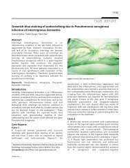
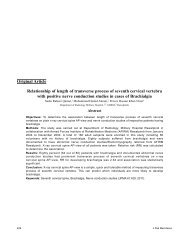
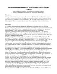
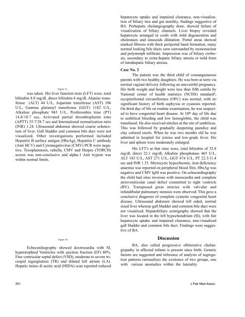
Figure 1a.<br />
was taken. His liver function tests (LFT) were, total<br />
bilirubin 8.8 mg/dl, direct bilirubin 6 mg/dl, Alanine transferase<br />
(ALT) 44 U/L, Aspartate transferase (AST) 106<br />
U/L, Gamma glutamyl transferase (GGT) 1182 U/L,<br />
Alkaline phosphate 943 U/L, Prothrombin time (PT)<br />
14.8/10.7 sec, Activated partial thromboplastin time<br />
(APTT) 35.7/28.7 sec and International normalization ratio<br />
(INR) 1.24. Ultrasound abdomen showed coarse echotexture<br />
of liver. Gall bladder and common bile duct were not<br />
visualized. Other investigations performed included<br />
Hepatitis B surface antigen (HbsAg), Hepatitis C antibody<br />
(<strong>An</strong>ti HCV) and Cytomegalovirus (CMV) PCR were negative.<br />
Toxoplasmosis, rubella, CMV and Herpes (TORCH)<br />
screen was non-conclusive and alpha-1 <strong>An</strong>ti trypsin was<br />
<strong>with</strong>in normal limits.<br />
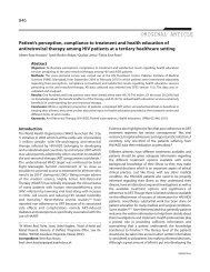
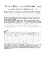
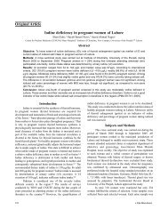
Figure 1b.<br />
Echocardiography showed dextrocardia <strong>with</strong> SI,<br />
hypertrophied Ventricles <strong>with</strong> ejection fraction (EF) 80%.<br />
Fine ventricular septal defect (VSD), moderate to severe tricuspid<br />
regurgitation (TR) and dilated left atrium (LA).<br />
Hepatic imino di acetic acid (HIDA) scan reported reduced<br />
hepatocyte uptake and impaired clearance, non-visualization<br />
of biliary tree and gut motility, findings suggestive of<br />
BA. Perhepatic cholangiography done, showed failure of<br />
visualization of biliary channels. Liver biopsy revealed<br />
hepatocyte arranged in cords <strong>with</strong> mild degeneration and<br />
cholestasis and sinusoids dilatation. Portal areas showed<br />
marked fibrosis <strong>with</strong> thick periportal band formation, many<br />
normal looking bile ducts seen surrounded by mononuclear<br />
and polymorph infiltrate. Impression was of biliary cirrhosis,<br />
secondary to extra-hepatic biliary atresia or mild form<br />
of intrahepatic biliary atresia.<br />
<strong>Case</strong> No. 2<br />
The patient was the third child of consanguineous<br />
parents <strong>with</strong> two healthy daughters. He was born at term via<br />
normal vaginal delivery following an uneventful pregnancy.<br />
His birth weight and height were less than fifth centile by<br />
National center of health statistics (NCHS) standard4, occipitofrontal circumference (OFC) was normal, <strong>with</strong> no<br />
significant history of birth asphyxia or cyanosis reported.<br />
On third day of life on routine examination, he was suspected<br />
to have congenital heart disease. At 10th day of life due<br />
to umbilical bleeding and low hemoglobin, the child was<br />
transfused. He also received stitches at the site of umbilicus.<br />
This was followed by gradually deepening jaundice and<br />
clay colored stools. When he was two months old he was<br />
admitted in hospital for icterus and low-grade fever. His<br />
liver and spleen were moderately enlarged.<br />
His LFT's at that time were, total bilirubin of 32.8<br />
mg/dl, direct 22.1 mg/dl, Alkaline phosphatase 465 U/L,<br />
ALT 183 U/L, AST 271 U/L, GGT 474 U/L, PT 22.3/11.4<br />
sec and INR 1.33. Microcytic hypochromic, iron deficiency<br />
anaemia was reported on peripheral blood film. HbsAg was<br />
negative and CMV IgM was positive. On echocardiography<br />
the child had situs inversus <strong>with</strong> mesocardia and complete<br />
atrioventricular canal defect committed to right ventricle<br />
(RV). Transposed great arteries <strong>with</strong> valvular and<br />
infundibular pulmonary stenosis were observed. This gave a<br />
conclusive diagnosis of complete cyanotic congenital heart<br />
disease. Ultrasound abdomen showed left sided, normal<br />
sized liver whereas gall bladder and common bile duct were<br />
not visualized. Hepatobiliary scintigraphy showed that the<br />
liver was located in the left hypochondrium (SI), <strong>with</strong> fair<br />
hepatocyte uptake and impaired clearance, non-visualized<br />
gall bladder and common bile duct. Findings were suggestive<br />
of BA.<br />
Discussion<br />
BA, also called progressive obliterative cholangiopathy<br />
in affected infants is present since birth. Genetic<br />
factors are suggested and inference of analysis of segregation<br />
patterns rationalizes the existence of two groups, one<br />
<strong>with</strong> various anomalies <strong>with</strong>in the laterality<br />
351 J Pak Med Assoc