Original Article Relationship of length of transverse process of ...
Original Article Relationship of length of transverse process of ...
Original Article Relationship of length of transverse process of ...

You also want an ePaper? Increase the reach of your titles
YUMPU automatically turns print PDFs into web optimized ePapers that Google loves.
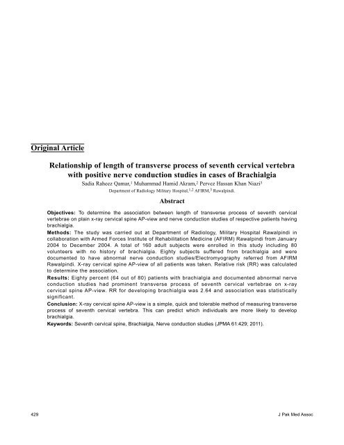
<strong>Original</strong> <strong>Article</strong><br />
<strong>Relationship</strong> <strong>of</strong> <strong>length</strong> <strong>of</strong> <strong>transverse</strong> <strong>process</strong> <strong>of</strong> seventh cervical vertebra<br />
with positive nerve conduction studies in cases <strong>of</strong> Brachialgia<br />
Sadia Raheez Qamar, 1 Muhammad Hamid Akram, 2 Pervez Hassan Khan Niazi 3<br />
Department <strong>of</strong> Radiology Military Hospital, 1,2 AFIRM, 3 Rawalpindi.<br />
Abstract<br />
Objectives: To determine the association between <strong>length</strong> <strong>of</strong> <strong>transverse</strong> <strong>process</strong> <strong>of</strong> seventh cervical<br />
vertebrae on plain x-ray cervical spine AP-view and nerve conduction studies <strong>of</strong> respective patients having<br />
brachialgia.<br />
Methods: The study was carried out at Department <strong>of</strong> Radiology, Military Hospital Rawalpindi in<br />
collaboration with Armed Forces Institute <strong>of</strong> Rehabilitation Medicine (AFIRM) Rawalpindi from January<br />
2004 to December 2004. A total <strong>of</strong> 160 adult subjects were enrolled in this study including 80<br />
volunteers with no history <strong>of</strong> brachialgia. Eighty subjects suffered from brachialgia and were<br />
documented to have abnormal nerve conduction studies/Electromyography referred from AFIRM<br />
Rawalpindi. X-ray cervical spine AP-view <strong>of</strong> all patients was taken. Relative risk (RR) was calculated<br />
to determine the association.<br />
Results: Eighty percent (64 out <strong>of</strong> 80) patients with brachialgia and documented abnormal nerve<br />
conduction studies had prominent <strong>transverse</strong> <strong>process</strong> <strong>of</strong> seventh cervical vertebrae on x-ray<br />
cervical spine AP-view. RR for developing brachialgia was 2.64 and association was statistically<br />
significant.<br />
Conclusion: X-ray cervical spine AP-view is a simple, quick and tolerable method <strong>of</strong> measuring <strong>transverse</strong><br />
<strong>process</strong> <strong>of</strong> seventh cervical vertebra. This can predict which individuals are more likely to develop<br />
brachialgia.<br />
Keywords: Seventh cervical spine, Brachialgia, Nerve conduction studies (JPMA 61:429; 2011).<br />
429 J Pak Med Assoc
Introduction<br />
Brachialgia is defined as "Pain in the arm." 1 Pain is<br />
hallmark <strong>of</strong> acute or chronic nerve root compression. Pain<br />
due to nerve root compression has certain characteristics:<br />
firstly, it tends to follow a dermatomal distribution; secondly<br />
it may be accompanied by paresthesia and thirdly it may be<br />
associated with loss <strong>of</strong> power in the muscles innervated by<br />
the root. 2<br />
Pain in the arm and symptoms caused by cervical<br />
spine (neck) disorders are a very common problem for many<br />
adults. Neck is composed <strong>of</strong> many different anatomic<br />
structures, including muscles, bones, ligaments, nerves and<br />
joints. 3 There are seven cervical vertebrae (C1- C7) in the<br />
vertebral column. The <strong>transverse</strong> <strong>process</strong>es <strong>of</strong> C7 are directed<br />
downward and outward, and are bifid at their extremity.<br />
These are usually <strong>of</strong> large size. Occasionally the <strong>transverse</strong><br />
<strong>process</strong> exists as a separate bone, and attains a large size<br />
(Figure-1). It is then known as "cervical rib." 4 One <strong>of</strong> the<br />
major causes <strong>of</strong> brachialgia at C8 and T1 nerve roots level is<br />
"enlarged seventh cervical <strong>transverse</strong> <strong>process</strong>" (Figure-2). 5<br />
Plain x-rays are still widely used in most units <strong>of</strong><br />
radiology worldwide. 6 Besides other anatomical landmarks;<br />
enlarged seventh cervical <strong>transverse</strong> <strong>process</strong> can be easily<br />
visualized by an antero-posterior view <strong>of</strong> the cervical spine.<br />
In addition to radiological examination, electro-diagnosis<br />
substantially alters the clinical impressions in a large<br />
percentage <strong>of</strong> patients with brachialgia. 7,8<br />
Electro diagnosis or nerve conduction studies/clinical<br />
electromyography (NCS/EMG) is the electrophysiological<br />
Figure-1: Left Cervical Rib and Right transversomegaly.<br />
Figure-2: Bilateral enlarged <strong>transverse</strong> <strong>process</strong>es <strong>of</strong> seventh cervical vertebra.<br />
study <strong>of</strong> nerve and muscle, which is used to diagnose nerve<br />
damage or destruction. 9-11 It is basically a test <strong>of</strong> speed <strong>of</strong><br />
conduction <strong>of</strong> impulses through an electrically stimulated<br />
nerve, usually with surface electrodes, placed on the skin<br />
over the nerve at various locations. 12 The results include<br />
tabulated nerve conduction studies (NCS) and needle EMG<br />
findings. 13-15<br />
The aim <strong>of</strong> this study was to measure the <strong>length</strong> <strong>of</strong> the<br />
<strong>transverse</strong> <strong>process</strong> <strong>of</strong> seventh cervical vertebrae on plain x-<br />
ray cervical spine AP-view and compare it with results <strong>of</strong><br />
nerve conduction studies/Electromyography in patients<br />
having brachialgia.<br />
Patients and Methods<br />
This prospective case-controlled study was carried out<br />
on 160 adult subjects from January 2004 to December 2004.<br />
Patients were divided into two groups. In Group 1 (Controls),<br />
80 subjects with no neurological symptoms were randomly<br />
included from those patients coming to Radiology<br />
Department, Military Hospital Rawalpindi for X-ray<br />
examination <strong>of</strong> regions other than cervical spine. Patients<br />
with trauma to cervical spine, inflammatory and degenerative<br />
conditions were excluded. In Group 2 (study group), 80<br />
patients with symptoms <strong>of</strong> brachialgia were included. These<br />
were those patients who presented in AFIRM, Rawalpindi for<br />
their nerve conduction studies. NCS were performed on<br />
peripheral nerves <strong>of</strong> upper limbs including Ulnar and Median<br />
nerves bilaterally. These nerves were examined across neck,<br />
arm, elbow and forearm using surface electrodes. Values <strong>of</strong><br />
peripheral nerve conduction velocities, amplitude and<br />
latencies were obtained. Motor and sensory velocities in<br />
symptomatic limbs were compared with other limb keeping<br />
50 m/sec as normal velocity <strong>of</strong> Median nerve and 65 m/sec in<br />
Ulnar nerve. Normal value for distal motor latency was 3.7<br />
Vol. 61, No. 5, May 2011 430
ms. EMG studies were done using concentric needle<br />
electrodes. Cervical paraspinal and 1st Dorsal Interossei<br />
muscles were sampled. Sampled muscles were evaluated for<br />
neuropathic patterns. These patients with abnormal nerve<br />
conduction studies and documented pro<strong>of</strong> <strong>of</strong> neurogenic<br />
thoracic outlet syndrome were referred to our department.<br />
Informed consent was obtained from all subjects included in<br />
the study. X-ray cervical spine-AP views <strong>of</strong> both groups were<br />
taken with same x-ray machine considering standard<br />
recommended position <strong>of</strong> patient. Film focus distance was<br />
kept 72 inches in every patient. Spinous <strong>process</strong> <strong>of</strong> cervical<br />
spine was taken as midpoint and the <strong>length</strong>s <strong>of</strong> both the<br />
<strong>transverse</strong> <strong>process</strong>es <strong>of</strong> cervical vertebrae were measured<br />
separately.<br />
Prominent <strong>transverse</strong> <strong>process</strong> rating scale was made in<br />
which, prominent <strong>transverse</strong> <strong>process</strong> was taken as a<br />
<strong>transverse</strong> <strong>process</strong> measuring > 4.5 cm on x-ray cervical<br />
spine AP view. Rating was done for both the <strong>transverse</strong><br />
<strong>process</strong>es separately. Keeping the nerve conduction studies as<br />
gold standard, results were compared between two groups.<br />
Data was expressed in frequencies or mean (standard<br />
deviation) unless stated otherwise. RR was calculated to<br />
determine the relative risk <strong>of</strong> an individual with enlarged<br />
<strong>transverse</strong> <strong>process</strong> for developing brachialgia as compared to<br />
those subjects whose <strong>transverse</strong> <strong>process</strong> was not enlarged.<br />
Chi-square test (c2 test) was used to compare different<br />
proportions among study and control subjects. p-value <strong>of</strong> <<br />
0.05 was considered significant.<br />
Results<br />
The patients included in this study had mean age <strong>of</strong><br />
41.58±11.69 years in group 1 and 39.69±12.12 years in<br />
group 2.<br />
In Group 1 (Controls), size range <strong>of</strong> <strong>transverse</strong><br />
<strong>process</strong>es was 3.2 to 4.9 cm on right side, and 3.1 to 5.2 cm<br />
on left side. In Group 2 (Cases), it was 3.7 to 6.0 cm on right<br />
side and 4.0 to 6.5 cm on left side. Taking 4.5 cm as cut<strong>of</strong>f<br />
point for prominent <strong>transverse</strong> <strong>process</strong>es, in Group1, 46.25%<br />
(32 out <strong>of</strong> 80) subjects had either prominent right or left<br />
<strong>transverse</strong> <strong>process</strong>es. In Group 2, 80% (64 out <strong>of</strong> 80) patients<br />
had prominent <strong>transverse</strong> <strong>process</strong> <strong>of</strong> one side or other.<br />
The relative risk for <strong>length</strong> <strong>of</strong> <strong>transverse</strong> <strong>process</strong><br />
indicating risk <strong>of</strong> development <strong>of</strong> brachialgia was<br />
calculated as 2.64 which means that a person with enlarged<br />
<strong>transverse</strong> <strong>process</strong> <strong>of</strong> seventh cervical vertebra is 2.64<br />
times more likely to develop brachialgia compared with a<br />
person whose <strong>transverse</strong> <strong>process</strong> is not enlarged (Table).<br />
The association between brachialgia and enlarged<br />
<strong>transverse</strong> <strong>process</strong> <strong>of</strong> seventh cervical vertebra, was<br />
statistically significant (p < 0.01).<br />
Results <strong>of</strong> nerve conduction studies (NCS) were<br />
Table: History <strong>of</strong> brachialgia with abnormal nerve conduction<br />
studies (NCS) and enlarged <strong>transverse</strong> <strong>process</strong> cross tabulation.<br />
Risk factor History <strong>of</strong> Brachialgia No History <strong>of</strong><br />
with positive NCS Brachialgia<br />
(Cases)<br />
(Controls)<br />
Enlarged <strong>transverse</strong> <strong>process</strong> 64 (a) 32 (b)<br />
Normal <strong>transverse</strong> <strong>process</strong> 16 (c) 48 (d)<br />
RISK ESTIMATE:<br />
Relative risk (RR) = a/ (a+b) / c/(c+d) = (64/96) / (16/64) = 0.66/0.25 = 2.64.<br />
compared with findings on x-ray cervical spine <strong>of</strong> patients<br />
in Group 2. Patients having symptoms in one limb with<br />
abnormal NCS had increase in size <strong>of</strong> ipsilateral <strong>transverse</strong><br />
<strong>process</strong> <strong>of</strong> seventh cervical vertebrae on plain x-ray cervical<br />
spine AP view. Fifty percent (40 out <strong>of</strong> 80) patients had<br />
increased size <strong>of</strong> left <strong>transverse</strong> <strong>process</strong> on x-ray cervical<br />
spine. Decreased motor and sensory velocities in left Ulnar<br />
and Median nerves were appreciated in 84.3% (33 out <strong>of</strong><br />
40) patients. Increased distal motor latency was observed in<br />
all these patients. Similarly 30% (24 out <strong>of</strong> 80) patients in<br />
Group 2 had increased size <strong>of</strong> right <strong>transverse</strong> <strong>process</strong> on x-<br />
ray cervical spine with 94.7 % (22 out <strong>of</strong> 24) <strong>of</strong> those having<br />
decreased conduction velocities and altered latencies in<br />
right sided Ulnar and Median nerves. Polyphasic giant<br />
potentials and discrete patterns suggestive <strong>of</strong> neuropathic<br />
EMG findings were appreciated in sampled cervical<br />
paraspinal and 1st Dorsal Interossei muscles in 80% (64 out<br />
<strong>of</strong> 80) patients in Group 2.<br />
Discussion<br />
The results <strong>of</strong> this study show that a cervical rib or C7<br />
transversomegaly is a strong predictor for diagnosis <strong>of</strong><br />
brachialgia in the adequate clinical context. Thoracic outlet<br />
region has been studied by advanced radiological modalities<br />
comparing both the affected and the contra lateral sides but<br />
the cervical spine is still best studied by conventional<br />
standard X-ray examination. 16,17<br />
In the presented study, the role <strong>of</strong> x-ray cervical spine<br />
was evaluated for presence <strong>of</strong> enlarged <strong>transverse</strong> <strong>process</strong> as<br />
a predictive risk factor for developing brachialgia. It was<br />
found that an individual with enlarged <strong>transverse</strong> <strong>process</strong> <strong>of</strong><br />
seventh cervical vertebra is 2.64 times more likely to develop<br />
brachialgia as compared to a person whose <strong>transverse</strong> <strong>process</strong><br />
is not enlarged. Thus, association between enlarged<br />
<strong>transverse</strong> <strong>process</strong> <strong>of</strong> seventh cervical vertebra and<br />
brachialgia was significant. This was in accordance to a study<br />
by Matsumoto M et al 18 which showed importance <strong>of</strong><br />
radiological examination especially x-ray cervical spine in<br />
patients having brachialgia.<br />
Like wise the patients having enlarged <strong>transverse</strong><br />
<strong>process</strong>es in our study were also assessed for the side <strong>of</strong> the<br />
lesion; 84.7% had increased size <strong>of</strong> one or other <strong>transverse</strong><br />
431 J Pak Med Assoc
<strong>process</strong> on x-ray cervical spine and had abnormal nerve<br />
conduction studies in the same limb. Marcaud et al 19 in a<br />
study conducted in France showed that in patients having<br />
brachialgia and suspected to have neurogenic thoracic outlet<br />
syndrome, plain radiography exhibited rudimentary bilateral<br />
cervical rib or an elongated ipsilateral C7 <strong>transverse</strong> <strong>process</strong><br />
in 100% <strong>of</strong> the cases.<br />
Similarly a case report from Spain, on 2 female<br />
patients with symptoms <strong>of</strong> brachialgia and positive nerve<br />
conduction studies, showed cervical ribs in one case and<br />
elongated <strong>transverse</strong> <strong>process</strong> <strong>of</strong> C7 in the other, on Xray<br />
cervical spine. This proved that association <strong>of</strong> enlarged C7<br />
<strong>transverse</strong> <strong>process</strong> in patients having brachialgia is always<br />
significant. 20<br />
This finding can be <strong>of</strong> help in the long term<br />
employment <strong>of</strong> manual workers. The employers can get an<br />
increased productivity from workers and lower their expense<br />
on medical treatment and claims. It can also help suffering<br />
individuals in selecting a suitable job.<br />
Conclusion<br />
Prominent <strong>transverse</strong> <strong>process</strong> <strong>of</strong> seventh cervical<br />
vertebra has a definite relationship with brachialgia in high<br />
percentage <strong>of</strong> cases. X-ray cervical spine AP-view is a<br />
simple, quick and tolerable method <strong>of</strong> measuring <strong>transverse</strong><br />
<strong>process</strong> <strong>of</strong> seventh cervical vertebra.<br />
References<br />
1. Dictionary definition <strong>of</strong> Brachialgia. [Homepage <strong>of</strong> Dictionary Barn, A medical<br />
dictionary]. (Online) 2002-2003 (Cited 2004 Dec 02]. Available from URL:<br />
http://www.dictionarybarn.com.<br />
2. Clinical features. [Homepage <strong>of</strong> GPnotebook]. (Online) 2004 (Cited 2004 Dec<br />
24). Available from URL: http://www.gpnotebook.co.uk/homepage.cfm.<br />
3. Symptoms. [Homepage <strong>of</strong> Neck Reference.com]. (Online) 2003 July 11 last<br />
update. (Cited 2004 Nov 28). Available from URL:<br />
http://www.neckreference.com.<br />
4. Gray H. Anatomy <strong>of</strong> the Human body, 20th ed. Vol.1. Philadelphia: Lea &<br />
Febiger, 1918.<br />
5. Wysocki J, Bubrowski M, Reymond J, Kwiatkowski J. Anatomical variants <strong>of</strong><br />
the cervical vertebrae and the first thoracic vertebra in man. Folia Morphol<br />
(Warsz) 2003; 62: 357-63.<br />
6. Sutton, D. Textbook <strong>of</strong> Radiology and Imaging. 7th Ed. Vol.1. New York:<br />
Churchill Livingston; 2003; pp 58.<br />
7. Haig AJ, Tzeng HM, LeBreck DB. The value <strong>of</strong> electro diagnostic<br />
consultation for patients with upper extremity nerve complaints: a prospective<br />
comparison with the history and physical examination. Arch Phys Med<br />
Rehabil 1999; 80: 1273-81.<br />
8. Investigations. [Homepage <strong>of</strong> GPnotebook]. (Online) 2004 (Cited 2004 Dec<br />
24). Available from URL: http://www.gpnotebook.co.uk/homepage.cfm.<br />
9. Katirji B. The clinical electromyography examination. An overview. Neurol<br />
Clin 2002; 20: 291-303.<br />
10. Le Forestier N, Mouton P, Maisonobe T, Fournier E, Moulonguet A, Willer JG,<br />
et al. True neurological thoracic outlet syndrome. Rev Neurol 2000; 156: 34-40.<br />
11. Aetna Inc. Nerve conduction velocity studies. Clinical Policy Bulletin.<br />
American Medical Association 2007; No: 0502.<br />
12. Azeem MA, Rakkah NIA, Mustafa MA, Ali A, Farroq N, Ilyas M. Evaluation<br />
<strong>of</strong> hyperpolarization potentials and nerve conduction parameters in axonal<br />
neuropathic patients. Pak J Physiol 2007; 3: 9.<br />
13. Dale WA. Thoracic outlet compression syndrome. Critique in 1982. Arch Surg<br />
1982; 117: 1437-45.<br />
14. Roos DB. New concepts <strong>of</strong> thoracic outlet syndrome that explain etiology,<br />
symptoms, diagnosis and treatment. J Vasc Surg 1979; 13: 313-21.<br />
15. Sanders RJ, Hammond SL. Management <strong>of</strong> cervical ribs and anomalous first<br />
ribs causing neurogenic thoracic outlet syndrome. J Vasc Surg 2002; 36: 51-6.<br />
16. Remy-Jardin M, Doyen J, Remy J, Artand P, Fribourg M, Ounamel A.<br />
Functional anatomy <strong>of</strong> thoracic outlet : evaluation with spiral CT. Radiology<br />
1997; 205: 843-51.<br />
17. Richter HP. Removal <strong>of</strong> the 1st rib in thoracic outlet syndrome. Is it helpful? Is<br />
it safe? Nervenarzt 1996; 67: 1034-7.<br />
18. Matsumoto M, Ishikawa M, Ishii K, Nishinzawa T, Maruiwa H, Nakamura M,<br />
et al. Usefulness <strong>of</strong> neurological examination for diagnosis <strong>of</strong> the affected level<br />
in patients with cervical compressive myelopathy: prospective comparative<br />
study with radiological evaluation. J Neurosurg Spine 2005; 2: 535-9.<br />
19. Marcaud V, Metral S. [Electrophysiological diagnosis <strong>of</strong> neurogenic thoracic<br />
outlet syndrome]. J Mal Vasc 2000; 25: 175-80.<br />
20. Cruz-Martinez A, Arpa J. Electrophysiological assessment in neurogenic<br />
thoracic outlet syndrome. Electromyogr Clin Neurophysiol 2001; 41: 253-6.<br />
Vol. 61, No. 5, May 2011 432