Greenish-blue staining of underclothing due to ... - Jpma.org.pk
Greenish-blue staining of underclothing due to ... - Jpma.org.pk
Greenish-blue staining of underclothing due to ... - Jpma.org.pk

You also want an ePaper? Increase the reach of your titles
YUMPU automatically turns print PDFs into web optimized ePapers that Google loves.
1192<br />
CASE REPORT<br />
<strong>Greenish</strong>-<strong>blue</strong> <strong>staining</strong> <strong>of</strong> <strong>underclothing</strong> <strong>due</strong> <strong>to</strong> Pseudomonas aeruginosa<br />
infection <strong>of</strong> intertriginous dermatitis<br />
Goknur Kalkan, 1 Fazilet Duygu, 2 Yalcin Bas 3<br />
Abstract<br />
Intertrigo (intertriginous dermatitis) is an<br />
inflamma<strong>to</strong>ry condition <strong>of</strong> the skin folds, induced or<br />
aggravated by heat, moisture, maceration, friction,<br />
and lack <strong>of</strong> air circulation. Intertrigo can become<br />
secondarily infected. Three cases <strong>of</strong> intertrigo are<br />
described herein which presented with greenish-<strong>blue</strong><br />
<strong>staining</strong> <strong>of</strong> <strong>underclothing</strong>. Cultures revealed<br />
Pseudomonas aeruginosa which is a gram-negative<br />
aerobic bacillus that produces the pigments<br />
pyocyanin and pyoverdin that responsible for this<br />
characteristic hue. All three patients responded <strong>to</strong> a<br />
course <strong>of</strong> oral cipr<strong>of</strong>loxacin with resolution <strong>of</strong> the<br />
intertriginous dermatitis. Therefore, greenish-<strong>blue</strong><br />
<strong>staining</strong> <strong>of</strong> clothing is an important indica<strong>to</strong>r for<br />
pseudomonal intertrigo.<br />
Keywords: Pseudomonas aeruginosa, Pyocyanin,<br />
Pyoverdin, Intertrigo.<br />
Introduction<br />
Intertrigo (intertriginous dermatitis) is an inflamma<strong>to</strong>ry<br />
condition <strong>of</strong> the skin folds, induced or aggravated by heat,<br />
moisture, maceration, friction, and lack <strong>of</strong> air circulation. It<br />
therefore has a predilection for moist areas such as the<br />
axilla, perineum, inframammary creases, neck and<br />
abdominal folds. Intertrigo can become colonised or<br />
secondarily infected, most <strong>of</strong>ten by Candida species, but<br />
also by bacterial, fungal, or viral pathogens. 1,2 Three<br />
patients with intertrigo are reported herein that<br />
presented with greenish-<strong>blue</strong> <strong>staining</strong> <strong>of</strong> their<br />
<strong>underclothing</strong>, and the culture <strong>of</strong> the affected skin grew<br />
Pseudomonas aeruginosa.<br />
Case Reports<br />
Case-1<br />
A 55-year-old woman presented with recurrent<br />
intertrigo with greenish-<strong>blue</strong> <strong>staining</strong> <strong>of</strong> her panties<br />
and bra since 2 years with no response <strong>to</strong> <strong>to</strong>pical<br />
antifungals and corticosteroids (Figure-1). On<br />
1,3Department <strong>of</strong> Derma<strong>to</strong>logy, 2 Department <strong>of</strong> Infectious Disease and Clinical<br />
Microbiology, Gaziosmanpasa University School <strong>of</strong> Medicine, Tokat, Turkey.<br />
Correspondence: Goknur Kalkan. Email: goknurkalkan@yahoo.com<br />
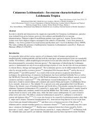
Figure-1: <strong>Greenish</strong>-<strong>blue</strong> stained bra <strong>of</strong> case 1.<br />
examination, a mild erythema<strong>to</strong>us appearance was<br />
detected in the inframammary and inguinal folds and<br />
the underclothes were stained a greenish-<strong>blue</strong> hue in<br />
the corresponding areas. Microscopic examination <strong>of</strong> a<br />
scraping from the inframammary region with 20%<br />
potassium hydroxide was negative for derma<strong>to</strong>phytes<br />
and yeasts. Culture <strong>of</strong> the area grew P. aeruginosa,<br />
Klebsiella pneumoniae and coagulase-negative<br />
staphylococci. The rash cleared after two weeks <strong>of</strong><br />
treatment with oral cipr<strong>of</strong>loxacin 500 mg twice daily<br />
and bacitracin-neomycin sulfate ointment 3 times daily.<br />
There was no further <strong>staining</strong> <strong>of</strong> <strong>underclothing</strong> after the<br />
completion <strong>of</strong> the treatment.<br />
Case-2<br />
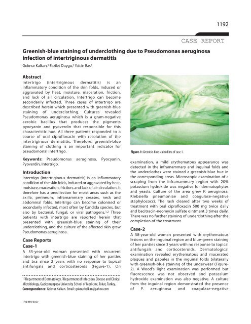
A 58-year-old woman presented with erythema<strong>to</strong>us<br />
lesions on the inguinal region and <strong>blue</strong>-green <strong>staining</strong><br />
<strong>of</strong> her panties since 3 years with no response <strong>to</strong> <strong>to</strong>pical<br />
antifungals and corticosteroids. Derma<strong>to</strong>logical<br />
examination revealed erythema<strong>to</strong>us and macerated<br />
plaques and papules in the inguinal folds bilaterally<br />
with greenish-<strong>blue</strong> <strong>staining</strong> <strong>of</strong> the underwear (Figure-<br />
2). A Wood's light examination was performed but<br />
fluorescence was not observed and potassium<br />
hydroxide examination was also negative. A culture<br />
from the inguinal region demonstrated the presence<br />
<strong>of</strong> P. aeruginosa and coagulase-negative<br />
J Pak Med Assoc
1193 <strong>Greenish</strong>-<strong>blue</strong> <strong>staining</strong> <strong>of</strong> <strong>underclothing</strong> <strong>due</strong> <strong>to</strong> Pseudomonas aeruginosa infection <strong>of</strong> intertriginous dermatitis<br />
and damaged skin associated with intertrigo.<br />
Staphylococcus aureus may present alone or with group<br />
A beta-haemolytic strep<strong>to</strong>coccus (GABHS). 3<br />
Pseudomonas aeruginosa, Proteus mirabilis, or Proteus<br />
vulgaris can also secondarily infect intertriginous areas<br />
as can a variety <strong>of</strong> fungi including yeasts, molds, and<br />
derma<strong>to</strong>phytes. 1,2,4 Since the patients presented here<br />
had all failed therapy with <strong>to</strong>pical antifungals and<br />
corticosteroids for their intertrigo, the affected area was<br />
cultured in search for bacterial pathogens and isolated P.<br />
aeruginosa.<br />
Figure-2: <strong>Greenish</strong>-<strong>blue</strong> discoloration <strong>of</strong> panties <strong>of</strong> case 2.<br />
staphylococcus. She received two weeks <strong>of</strong> treatment<br />
with oral cipr<strong>of</strong>loxacin, <strong>to</strong>pical 1% gentian violet and<br />
bacitracin-neomycin sulfate ointment with complete<br />
resolution.<br />
Case-3<br />
A 48-year-old woman presented with erythema<strong>to</strong>us<br />
follicular and papulopustular lesions on the front and<br />
nape <strong>of</strong> the neck and inframammary folds. On<br />
derma<strong>to</strong>logical examination, a mild erythema<strong>to</strong>us and<br />
macerated appearance was observed in addition <strong>to</strong><br />
papulopustular lesions in the inframammary region. The<br />
neighbouring parts <strong>of</strong> underclothes were observed <strong>to</strong> be<br />
greenish-<strong>blue</strong> stained. There was greenish- <strong>blue</strong> <strong>staining</strong><br />
<strong>of</strong> her bra which she described as turquoise-coloured.<br />
Culture <strong>of</strong> the inframammary skin produced a heavy<br />
growth <strong>of</strong> P. Aeruginosa, Klebsiella pneumoniae and<br />
coagulase-negative staphylococci. The lesions and<br />
inframammary dermatitis cleared with 1 week <strong>of</strong><br />
treatment with oral cipr<strong>of</strong>loxacin 500 mg twice daily and<br />
gentamicin sulfate ointment. The follicular and<br />
papulopustular lesions were probably because <strong>of</strong> P.<br />
aeruginosa infection since they also responded <strong>to</strong> the<br />
treatment.<br />
Discussion<br />
Intertrigo is a superficial inflamma<strong>to</strong>ry skin disorder<br />
involving any area <strong>of</strong> the body where two opposing skin<br />
surfaces can <strong>to</strong>uch, rub or chaff. 1,2 Secondary cutaneous<br />
infections are commonly observed because <strong>of</strong> the moist<br />
Primary cutaneous manifestations <strong>of</strong> P. aeruginosa<br />
infection include pyoderma, infection <strong>of</strong> wounds,<br />
ulcers, and burns, omphalitis, digital intertrigo, green<br />
nail syndrome, folliculitis associated with the use <strong>of</strong><br />
swimming pool, whirlpool, and hot tubs and<br />
exacerbation <strong>of</strong> acne vulgaris. 4 The presence <strong>of</strong> a<br />
greenish exudate, which is fluorescent under Wood's<br />
light and smells <strong>of</strong> ripe fruit, is very characteristic <strong>of</strong><br />
Pseudomonas infection. 5 P. aeruginosa produces<br />
multiple hydrosoluble pigments, the most important<br />
<strong>of</strong> which are pyocyanin and pyoverdin which are<br />
responsible for the characteristic greenish <strong>blue</strong><br />
discoloration <strong>of</strong> Pseudomonas infection. Pyocyanin is<br />
a non-fluorescent <strong>blue</strong> redox-active secondary<br />
metabolite, greenish-<strong>blue</strong> in colour and has an<br />
essential role in the virulence <strong>of</strong> P. aeruginosa.<br />
Pyoverdin is a greenish yellow coloured pigment<br />
which is fluorescent under Wood's light and can be<br />
seen in all pseudomonads and represents a marker for<br />
bacterial differentiation. Some species produce<br />
trimethylamine, an <strong>org</strong>anic metabolite responsible for<br />
the sweet, fruity, grape-like odour so characteristic <strong>of</strong><br />
these infections. 5,6 All three <strong>of</strong> our cases had greenish<strong>blue</strong><br />
<strong>staining</strong> <strong>of</strong> their <strong>underclothing</strong> which can be<br />
attributed <strong>to</strong> pyocyanin and pyoverdin production by<br />
P. aeruginosa.<br />
Yilmaz et al reported a patient with erythroderma and<br />
anticonvulsant hypersensitivity syndrome who<br />
developed pseudomonal intertrigo in the inguinal<br />
region. Green <strong>staining</strong> <strong>of</strong> the underwear was thought <strong>to</strong><br />
be a diagnostic sign for pseudomonal infection. 7 Kaya<br />
et al also reported a similar case <strong>of</strong> pseudomonal<br />
inguinal intertrigo with <strong>blue</strong> discoloration <strong>of</strong> the<br />
underpants. 8<br />
Not only can P. aeruginosa secondarily infect<br />
intertriginous areas, it can additionally cause folliculitis as<br />
occurred in our third patient. Outbreaks <strong>of</strong> pseudomonas<br />
folliculitis associated with the use <strong>of</strong> under-chlorinated<br />
whirlpools, hot tubs, swimming pools, saunas, and<br />
Vol. 63, No. 9, September 2013
G. Kalkan, F. Duygu, Y. Bas 1194<br />
hydrotherapy pools have also been reported. Patients<br />
<strong>of</strong>ten present with follicular, macular, papular, vesicular, or<br />
pustular lesions on any part <strong>of</strong> the body usually starting 8-<br />
48 h after exposure. 9<br />
Conclusion<br />
We need <strong>to</strong> be aware that intertrigo can result in greenish<strong>blue</strong><br />
<strong>staining</strong> <strong>of</strong> <strong>underclothing</strong> if secondarily infected with<br />
P. aeruginosa and consider empirical anti-pseudomonal<br />
treatment in a patient that presents with this condition.<br />
Cipr<strong>of</strong>loxacin appears <strong>to</strong> be a good choice in the<br />
outpatient setting for management <strong>of</strong> uncomplicated<br />
primary pseudomonal cutaneous infections.<br />
References<br />
1. Guitart J, Woodley DT. Intertrigo: a practical approach. Compr Ther<br />
1994; 20: 402-9.<br />
2. Mistiaen P, Poot E, Hickox S, Jochems C, Wagner C. Preventing and<br />
treating intertrigo in the large skin folds <strong>of</strong> adults: a literature<br />
overview. Derma<strong>to</strong>l Nurs 2004; 16: 43-6, 49-57.<br />
3. Honig PJ, Frieden IJ, Kim HJ, Yan AC. Strep<strong>to</strong>coccal intertrigo: an<br />
underrecognized condition in children. Pediatrics 2003; 112:<br />
1427-9.<br />
4. Silvestre JF, Betlloch MI. Cutaneous manifestations <strong>due</strong> <strong>to</strong><br />
Pseudomonas infection. Int J Derma<strong>to</strong>l 1999; 38: 419-31.<br />
5. García-Pa<strong>to</strong>s V, Castells A. Skin infections by Pseudomonas<br />
aeruginosa. Piel 1995; 10: 87-98.<br />
6. Pollack M. Pseudomonas aeruginosa. In: Mandell GL, Douglas<br />
RG Jr., Bennett JE (eds.). Principles and Practice <strong>of</strong> Infectious<br />
Diseases. 3rd ed. New York: Churchill Livings<strong>to</strong>ne; 1990; pp<br />
1673-91.<br />
7. Yilmaz E, Savk E, Oncü S, Güleç GU, Ertugrul B, Sakarya S, et al.<br />
Green <strong>staining</strong> <strong>of</strong> clothing: a signal for pseudomonal infection.<br />
Cutis 2010; 86: 305-8.<br />
8. Kaya TI, Delialioglu N, Yazici AC, Tursen U, Ikizoglu G. Medical pearl:<br />
<strong>blue</strong> underpants sign — a diagnostic clue for Pseudomonas<br />
aeruginosa intertrigo <strong>of</strong> the groin. J Am Acad Derma<strong>to</strong>l 2005; 53:<br />
869-71.<br />
9. Fox AB, Hambrick GW Jr. Recreationally associated Pseudomonas<br />
aeruginosa folliculitis. Report <strong>of</strong> an epidemic. Arch Derma<strong>to</strong>l 1984;<br />
120: 1304-7.<br />
J Pak Med Assoc