A Randomized Clinical Trial Comparing Intralesional Bone Marrow ...
A Randomized Clinical Trial Comparing Intralesional Bone Marrow ...
A Randomized Clinical Trial Comparing Intralesional Bone Marrow ...
Create successful ePaper yourself
Turn your PDF publications into a flip-book with our unique Google optimized e-Paper software.
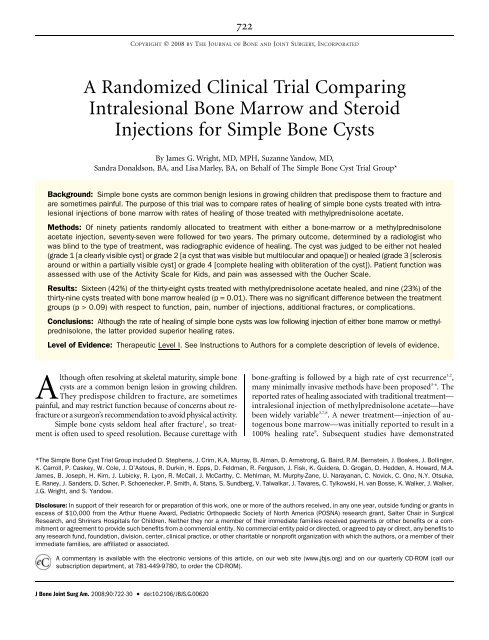
A <strong>Randomized</strong> <strong>Clinical</strong> <strong>Trial</strong> <strong>Comparing</strong><br />
<strong>Intralesional</strong> <strong>Bone</strong> <strong>Marrow</strong> and Steroid<br />
Injections for Simple <strong>Bone</strong> Cysts<br />
By James G. Wright, MD, MPH, Suzanne Yandow, MD,<br />
Sandra Donaldson, BA, and Lisa Marley, BA, on Behalf of The Simple <strong>Bone</strong> Cyst <strong>Trial</strong> Group*<br />
Background: Simple bone cysts are common benign lesions in growing children that predispose them to fracture and<br />
are sometimes painful. The purpose of this trial was to compare rates of healing of simple bone cysts treated with intralesional<br />
injections of bone marrow with rates of healing of those treated with methylprednisolone acetate.<br />
Methods: Of ninety patients randomly allocated to treatment with either a bone-marrow or a methylprednisolone<br />
acetate injection, seventy-seven were followed for two years. The primary outcome, determined by a radiologist who<br />
was blind to the type of treatment, was radiographic evidence of healing. The cyst was judged to be either not healed<br />
(grade 1 [a clearly visible cyst] or grade 2 [a cyst that was visible but multilocular and opaque]) or healed (grade 3 [sclerosis<br />
around or within a partially visible cyst] or grade 4 [complete healing with obliteration of the cyst]). Patient function was<br />
assessed with use of the Activity Scale for Kids, and pain was assessed with the Oucher Scale.<br />
Results: Sixteen (42%) of the thirty-eight cysts treated with methylprednisolone acetate healed, and nine (23%) of the<br />
thirty-nine cysts treated with bone marrow healed (p = 0.01). There was no significant difference between the treatment<br />
groups (p > 0.09) with respect to function, pain, number of injections, additional fractures, or complications.<br />
Conclusions: Although the rate of healing of simple bone cysts was low following injection of either bone marrow or methylprednisolone,<br />
the latter provided superior healing rates.<br />
Level of Evidence: Therapeutic Level I. See Instructions to Authors for a complete description of levels of evidence.<br />
Although often resolving at skeletal maturity, simple bone<br />
cysts are a common benign lesion in growing children.<br />
They predispose children to fracture, are sometimes<br />
painful, and may restrict function because of concerns about refracture<br />
or a surgeon’s recommendation to avoid physical activity.<br />
Simple bone cysts seldom heal after fracture 1 , so treatment<br />
is often used to speed resolution. Because curettage with<br />
722<br />
COPYRIGHT Ó 2008 BY THE JOURNAL OF BONE AND JOINT SURGERY, INCORPORATED<br />
bone-grafting is followed by a high rate of cyst recurrence 1,2 ,<br />
many minimally invasive methods have been proposed 3-6 . The<br />
reported rates of healing associated with traditional treatment—<br />
intralesional injection of methylprednisolone acetate—have<br />
been widely variable 3,7,8 . A newer treatment—injection of autogenous<br />
bone marrow—was initially reported to result in a<br />
100% healing rate 9 . Subsequent studies have demonstrated<br />
*The Simple <strong>Bone</strong> Cyst <strong>Trial</strong> Group included D. Stephens, J. Crim, K.A. Murray, B. Alman, D. Armstrong, G. Baird, R.M. Bernstein, J. Boakes, J. Bollinger,<br />
K. Carroll, P. Caskey, W. Cole, J. D’Astous, R. Durkin, H. Epps, D. Feldman, R. Ferguson, J. Fisk, K. Guidera, D. Grogan, D. Hedden, A. Howard, M.A.<br />
James, B. Joseph, H. Kim, J. Lubicky, R. Lyon, R. McCall, J. McCarthy, C. Mehlman, M. Murphy-Zane, U. Narayanan, C. Novick, C. Ono, N.Y. Otsuka,<br />
E. Raney, J. Sanders, D. Scher, P. Schoenecker, P. Smith, A. Stans, S. Sundberg, V. Talwalkar, J. Tavares, C. Tylkowski, H. van Bosse, K. Walker, J. Walker,<br />
J.G. Wright, and S. Yandow.<br />
Disclosure: In support of their research for or preparation of this work, one or more of the authors received, in any one year, outside funding or grants in<br />
excess of $10,000 from the Arthur Huene Award, Pediatric Orthopaedic Society of North America (POSNA) research grant, Salter Chair in Surgical<br />
Research, and Shriners Hospitals for Children. Neither they nor a member of their immediate families received payments or other benefits or a commitment<br />
or agreement to provide such benefits from a commercial entity. No commercial entity paid or directed, or agreed to pay or direct, any benefits to<br />
any research fund, foundation, division, center, clinical practice, or other charitable or nonprofit organization with which the authors, or a member of their<br />
immediate families, are affiliated or associated.<br />
A commentary is available with the electronic versions of this article, on our web site (www.jbjs.org) and on our quarterly CD-ROM (call our<br />
subscription department, at 781-449-9780, to order the CD-ROM).<br />
J <strong>Bone</strong> Joint Surg Am. 2008;90:722-30 d doi:10.2106/JBJS.G.00620


