Interlocking Contoured Intramedullary Nail Fixation for Selected ...
Interlocking Contoured Intramedullary Nail Fixation for Selected ...
Interlocking Contoured Intramedullary Nail Fixation for Selected ...

You also want an ePaper? Increase the reach of your titles
YUMPU automatically turns print PDFs into web optimized ePapers that Google loves.
d THE J OURNAL OF B ONE &JOINT S URGERY JBJS. ORG<br />
d d VOLUME 90-A N UMBER 9 S EPTEMBER 2008<br />
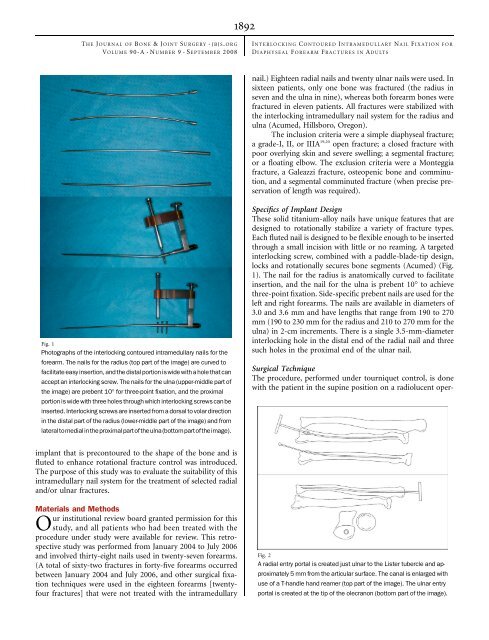
Fig. 1<br />
Photographs of the interlocking contoured intramedullary nails <strong>for</strong> the<br />
<strong>for</strong>earm. The nails <strong>for</strong> the radius (top part of the image) are curved to<br />
facilitate easy insertion,and the distal portion iswide with a hole thatcan<br />
accept an interlocking screw. The nails <strong>for</strong> the ulna (upper-middle part of<br />
the image) are prebent 10° <strong>for</strong> three-point fixation, and the proximal<br />
portion is wide with three holes through which interlocking screws can be<br />
inserted. <strong>Interlocking</strong> screws are inserted from a dorsal to volar direction<br />
in the distal part of the radius (lower-middle part of the image) and from<br />
lateraltomedialintheproximalpartoftheulna(bottompartoftheimage).<br />
implant that is precontoured to the shape of the bone and is<br />
fluted to enhance rotational fracture control was introduced.<br />
The purpose of this study was to evaluate the suitability of this<br />
intramedullary nail system <strong>for</strong> the treatment of selected radial<br />
and/or ulnar fractures.<br />
Materials and Methods<br />
Our institutional review board granted permission <strong>for</strong> this<br />
study, and all patients who had been treated with the<br />
procedure under study were available <strong>for</strong> review. This retrospective<br />
study was per<strong>for</strong>med from January 2004 to July 2006<br />
and involved thirty-eight nails used in twenty-seven <strong>for</strong>earms.<br />
(A total of sixty-two fractures in <strong>for</strong>ty-five <strong>for</strong>earms occurred<br />
between January 2004 and July 2006, and other surgical fixation<br />
techniques were used in the eighteen <strong>for</strong>earms [twentyfour<br />
fractures] that were not treated with the intramedullary<br />
1892<br />
INTERLOCKING C ONTOURED I NTRAMEDULLARY NAIL F IXATION FOR<br />
DIAPHYSEAL F OREARM FRACTURES IN A DULTS<br />
nail.) Eighteen radial nails and twenty ulnar nails were used. In<br />
sixteen patients, only one bone was fractured (the radius in<br />
seven and the ulna in nine), whereas both <strong>for</strong>earm bones were<br />
fractured in eleven patients. All fractures were stabilized with<br />
the interlocking intramedullary nail system <strong>for</strong> the radius and<br />
ulna (Acumed, Hillsboro, Oregon).<br />
The inclusion criteria were a simple diaphyseal fracture;<br />
a grade-I, II, or IIIA 19,20 open fracture; a closed fracture with<br />
poor overlying skin and severe swelling; a segmental fracture;<br />
or a floating elbow. The exclusion criteria were a Monteggia<br />
fracture, a Galeazzi fracture, osteopenic bone and comminution,<br />
and a segmental comminuted fracture (when precise preservation<br />
of length was required).<br />
Specifics of Implant Design<br />
These solid titanium-alloy nails have unique features that are<br />
designed to rotationally stabilize a variety of fracture types.<br />
Each fluted nail is designed to be flexible enough to be inserted<br />
through a small incision with little or no reaming. A targeted<br />
interlocking screw, combined with a paddle-blade-tip design,<br />
locks and rotationally secures bone segments (Acumed) (Fig.<br />
1). The nail <strong>for</strong> the radius is anatomically curved to facilitate<br />
insertion, and the nail <strong>for</strong> the ulna is prebent 10° to achieve<br />
three-point fixation. Side-specific prebent nails are used <strong>for</strong> the<br />
left and right <strong>for</strong>earms. The nails are available in diameters of<br />
3.0 and 3.6 mm and have lengths that range from 190 to 270<br />
mm (190 to 230 mm <strong>for</strong> the radius and 210 to 270 mm <strong>for</strong> the<br />
ulna) in 2-cm increments. There is a single 3.5-mm-diameter<br />
interlocking hole in the distal end of the radial nail and three<br />
such holes in the proximal end of the ulnar nail.<br />
Surgical Technique<br />
The procedure, per<strong>for</strong>med under tourniquet control, is done<br />
with the patient in the supine position on a radiolucent oper-<br />
Fig. 2<br />
A radial entry portal is created just ulnar to the Lister tubercle and approximately<br />
5 mm from the articular surface. The canal is enlarged with<br />
use of a T-handle hand reamer (top part of the image). The ulnar entry<br />
portal is created at the tip of the olecranon (bottom part of the image).