Measuring and segmentation in CT data using deformable models
Measuring and segmentation in CT data using deformable models
Measuring and segmentation in CT data using deformable models

Create successful ePaper yourself
Turn your PDF publications into a flip-book with our unique Google optimized e-Paper software.
<strong>Measur<strong>in</strong>g</strong> <strong>and</strong> <strong>segmentation</strong> <strong>in</strong> <strong>CT</strong> <strong>data</strong> us<strong>in</strong>g <strong>deformable</strong><br />
<strong>models</strong><br />
Václav Krajíček<br />
Faculty of Mathematics <strong>and</strong> Physics<br />
Charles University, Prague<br />
Malostranské nám. 25, 11800 Praha 1,<br />
Czech Republic<br />
vajicek@volny.cz<br />
Josef Pelikán<br />
Faculty of Mathematics <strong>and</strong> Physics<br />
Charles University, Prague<br />
Malostranské nám. 25, 11800 Praha 1,<br />
Czech Republic<br />
Josef.Pelikan@mff.cuni.cz<br />
Abstract<br />
Mart<strong>in</strong> Horák<br />
Department of Radiology,<br />
Bulovka Hospital, Prague<br />
Budínova 2, 180 81 Praha 8 - Libeň,<br />
Czech Republic<br />
mart<strong>in</strong>.horak@volny.cz<br />
Accurate measur<strong>in</strong>g of physical properties of human body has great importance for determ<strong>in</strong><strong>in</strong>g the best treatment. Our work<br />
aims at measur<strong>in</strong>g volumes of organs such as kidney or liver <strong>in</strong> image <strong>data</strong> obta<strong>in</strong>ed from computed tomography (<strong>CT</strong>). We take<br />
advantage of long-time research <strong>in</strong> the area of <strong>deformable</strong> <strong>models</strong>. We have developed parametric model us<strong>in</strong>g closed B-Spl<strong>in</strong>e<br />
curves <strong>and</strong> have formulated energetic equation for their iterative evolution. Interior <strong>and</strong> exterior <strong>in</strong>tensity distributions are taken<br />
<strong>in</strong>to account, together with upcom<strong>in</strong>g shape <strong>and</strong> position of regions <strong>in</strong> neighbour<strong>in</strong>g slices of multi-slice <strong>CT</strong> <strong>data</strong>. This approach<br />
does not require gradient <strong>in</strong>formation, which is unreliable <strong>in</strong> medical images.<br />
Keywords: Deformable Models, Active Snakes, <strong>CT</strong>, Medical Segmentation, Volume Measurement.<br />
1 INTRODU<strong>CT</strong>ION<br />
Computed tomography is a common tool for medical<br />
diagnostics. It simply produces images of a human<br />
body <strong>in</strong>teriors. They serve <strong>in</strong> the same way as X-Ray<br />
images have done for a century. They are as badly readable<br />
as X-Rays for untra<strong>in</strong>ed observer. On the contrary,<br />
<strong>CT</strong> images offer much more <strong>in</strong>formation. They<br />
are based on calibrated 3D <strong>data</strong> (position <strong>and</strong> density),<br />
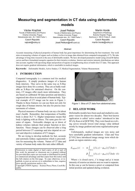
organized <strong>in</strong>to slices <strong>in</strong> axial plane of human body. Typical<br />
example of <strong>CT</strong> image can be seen <strong>in</strong> Figure 1.<br />
Thanks to these features we can use them not only for<br />
rough idea of human <strong>in</strong>terior, but also for precise measurements<br />
of volume.<br />
Physical measures of human body can say a lot about<br />
man’s health. Remember, that temperature of healthy<br />
body is about 36,5 ◦ C. Higher temperature means that<br />
body is fight<strong>in</strong>g with an illness. The same goes for volumes<br />
of organs. Noticeable changes up or down of<br />
kidney-volumes <strong>in</strong>dicate their proper function or dysfunction.<br />
These changes can be observed after a long<br />
period between <strong>CT</strong> scann<strong>in</strong>gs <strong>and</strong> also depend on correct<br />
<strong>and</strong> objective evaluation of <strong>CT</strong> images.<br />
We are try<strong>in</strong>g to develop methods for fast, accurate<br />
<strong>and</strong> automatic kidney <strong>segmentation</strong> which is precondition<br />
for measur<strong>in</strong>g the volume. Nature of <strong>CT</strong> <strong>data</strong> <strong>and</strong><br />
variety of human body make this task rather difficult.<br />
Permission to make digital or hard copies of all or part of<br />
this work for personal or classroom use is granted without<br />
fee provided that copies are not made or distributed for profit<br />
or commercial advantage <strong>and</strong> that copies bear this notice <strong>and</strong><br />
the full citation on the first page. To copy otherwise, or republish,<br />
to post on servers or to redistribute to lists, requires<br />
prior specific permission <strong>and</strong>/or a fee.<br />
Copyright UNION Agency – Science Press, Plzen, Czech Republic<br />
Figure 1: Slice of <strong>CT</strong> <strong>data</strong> from abdom<strong>in</strong>al area.<br />
2 RELATED WORK<br />
Deformable <strong>models</strong> have been used <strong>and</strong> studied <strong>in</strong> computer<br />
vision for almost two decades. Their best known<br />
application is called ’active snakes’ <strong>in</strong>troduced <strong>in</strong> late<br />
80’s by Kass et al [KWT88]. They were based on evolv<strong>in</strong>g<br />
curves towards lowest total energy value def<strong>in</strong>ed<br />
mostly by image gradient <strong>and</strong> some regularization properties.<br />
Unfortunately, medical images are very noisy <strong>and</strong><br />
have unreliable gradient <strong>in</strong>formation. Chan <strong>and</strong> Vese<br />
[CV01] have <strong>in</strong>troduced image energy term which depends<br />
on region statistics only.<br />
Eregion1(s) =<br />
<br />
α<br />
<strong>in</strong>t(s)<br />
<br />
β<br />
ext(s)<br />
( f (x,y) − µext(s)) 2 dxdy +<br />
( f (x,y) − µext(s)) 2 dxdy (1)<br />
Where s is closed curve, f is image <strong>and</strong> µ is mean<br />
<strong>in</strong>tensity of exterior an <strong>in</strong>terior area we want to separate.<br />
In this case µ can be known a priori or computed from<br />
<strong>in</strong>itial position <strong>and</strong> updated dur<strong>in</strong>g evolution.
Figure 2: left: Results of converged B-Spl<strong>in</strong>e snakes without similarity force <strong>in</strong>cluded. right: The same example<br />
of converged snakes with similarity forces <strong>in</strong>cluded us<strong>in</strong>g parameter γ = 0.3.<br />
We are us<strong>in</strong>g another improvement - classical curve<br />
representation is replaced by set of so called ’snaxels’<br />
with closed B-Spl<strong>in</strong>e curve. B-Spl<strong>in</strong>e curve as perfect<br />
tool for contour detection was <strong>in</strong>troduced <strong>in</strong> [BHU00].<br />
This approach reduces number of optimized parameters<br />
to coord<strong>in</strong>ates of control po<strong>in</strong>ts only. Us<strong>in</strong>g B-<br />
Spl<strong>in</strong>e implicitly br<strong>in</strong>gs benefit of <strong>in</strong>tr<strong>in</strong>sic regularization<br />
property. B-Spl<strong>in</strong>e snakes with a very efficient<br />
computational scheme are presented <strong>in</strong> work of Jacob<br />
et al [JBU04].<br />
3 ENERGY MODEL<br />
We utilize a flexibility of snakes framework <strong>and</strong> we<br />
tried to f<strong>in</strong>d new terms which can be <strong>in</strong>corporated <strong>in</strong>to<br />
it. We based our energy model based on regions. Contours<br />
<strong>in</strong> our model are represented by B-Spl<strong>in</strong>e curves.<br />
In our first attempt we extend formula by an additional<br />
term which describes relation to the adjacent<br />
slices <strong>in</strong> <strong>CT</strong> <strong>data</strong>set. This relation is based on a shape<br />
similarity measure. We are try<strong>in</strong>g to f<strong>in</strong>d the best measure<br />
which is able to reduce unwanted behavior of the<br />
evolv<strong>in</strong>g curve.<br />
E(s) = Eregion1(s) + γ · similarity(s,sneigbour) (2)<br />
We tried to f<strong>in</strong>d how to express local high frequency<br />
changes of a shape <strong>in</strong> the similarity measure. This criterion<br />
can <strong>in</strong>dicate overflow of the contour to a adjacent<br />
area through ’bridges’ of similar <strong>in</strong>tensity. These<br />
bridges should be narrow enough to be rendered as<br />
high frequency. High frequency can rise the similarity<br />
term <strong>in</strong> our formulation (2). M<strong>in</strong>imization scheme<br />
can isolate <strong>and</strong> elim<strong>in</strong>ate them because of their significant<br />
differences. We construct shape describ<strong>in</strong>g vector<br />
as Fourier power spectrum of boundary orientation<br />
angle changes. Similarity of two shapes is equal to l2distance<br />
of their shape vectors. It is used as similarity<br />
term <strong>in</strong> (2). In [LAL03] it was shown that this similarity<br />
measure is sensitive to significant differences of<br />
correspond<strong>in</strong>g parts of curves.<br />
Simple Chan <strong>and</strong> Vese region energy scheme (1) <strong>in</strong>corporates<br />
square distance from mean, which is very<br />
rough approximation of <strong>in</strong>verted probability. Instead of<br />
this we are directly us<strong>in</strong>g probability of element classification<br />
to exterior or <strong>in</strong>terior region <strong>in</strong> fashion of<br />
[JBU04]. Furthermore we simplify weight coefficients<br />
α <strong>and</strong> β to α <strong>and</strong> (1 − α) express<strong>in</strong>g balance between<br />
exterior <strong>and</strong> <strong>in</strong>terior. It has only little importance <strong>in</strong> the<br />
new scheme.<br />
<br />
Eregion2(s) = −α<br />
<strong>in</strong>t(s)<br />
<br />
(1 − α)<br />
log(P<strong>in</strong>t( f )) −<br />
ext(s)<br />
log(Pext( f )) (3)<br />
Probability <strong>in</strong> (3) can be approximated by normal distribution<br />
from analysis of <strong>in</strong>itial shape position. We<br />
can also completely rely on user’s <strong>in</strong>itial <strong>in</strong>put <strong>and</strong> use<br />
normalized <strong>and</strong> optionally smoothed histogram of areas<br />
(<strong>in</strong>terior, exterior) as our probability distributions.<br />
There are other <strong>in</strong>terest<strong>in</strong>g region-based energy <strong>models</strong><br />
<strong>in</strong>troduced <strong>in</strong> [JTW99].<br />
Eregion3(s) = − 1<br />
2 (ρ2 <strong>in</strong>t − ρ 2 ext) 2<br />
Eregion4(s) = − 1<br />
2 (µ2 <strong>in</strong>t − µ 2 ext) 2<br />
(4)<br />
(5)<br />
M<strong>in</strong>imization itself can be achieved by the gradient<br />
descent method. Partial derivatives of functional E by<br />
parameters x of the contour are the only terms to compute.<br />
sn+1 = sn + δ · ∇xE(s(x)) (6)<br />
These parameters are <strong>in</strong> our case identical to control<br />
po<strong>in</strong>ts of B-Spl<strong>in</strong>e curve. The most important feature<br />
of the region-based snake <strong>segmentation</strong> is possibility<br />
of reduc<strong>in</strong>g computational complexity from comput<strong>in</strong>g<br />
an area <strong>in</strong>tegral to comput<strong>in</strong>g a curve <strong>in</strong>tegral us<strong>in</strong>g<br />
Green’s theorem.<br />
We did not follow an example of [CV01] add<strong>in</strong>g more<br />
then two types of energy functionals. It encounters
problem of energy equivalence which can not be always<br />
completely solved by parameters tun<strong>in</strong>g. We can not<br />
simply (<strong>in</strong> mean<strong>in</strong>g of l<strong>in</strong>ear relation) answer a question<br />
of how much region energy is equivalent to a unit<br />
of gradient based edge energy, length or area energy.<br />
4 B-SPLINE CURVES<br />
B-Spl<strong>in</strong>e curves are well known spl<strong>in</strong>es us<strong>in</strong>g polygonal<br />
base functions with limited support, which gives<br />
them great local control property. There are few problems<br />
we should h<strong>and</strong>le with.<br />
First is that we must use B-Spl<strong>in</strong>es <strong>in</strong> all energy functionals<br />
<strong>in</strong> place of a contour s. If we work with po<strong>in</strong>ts<br />
on the contour <strong>and</strong> their displacement dur<strong>in</strong>g the m<strong>in</strong>imization<br />
step we must use control po<strong>in</strong>ts only. It is<br />
a big advantage because there are fewer of them then<br />
contour po<strong>in</strong>ts even if sampl<strong>in</strong>g is very rough .<br />
First one - we must use B-Spl<strong>in</strong>es <strong>in</strong> all energy functionals<br />
<strong>in</strong> place of a contour s. If we are work<strong>in</strong>g with<br />
po<strong>in</strong>ts on the contour <strong>and</strong> their displacement dur<strong>in</strong>g the<br />
m<strong>in</strong>imization step we must use control po<strong>in</strong>ts only. This<br />
is a big advantage because there are fewer of them then<br />
contour po<strong>in</strong>ts, even if sampl<strong>in</strong>g is very rough.<br />
Order of B-Spl<strong>in</strong>e is another question. Cubic B-<br />
Spl<strong>in</strong>e is good enough for our purpose.<br />
Number of control po<strong>in</strong>ts or number of B-Spl<strong>in</strong>e sections<br />
depends on length of the whole closed contour.<br />
We def<strong>in</strong>e m<strong>in</strong>imal <strong>and</strong> maximal length of segment <strong>and</strong><br />
dur<strong>in</strong>g iterations we check the length. If our limits are<br />
exceeded we split segment or merge adjacent segments<br />
as necessary.<br />
The last problem relates to <strong>in</strong>tersections. Sometimes<br />
evolv<strong>in</strong>g curve happens to be self-<strong>in</strong>tersect<strong>in</strong>g. For example<br />
if we are segment<strong>in</strong>g shape with holes, curve always<br />
<strong>in</strong>tertw<strong>in</strong>es around the hole <strong>and</strong> cycles forever.<br />
One solution consists <strong>in</strong> detect<strong>in</strong>g <strong>in</strong>tersections <strong>and</strong><br />
break<strong>in</strong>g off parts of the curve. Free ends of the bigger<br />
part of the curve will be sticked together <strong>and</strong> the rest<br />
can be thrown away. We could also use the rest of the<br />
curve as an <strong>in</strong>ner structure <strong>segmentation</strong>. This can be<br />
seen when segment<strong>in</strong>g higher or lower parts of kidney,<br />
which has pelvis <strong>in</strong> the middle(pelvis has very different<br />
tissue density). We might ignore it first time <strong>and</strong> later<br />
we can segment it by simple treshold<strong>in</strong>g. Intersection<br />
detection can use convex wrapper property of B-Spl<strong>in</strong>e<br />
segments for computation speedup.<br />
In [JBU04] they use <strong>in</strong>tegration of angel changes<br />
which gives various multiples of 2π depend<strong>in</strong>g on number<br />
<strong>and</strong> direction of loops. This method is detect<strong>in</strong>g<br />
loops which implicate self-<strong>in</strong>tersections, but is <strong>in</strong>effective<br />
<strong>in</strong> cases described above.<br />
5 SEGMENTATION PROCEDURE<br />
We are try<strong>in</strong>g to segment 3D volume us<strong>in</strong>g separate <strong>segmentation</strong><br />
of each slice. Shape similarity measure is<br />
used as <strong>in</strong>ter-slice relation which b<strong>in</strong>ds adjacent slices<br />
together. Reasons for our preference to this scheme<br />
(compared to pure 3D <strong>deformable</strong> model technique like<br />
Active Surfaces) are <strong>in</strong>homogeous <strong>data</strong> <strong>and</strong> computational<br />
efficiency which is far better <strong>in</strong> the case of 2D<br />
techniques. We are work<strong>in</strong>g with <strong>data</strong>sets of voxels<br />
which are larger <strong>in</strong> one dimension then <strong>in</strong> other two.<br />
In case of pure 3D technique, evolution <strong>in</strong> two different<br />
directions could be <strong>in</strong>comparable. This approach<br />
offers almost <strong>in</strong>teractive response <strong>and</strong> better user control(better<br />
then edit<strong>in</strong>g 3D surface control po<strong>in</strong>ts).<br />
Our <strong>segmentation</strong> procedure starts specify<strong>in</strong>g top <strong>and</strong><br />
bottom end po<strong>in</strong>ts of segmented organ by a user. Optionally<br />
user can set other po<strong>in</strong>ts <strong>in</strong> slices between top<br />
<strong>and</strong> bottom ends, which can help to determ<strong>in</strong>e start<strong>in</strong>g<br />
positions. Start<strong>in</strong>g positions on other slices are placed<br />
automatically between these which were set manually<br />
by the user. Initial shape is def<strong>in</strong>ed as small circle<br />
around these po<strong>in</strong>ts. Probability distribution is estimated<br />
from area <strong>in</strong>side these circles. It is important to<br />
put them <strong>in</strong> the right place <strong>in</strong>side segmented organ. The<br />
user can optionally supply probability distribution from<br />
manually segmented shape <strong>in</strong> one slice.<br />
Iteration starts after shape <strong>in</strong>itialization <strong>and</strong> placement<br />
on each slice. In each iteration of the procedure<br />
we perform one step of snake energy m<strong>in</strong>imization.<br />
Thanks to this scheme the whole object is chang<strong>in</strong>g<br />
its shape dur<strong>in</strong>g <strong>segmentation</strong> process <strong>and</strong> thus can<br />
be observed <strong>and</strong> possibly corrected by the user. After<br />
one of slices converges it became steady po<strong>in</strong>t for<br />
its neighbors <strong>and</strong> is available for use <strong>in</strong> similarity term<br />
<strong>in</strong> equation (2). Neighbors of already converged slices<br />
are restricted by similarity <strong>in</strong> their further evolution.<br />
This scheme works well for rounded organs whose axial<br />
slices do not change topology <strong>and</strong> shape variation<br />
too rapidly.<br />
6 RESULTS<br />
Convergence of the basic model (2) is strongly dependent<br />
on parameters α,β,γ. The model (3), if supplied<br />
with proper probability distribution, gives very promis<strong>in</strong>g<br />
results. Our improvement with similarity measure<br />
can help to hold a curve <strong>in</strong> correct shape as can be seen<br />
<strong>in</strong> Figure 2.<br />
We are work<strong>in</strong>g with native images of a kidney<br />
checkup <strong>and</strong> also with images of the same checkup with<br />
an contrast agent <strong>in</strong> several phases of satiation. In each<br />
case different strategy <strong>in</strong> parameter sett<strong>in</strong>g <strong>and</strong> probability<br />
distribution should be used to obta<strong>in</strong> optimal result.<br />
Native images work well with approximation by<br />
normal distribution. But <strong>in</strong> a case of th<strong>in</strong> patient lack<strong>in</strong>g<br />
of contrast <strong>in</strong>ter-organ fat, it is almost impossible to f<strong>in</strong>d<br />
a distribution which prevents overflow<strong>in</strong>g to neigbour<strong>in</strong>g<br />
organs without los<strong>in</strong>g too much tissue on the surface<br />
of segmented organ. Us<strong>in</strong>g this scheme on slightly satiated<br />
kidney we are able to segment the whole organ <strong>and</strong><br />
determ<strong>in</strong>e its volume well. Figure 4 shows f<strong>in</strong>al results
Figure 3: Histograms of segmented shapes. First<br />
graph from native <strong>CT</strong> corresponds to normal distribution.<br />
Second graph from an image with medium satiation<br />
of the contrast agent demonstrates slightly skewed<br />
distribution.<br />
of our efforts. The approximation by normal distribution<br />
fails on a highly satiated kidney because it does<br />
not correspond to reality as shown <strong>in</strong> Figure 6. On the<br />
other side f<strong>in</strong>e-tuned probability distribution works well<br />
on majority of patients.<br />
A speed of the <strong>segmentation</strong> procedure is satisfy<strong>in</strong>g.<br />
Process<strong>in</strong>g of a five-millimeter thick slices <strong>data</strong>set takes<br />
about 80-90 seconds (<strong>in</strong>tersection check is performed<br />
after each 40 iterations, convergence is achieved after<br />
about 400 iterations). On a two-millimeter slices<br />
<strong>data</strong>set it takes 160 seconds, but the measurement<br />
is more precise. Time measurements were done on<br />
AthlonXP 2600+ computer.<br />
7 CONCLUSION AND FUTURE<br />
WORK<br />
We proved that our task can be effectively solved by<br />
<strong>segmentation</strong> us<strong>in</strong>g <strong>deformable</strong> <strong>models</strong>. Build<strong>in</strong>g efficient<br />
scheme for faster automatic <strong>segmentation</strong> will be<br />
our next goal. We have improved our framework by <strong>in</strong>corporat<strong>in</strong>g<br />
methods based on Green’s theorem which<br />
speed up computations of region <strong>in</strong>tegral <strong>and</strong> its derivative.<br />
Detect<strong>in</strong>g self-<strong>in</strong>tersection is the slowest component<br />
of our algorithm, but also <strong>in</strong>troduces simple topol-<br />
Figure 4: Schematic model(left) <strong>and</strong> 3D model(right) of<br />
segmented kidney.<br />
ogy control (detection of holes). We are go<strong>in</strong>g to implement<br />
other enhancements like parallel computation<br />
of distant slices or multi-scale computations. Results<br />
of our measurements will be evaluated <strong>and</strong> assessed by<br />
medical specialist to determ<strong>in</strong>e its accuracy <strong>and</strong> possible<br />
usability <strong>in</strong> practice.<br />
ACKNOWLEDGEMENTS<br />
This work is supported by GAUK grant No. 359/2006<br />
(Charles University <strong>in</strong> Prague). MUDr. Mart<strong>in</strong> Horák<br />
from Bulovka Hospital <strong>in</strong> Prague is provid<strong>in</strong>g us with<br />
all necessary <strong>data</strong>, medical knowledge <strong>and</strong> experience.<br />
REFERENCES<br />
[BHU00] Brigger, P., Hoeg, J., <strong>and</strong> Unser, M. (2000).<br />
B-Spl<strong>in</strong>e snakes: A flexible tool for parametric contour<br />
detection. IEEE Transactions on Image Process<strong>in</strong>g,<br />
9(9):1484–1496.<br />
[CV01] Chan, T. <strong>and</strong> Vese, L. (2001). Active contours<br />
without edges. IEEE Trans. Image Process<strong>in</strong>g,<br />
10(2):266–277.<br />
[JBU04] Jacob, M., Blu, T., <strong>and</strong> Unser, M. (2004).<br />
Efficient energies <strong>and</strong> algorithms for parametric<br />
snakes. IEEE Transactions on Image Process<strong>in</strong>g,<br />
13(9):1231–1244.<br />
[JTW99] Jr, A. Y., Tsai, A., <strong>and</strong> Willsky, A. (1999).<br />
A statistical approach to snakes for bimodal <strong>and</strong> trimodal<br />
imagery. In ICCV ’99: Proceed<strong>in</strong>gs of the International<br />
Conference on Computer Vision-Volume<br />
2, page 898, Wash<strong>in</strong>gton, DC, USA. IEEE Computer<br />
Society.<br />
[KWT88] Kass, M., Witk<strong>in</strong>, A., <strong>and</strong> Terzopoulos, D.<br />
(1988). Snakes: Active contour <strong>models</strong>. International<br />
Journal of Computer Vision, 1(4):321–331.<br />
[LAL03] Lee, D., Antani, S., <strong>and</strong> Long, L. R. (2003).<br />
Similarity measurement us<strong>in</strong>g polygon curve representation<br />
<strong>and</strong> fourier descriptors for shape-based<br />
vertebral image retrieval.