Common pediatric surgery problems.pdf
Common pediatric surgery problems.pdf
Common pediatric surgery problems.pdf

You also want an ePaper? Increase the reach of your titles
YUMPU automatically turns print PDFs into web optimized ePapers that Google loves.
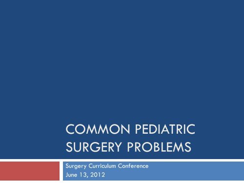
COMMON PEDIATRIC<br />
SURGERY PROBLEMS<br />
Surgery Curriculum Conference<br />
June 13, 2012
Case 1<br />
39 week gestational age<br />
Normal pregnancy and vaginal delivery<br />
Apgars 9 1, 10 5<br />
Started breastfeeding and started to have multiple<br />
episodes of bright yellow/green emesis
Prenatal work-up<br />
Trisomy 21<br />
Normal ultrasound at 18weeks<br />
Mom has negative serologies
Case 1<br />
Clinical examination<br />
HR 160, RR 40, BP 80/50<br />
O2 sats 100% room air<br />
HEENT: macroglossia, epicanthic fold of the eyelid,<br />
upslanting palpebral fissures<br />
Chest: Good AE=AE<br />
Cardiac: holosystolic III/VI murmur, normals S1, S2<br />
Abdomen: non distended, soft, nontender, no erythema, no<br />
HSM<br />
Normal female genitalia<br />
MSK/Neuro: Simian crease, slight decreased muscle tone.
Case 1<br />
Investigations<br />
Bloodwork<br />
Imaging<br />
Any other diagnostic tests?
ECHO<br />
VSD<br />
Normal BMP, CBC, neg cultures
Neonatal Emesis DDx<br />
Upper GI<br />
Duodenal atresias/webs<br />
small bowel atresias<br />
malrotation/midgut volvulus<br />
GERD<br />
Meconium ileus<br />
pyloric stenosis<br />
Inguinal hernia<br />
NEC
Neonatal Emesis DDx<br />
Lower GI<br />
Colonic atresia<br />
Meconium plug<br />
Hirschsprung’s<br />
Small Left Colon Syndrome<br />
Microcolon-Intestinal Hypoperistalsis Syndrome<br />
Imperforate anus
Neonatal Emesis DDx<br />
Medical causes<br />
Sepsis<br />
Metabolic disorders<br />
Hypothyroidism<br />
Electrolyte disturbances<br />
GERD
Radiological workup<br />
KUB/Cross-table lateral<br />
Contrast enemas for distal obstructions<br />
UGI for malrotation/proximal atresias
Duodenal atresia<br />
Management<br />
NGT<br />
Resuscitate<br />
Surgical approach<br />
duodenoduodenostomy
Which of the following is TRUE regarding duodenal<br />
atresia?<br />
A. It is associated with trisomy 21 in 10% cases.<br />
B. Abdominal X-ray is usually normal.<br />
C. Results from disruption of fetal blood supply.<br />
D. Operative repair involves duodenal resection.<br />
E. Concomitant abnormalities can include annular pancreas,<br />
esophageal atresia, or VACTERL lesions.
Duodenal atresia
Duodenal Atresia<br />
Failure to recanalize lumen of duodenum after<br />
solid phase of embryologic development<br />
Distal atresias are due to vascular events<br />
Associated with Down’s syndrome in 30%<br />
Atresia seen in 10% of Down’s patients<br />
Vomiting can be bilious or non-bilious<br />
Abdominal X-ray shows “double-bubble”<br />
Best repaired by bypass -><br />
duodenoduodenostomy or duodenojejunostomy<br />
no indication to divide annular pancreas
Case 2<br />
2 day old infant in newborn nursery<br />
Sent to NICU for evaluation of bilious emesis
Physical Examination<br />
HR 165, RR 50, O2 sats 98 RA<br />
HEENT<br />
Normal oropharynx<br />
Chest<br />
Clear, AE=AE<br />
Cardiac<br />
Normal HS, good peripheral pulses<br />
Abdo<br />
Nondistended, generalized tenderness<br />
Soft, no discoloration, no masses, no HSM<br />
No inguinal hernias
Work-up<br />
Bloodwork<br />
Imaging
Normal embryologic rotation
Abnormal rotation and nonfixation
Management of Malrotation/volvulus<br />
Resuscitate<br />
Urgent <strong>surgery</strong>
Steps to correcting malrotation<br />
1. Entry into abdominal cavity and evisceration (open)<br />
2. Counterclockwise detorsion of the bowel (acute<br />
cases)<br />
3. Division of Ladd’s cecal bands<br />
4. Broadening of the small intestine mesentery<br />
5. Incidental appendectomy<br />
6. Placement of small bowel along the right lateral<br />
gutter and colon along the left lateral gutter
Ladd Procedure
Malrotation<br />
Occurs in 1/200 – 1/500 live births<br />
Symptomatic in 1/6000 live births<br />
30-62% have associated anomaly<br />
Up to 75% present w/in 1st month of life<br />
Classic presentation is infant with bilious emesis<br />
May present as pain, duodenal obstruction,<br />
malnutrition, acute abdomen/shock
Malrotation<br />
Due to abnormal fixation of midgut to<br />
retroperitoneum – leads to narrow base of<br />
mesentery which can easily twist<br />
Ladd Procedure<br />
Reduce volvulus by rotating counterclockwise<br />
Division of Ladd’s bands between cecum and<br />
duodenum/right gutter<br />
Division of adhesions to widen mesentery<br />
Run bowel to r/o obstructions<br />
Appendectomy<br />
Place bowel in nonrotated position
Case 3<br />
2 day old infant in NICU 3<br />
Consulted for abdominal distention and bilious<br />
emesis<br />
Work-up and differential
Pathophysiology of intestinal atresias<br />
How would you confirm diagnosis and what would<br />
you see<br />
Classification scheme
Case 4<br />
3 day old infant<br />
Failure to pass stools, abdominal distention and<br />
bilious emesis<br />
Work-up and differential
What other tests should be done<br />
Sweat test for CF<br />
Genetic testing for CFTR gene mutation
Which of the following is FALSE regarding meconium<br />
ileus?<br />
A. Underlying diagnosis is usually cystic fibrosis.<br />
B. Most often requires operative intervention.<br />
C. Presents as a neonatal bowel obstruction.<br />
D. X-rays may reveal a stippled pattern in the RLQ (“soap<br />
bubble” sign).<br />
E. May be relieved by water-soluble contrast enema.
Meconium Ileus<br />
Newborn bowel obstruction secondary to<br />
inspissated meconuim in distal ileum<br />
Enema reveals microcolon -> may be therapeutic<br />
Non-operative management successful in 2/3<br />
OR required for perforation or failed enema<br />
may flush bowel with N-acetylcysteine in saline
Management<br />
Fluid resuscitaion<br />
Gastric decompression<br />
Pulmonary support as needed<br />
Contrast enema with water soluble contrast<br />
Failure of nonoperative management<br />
Surgery<br />
2-4% NAC, 50% hyperosmolar agent via appendix<br />
Alternative surgical techniques involve resection, anastomosis, and<br />
temporary enterostomy through which postoperative irrigations<br />
may be delivered
Simple vs Complicated meconium ileus<br />
Complicated<br />
Volvulus<br />
Perforation resulting in meconium peritonitis<br />
adhesive meconium peritonitis<br />
giant cystic meconium peritonitis or pseudocyst<br />
meconium ascites<br />
infected meconium peritonitis
Case 5<br />
An 8 hr old infant drools and spits up his first feed. A<br />
tube is passed into the esophagus and a film is<br />
obtained.<br />
What is the diagnosis?
Esophageal Atresia and<br />
Tracheoesophageal Fistula<br />
Incomplete partitioning of primitive foregut<br />
5 types of atresias<br />
Esophageal atresia with distal TEF most common<br />
8% 1% 85% 2% 4%
Esophageal Atresia and<br />
Tracheoesophageal Fistula<br />
Can be part of VACTERL anomalies<br />
vertebral, anal, cardiac, TEF, renal, limb<br />
Atresias detected by inability to pass NGT/OGT<br />
TEF w/o atresia presents with recurrent aspiration<br />
Low-risk infants should get primary repair<br />
long gap (>3 vertebral bodies) repair is delayed<br />
high-risk babies get gastrostomy<br />
Post-op complications include esophageal leak,<br />
dysmotility, GE reflux, strictures
Case 6<br />
A listless 9-month-old boy presents with acute onset<br />
of severe intermittent abdominal pain. Rectal<br />
exam is guaiac positive. What is the most likely<br />
diagnosis?<br />
A. Meckel’s diverticulum.<br />
B. Acute appendicitis.<br />
C. Intussusception.<br />
D. Intestinal polyp.<br />
E. Gastritis.
A. Meckel’s diverticulum.<br />
B. Acute appendicitis.<br />
C. Intussusception.<br />
D. Intestinal polyp.<br />
E. Gastritis.
Intussusception<br />
<strong>Common</strong>ly affects children 3 months to 2 yrs<br />
severe crampy abdominal pain (every 10-20 minutes)<br />
vomiting, “currant jelly” stools<br />
tender, sausage-like mass in RUQ<br />
Telescoping of terminal ileum into large intestine<br />
Contrast enema for diagnosis will reduce 80%<br />
air pressure to 120 mmHg, barium to 100 cm H 2O<br />
10% recurrence, often within hours<br />
OR reduction if not reduced radiographically<br />
5% of patients need resection
Intussusception<br />
Plain AXR<br />
Look for gas in cecum<br />
Abdominal ultrasound – look for target
Which of the following statements is TRUE with respect<br />
to neonatal abdominal wall defects?<br />
A. The bowel in omphalocele is covered by a sac.<br />
B. Gastroschisis is frequently associated with other anomalies.<br />
C. A Silastic silo is rarely employed in management of these<br />
defects.<br />
D. Mortality is higher in gastroschisis.<br />
E. Operative management of omphalocele usually requires<br />
bowel resection.
Omphalocele<br />
Occur 1 in 5000 live births, more common in boys<br />
over 50% have associated cardiac, GI, GU,<br />
musculoskeletal, or CNS anomalies<br />
Herniation of abdominal contents through defective<br />
umbilical ring<br />
overlying sac of outer amnion and peritoneum<br />
umbilical cord in continuity with sac<br />
liver involved in larger defects<br />
High mortality (30-60%) due to other anomalies
Omphalocele
Omphalocele<br />
Non-operative management with escharotic agent<br />
OR for reduction and closure of abdominal wall<br />
keep intra-abdominal pressure < 20 mmHg<br />
large defects require skin flap or prosthetic<br />
Silastic silo most common, reduce daily for 3-10 days<br />
Post-op complications include sepsis, GE reflux,<br />
inguinal hernias, abdominal wall hernia
Gastroschisis<br />
Anterior abdominal wall defect (“belly cleft”)<br />
usually to right of umbilical cord<br />
no sac or membrane covering contents<br />
exposed bowel thick, edematous, exudative peel<br />
associated intestinal atresias in 10%<br />
Initial management<br />
aggressive fluid replacement (2-3X normal)<br />
protection of exposed bowel w/occlusive dressing
Uterus +<br />
Fallopian Tube<br />
Bladder<br />
Small bowel<br />
Colon<br />
Stomach
Gastroschisis<br />
Primary reduction and closure in 80-90% cases<br />
Silastic silo if high intra-abdominal pressure<br />
may require resection if exposed bowel non-viable<br />
Post-op complications:<br />
abdominal compartment syndrome<br />
sepsis<br />
necrotizing enterocolitis<br />
abdominal wall cellulitis<br />
prolonged ileus<br />
short gut syndrome w/ TPN dependence
Case 7<br />
3. A 1.5 kg, 30-wk preemie develops abdominal distention<br />
and bloody stool after 1st feedings. Which of the following is<br />
TRUE regarding his condition?<br />
A. Supportive treatment includes stopping all feeds, NGT<br />
drainage, IVF, serial abdominal exams and radiographs.<br />
B. IV antibiotics not indicated unless pathogen identified.<br />
C. Barium enema is the imaging modality of choice.<br />
D. Overall mortality reported as 50-60%.<br />
E. Intestinal stricture formation is rare.
Case 7<br />
A. Supportive treatment includes stopping all feeds, NGT<br />
drainage, IVF, serial abdominal exams and radiographs.<br />
B. IV antibiotics not indicated unless pathogen identified.<br />
C. Barium enema is the imaging modality of choice.<br />
D. Overall mortality reported as 50-60%.<br />
E. Intestinal stricture formation is rare.
Necrotizing Entercolitis (NEC)<br />
Idiopathic mucosal intestinal injury, may progress to<br />
transmural necrosis<br />
1/2 patients < 1500 g (7% incidence), 80% < 2500 g<br />
at birth<br />
90% in premature neonates
Necrotizing Entercolitis (NEC)<br />
Signs:<br />
feeding intolerance<br />
vomiting<br />
abdominal distention<br />
progressive sepsis<br />
autonomic instability (Apneas and Bradys)<br />
abdominal wall erythema +/- mass<br />
Labs:<br />
metabolic acidosis<br />
thrombocytopenia
X-rays:<br />
Necrotizing Enterocolitis (NEC)<br />
distended loops c/w ileus,<br />
pneumatosis intestinalis<br />
May appear normal or<br />
mild ileus at first<br />
Progression demonstrates<br />
portal venous air<br />
(pathognomonic)
Necrotizing Enterocolitis (NEC)<br />
Pathogenesis<br />
No single predisposing factor<br />
Prevention<br />
Breast milk
Necrotizing Enterocolitis (NEC)<br />
Medical Treatment<br />
NPO, NGT, TPN<br />
AXR q 8 hr<br />
Usually necessitates <strong>surgery</strong> within 24 hr or not at all<br />
NPO for 10 to 14 days after radiographic evidence of<br />
disease has abated<br />
Broad spectrum Abx<br />
Bacterial translocation<br />
Amp/Gent/Clinda or Flagyl
Necrotizing Enterocolitis (NEC)<br />
Indications for OR are free air (absolute), fixed<br />
abdominal mass, abdominal wall erythema, failure<br />
to improve (controversial)<br />
OR for resection of dead bowel, formation of stomas<br />
“second-look laparotomy” 24-48 hrs if needed<br />
Peritoneal drainage<br />
Overall mortality 20-40%<br />
Long term complications of strictures, short bowel<br />
syndrome
Case 8<br />
4. A full-term newborn has not passed meconuim by DOL 2.<br />
Which of the following is FALSE regarding his likely diagnosis?<br />
A. It is more common in males.<br />
B. Suction rectal biopsy is rarely adequate for diagnosis.<br />
C. Enterocolitis is a significant cause of mortality.<br />
D. Disease is most often confined to the distal colon.<br />
E. Barium enema may be normal.
Case 8<br />
A. It is more common in males.<br />
B. Suction rectal biopsy is rarely adequate for diagnosis.<br />
C. Enterocolitis is a significant cause of mortality.<br />
D. Disease is most often confined to the distal colon.<br />
E. Barium enema may be normal.
Hirschsprung’s Disease<br />
Absence of ganglia in submucosal and myenteric<br />
plexuses<br />
variable proximal extension of aganglionosis<br />
lack of peristalsis and failure of sphincter relaxation<br />
rectosigmoid only in 75%, entire colon in 8%<br />
1:5000 births<br />
70 – 80% boys<br />
4X greater in Down’s babies
Hirschsprung’s Disease<br />
Presents as failure to pass meconium w/in 24 hrs or<br />
constipation in older child<br />
Diagnosis best made by rectal biopsy<br />
suction adequate if submucosa present<br />
Rectal biopsy<br />
Anorectal manometry
Hirschsprung’s Disease<br />
OR requires biopsies to confirm ganglion cells in<br />
normal bowel<br />
“Pull-through” operations<br />
Swenson: complete excision, anastamosis to proximal<br />
anal canal at columns of Morgagni<br />
Soave: endorectal mucosal excision, pull through rectal<br />
muscular sleeve<br />
Duhamel: retains portion of aganglionic bowel<br />
anteriorly using GIA stapler
Hirschsprung’s Disease
Hirschsprung’s Disease<br />
Ganglion cells
Hirschsprung’s Disease<br />
1. Absence of<br />
ganglion cells<br />
2. Hypertrophic nerve<br />
trunks
Hirschsprung’s Disease<br />
Swenson Soave Duhamel
Hirschsprung’s Disease<br />
Enterocolitis<br />
12 – 58%<br />
? Fecal stasis<br />
Life threatening<br />
Treat with rectal irrigation and flagyl
Case 9<br />
Newborn infant, 36 week gestational age,<br />
delivered for PROM<br />
No prenatal care<br />
Significant respiratory distress at birth requiring<br />
emergent intubation<br />
Apgars 2 and 5
Case 9<br />
Decreased breath sounds on the left side<br />
Scaphoid abdomen<br />
Workup?
Congenital Diaphragmatic Hernia
CDH<br />
Primary physiologic<br />
disturbance:<br />
pulmonary hypoplasia<br />
Pulmonary hypertension<br />
most important (reversible)<br />
Prenatal:<br />
Polyhydramnios<br />
Interventions<br />
Not proven to improve<br />
outcomes
CDH – Post natal Treatment<br />
Gentle ventilation<br />
nitric oxide<br />
surfactant<br />
high frequency, oscillating ventilation<br />
muscle paralysis, induced alkalosis<br />
spontaneous respiration, permissive hypercapnea<br />
perfluorocarbon ventilation<br />
combinations of the above<br />
extracorporeal life support<br />
SURGERY – once physiolgically stable
ECMO CANNULATION
ECMO CANNULATION<br />
VENO-ARTERIAL CANNULATION
ECMO CANNULATION<br />
VENO-VENOUS CANNULATION
ECMO Circuit
CDH - Survival<br />
Prognosis:<br />
Pulmonary recovery: Overall reported survival varies<br />
among institutions. When all resources, including<br />
ECMO, are provided, survival rates range from 40-<br />
69%.<br />
Long-term morbidity: Significant long-term morbidity,<br />
including chronic lung disease, growth failure,<br />
gastroesophageal reflux, and neurodevelopmental<br />
delay, may occur in survivors.
Case 10<br />
A 5-wk-old boy presents with 3 days of non-bilious projectile<br />
vomiting and dehydration. Which of the following is TRUE<br />
about his condition?<br />
A. Immediate laparotomy is warranted.<br />
B. UGI series is the diagnostic procedure of choice.<br />
C. Delay in diagnosis leads to metabolic acidosis.<br />
D. Most commonly seen in females.<br />
E. Fluid replacement consists of ½ NS + KCL
Pyloric Stenosis<br />
1 in 600 births, male: female ratio 4:1, 3-12<br />
weeks<br />
Gastric outlet obstruction due to hypertrophy of<br />
pyloric muscle<br />
Progressive, projectile non-bilious vomiting<br />
Hypochloremic, hypokalemic metabolic alkalosis<br />
renal compensation for hypovolvemia<br />
Ultrasound is diagnostic procedure of choice<br />
thickness > 5 mm, channel length > 15 mm<br />
Repair via Fredet-Ramstedt pyloromyotomy
Pyloromyotomy
Case 11<br />
A 6-wk-old infant presents with jaundice. A sonogram<br />
appears normal. HIDA scan fails to demonstrate emptying<br />
into the duodenum. What is the next best step in<br />
management?<br />
A. List for liver transplant.<br />
B. Follow closely until 3 months of age, then do Kasai.<br />
C. Percutaneous liver biopsy.<br />
D. Initiate anti-inflammatory therapy.<br />
E. Laparotomy with operative cholangiogram and liver<br />
biopsy, then Kasai if warranted.
A 6-wk-old infant presents with jaundice. An abdominal<br />
USG appears normal. HIDA scan fails to demonstrate<br />
emptying into the duodenum. What is the next best step in<br />
management?<br />
A. List for liver transplant.<br />
B. Follow closely until 3 months of age, then do Kasai.<br />
C. Percutaneous liver biopsy.<br />
D. Initiate anti-inflammatory therapy.<br />
E. Laparotomy with operative cholangiogram and liver<br />
biopsy, then Kasai if warranted.
Biliary Atresia<br />
Fibrous obliteration of extrahepatic bile ducts<br />
1 in 10-15 thousand births<br />
Jaundice, conjugated hyperbilirubinemia, firm<br />
hepatomegaly due to biliary cirrhosis<br />
Lab work up should include LFTs, Alpha-1 antitrypsin,<br />
TORCH infections, sweat test, hepatitis<br />
Sono shows no extrahepatic ducts, tiny gallbladder<br />
HIDA scan reveals no emptying into the duodenum<br />
Liver biopsy reveals cholestasis and bile duct<br />
proliferation
Kasai Portoenterostomy<br />
Roux-en-Y limb of jejenum sutured to porta where<br />
atretic bile ducts exit hepatic parenchyma<br />
Results depend on age (10 weeks), anatomy and<br />
histology of atretic bile ducts, ? degree of cirrhosis<br />
overall:<br />
1/3 fail immediately<br />
Long term survival in 25% of those that have drainage<br />
Results of liver transplantation not affected by Kasai<br />
procedure
Biliary Atresia
Biliary Atresia
Kasai Portoenterostomy
Congenital Lung lesions<br />
Which statement is FALSE regarding extrapulmonary<br />
sequestration?<br />
• A. The parenchyma is not connected to the<br />
tracheobronchial tree<br />
• B. Arterial blood supply is systemic<br />
• C. Venous blood supply is pulmonary<br />
• D. Most frequently in males<br />
• E. <strong>Common</strong>ly associated with other anomalies
Congenital Pulmonary Airway<br />
Malformation
Pulmonary Sequestration<br />
Cystic mass of nonfuctioning primitive lung tissue<br />
not connected to tracheobronchial tree<br />
Extrapulmonary<br />
usually diagnosed in first year due to other anomalies<br />
Intrapulmonary (90%)<br />
Usually diagnosed later childhood/adolescence<br />
Males 3-4:1<br />
Systemic arterial supply – 95%<br />
Systemic venous drainage – >80%
Pulmonary Sequestration<br />
Usually located b/w LLL and diaphragm<br />
Extrapulmonary may also be found connected to gi<br />
tract<br />
Associated anomalies – 65%<br />
Pulmonary hypoplasia 25%, CDH 16%
Congenital Lobar Emphysema<br />
Air trapped in the lobe<br />
Leads to adjacent lobe atelectasis<br />
Shifts mediastinum to opposite side<br />
More common in the upper lobes<br />
CXR for diagnosis<br />
Nonop management – low vent pressure/volume,<br />
positioning<br />
Resection provides definitive treatment
PEDIATRIC HEAD AND<br />
NECK MASSES
Case 1<br />
18mos old female<br />
Presents to your office with a mass above her left<br />
eyebrow<br />
What next?<br />
Differential diagnosis
Evaluation of mass<br />
H&P<br />
Age<br />
Onset<br />
Rapidity of growth<br />
Fluctuation in size<br />
Pain<br />
Infection<br />
Trauma<br />
Travel<br />
Exposure<br />
PE<br />
Size<br />
Multiplicity<br />
Laterality<br />
Consistency<br />
Color<br />
Mobility<br />
Tenderness<br />
Fluctuation
Case 1
Differential diagnosis
Differential Diagnosis<br />
Congenital<br />
Branchial cleft cysts<br />
Thyroglossal duct cyst<br />
Dermoid cyst<br />
Vascular malformation<br />
Lymphatic<br />
Hemangioma<br />
Teratoma<br />
Bronchogenic cyst<br />
Thymic cyst<br />
Myelomeningocele<br />
Inflammatory lesions<br />
Reactive lymphadenopathy<br />
Granulomatous disease<br />
Atypical mycobacteria<br />
Cat scratch disease<br />
Toxoplasmosis<br />
Sarcoid<br />
Suppurative lymphadenitis<br />
Noninflammatory benign<br />
Inclusion cyst<br />
Fibromatosis<br />
Keloid
Differential Diagnosis<br />
Benign neoplasms<br />
Neurofibroma<br />
Lipoma<br />
Paraganglioma<br />
Goiter<br />
Thyroid nodule<br />
Malignant Neoplasm<br />
Lymphoma<br />
Hodgkins<br />
NonHodgkins<br />
Thyroid Carcinoma<br />
Sarcoma<br />
Neuroblastoma
Case 2<br />
2 year old male<br />
Mass on side of neck<br />
Noticed recently and slowly has increased in size<br />
One episode where it was erythematous and tender<br />
Treated with antibiotics and resolved
Case 2<br />
Mass is anterior to sternoclavicular musle<br />
Less than 5 mm<br />
Small skin opening
Branchial cleft anomalies
Branchial cleft anomalies
Branchial arches
Case 3<br />
12 year old girl<br />
Mass in the anterior neck
Case 3
An 8 y.o. boy has a recurrent painful swelling in a 2cm<br />
mass in the midline of his neck below the hyoid bone.<br />
Which is TRUE?<br />
A. Ectopic thyroid is present in 50% of cases<br />
B. surgical excision includes the pyramidal lobe of the thyroid<br />
C. the structure originates at the foramen cecum<br />
D. Fistula tracts drain laterally at the inferior border of the<br />
sternoclaidomastoid<br />
E. Simple excision can be done with local anesthesia
Thyroglossal Duct Cyst<br />
Arise from duct formed when developing thyroid<br />
passes from lingual foramen cecum through/near<br />
hyoid bone to neck<br />
Most common midline neck mass in kids<br />
May be lateral (within 2cm) in 25% of cases<br />
Can extend to pyramidal lobe<br />
Contain aberrant thyroid tissue in 1%
Thyroglossal Duct Cyst<br />
May contain papillary or mixed papillary/follicular<br />
adenocarcinoma in 1%<br />
Sistrunk procedure<br />
Excise entire duct to level of foramen cecum, including<br />
part of hyoid bone to prevent recurrence<br />
Periop antibiotics unnecessary, 4% infection rate
Sistrunk
Sistrunk