echocardiography: basic principles Two dimensional speckle tracking
echocardiography: basic principles Two dimensional speckle tracking
echocardiography: basic principles Two dimensional speckle tracking

You also want an ePaper? Increase the reach of your titles
YUMPU automatically turns print PDFs into web optimized ePapers that Google loves.
Downloaded from<br />
heart.bmj.com on May 1, 2010 - Published by group.bmj.com<br />
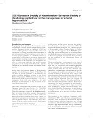
Figure 9 Semi-automated definition of left ventricular<br />
endocardial and epicardial borders (ROI) in an apical four<br />
chamber view.<br />
rate affect the accuracy of STE. This shortcoming<br />
could be overcome by the use of 3D<br />
<strong>speckle</strong> <strong>tracking</strong> technology.<br />
< Unknown software algorithms: To track<br />
<strong>speckle</strong>s and compute strain and SR values,<br />
filtering algorithms are used. The effect of this<br />
filtering on the results represents a ‘black box’<br />
and may vary from vendor to vendor. It is, thus,<br />
unclear how values from different scanners and<br />
software versions compare. Cross platform<br />
comparisons and a clear definition of global<br />
and regional norm values are essential for<br />
a broad application of STE.<br />
< Correct tracing of myocardial region of<br />
interest: One of the major limitations is the<br />
exact detection of borders. Even though <strong>speckle</strong><br />
<strong>tracking</strong> itself seems to enhance the capabilities<br />
of endocardial delineation, it is still necessary to<br />
correct contours manually. In addition, assessment<br />
of strain and SR also requires definition of<br />
the epicardial borders. In most software versions<br />
a uniform thickness of the myocardium is<br />
assumeddan assumption which is not true.<br />
< Size of left ventricle: A further limitation,<br />
encountered in large ventricles, is that it is often<br />
difficult to image the entire myocardium,<br />
especially the apical segments.<br />
Education in Heart<br />
FUTURE PERSPECTIVES<br />
STE is rapidly evolving both on the investigational<br />
and the technological front. It will be necessary to<br />
clearly define normal values and clinical settings<br />
where STE is useful. A primary goal will be the<br />
definition of cut-off values for medical decision<br />
making and to correlate these with hard end points.<br />
This will also require standardisation among<br />
different scanners to assure cross platform reproducibility<br />
and clear guidelines for the integration of<br />
STE into routine <strong>echocardiography</strong>.<br />
Optimisation of the algorithms for strain and SR<br />
assessment will certainly occur. This will include<br />
a more flexible endo- and epicardial border detection<br />
algorithm that accounts for differences in<br />
myocardial thickness and enhanced mathematical<br />
models and filtering techniques.<br />
Figure 10 Speckle <strong>tracking</strong> derived apical rotation rate of a subject with normal diastology (A) and with pseudonormal<br />
filling pattern. White lines indicate mitral valve opening, arrows indicate peak rotation rate during early diastole.<br />
Reproduced with permission from Perry et al. 10<br />
Three <strong>dimensional</strong> STE applications will help to<br />
improve the understanding of myocardial motion.<br />
The current practice of just measuring longitudinal,<br />
radial, and circumferential strain is a simplification<br />
of the complex myocardial fibre contraction<br />
pattern, and neglects ‘shear strains’ and out-ofplane<br />
motion. These problems may be overcome<br />
with three <strong>dimensional</strong> technology. 32e35<br />
Finally, more advanced technologies will allow<br />
LV rotation/torsion and strain/SR measurement of<br />
the endocardial, midwall, and epicardial myocardial<br />
layers and thus deliver a deeper insight into the<br />
physiology of myocardial mechanics, and permit<br />
the study of global and local processes within the<br />
LV wall. 36<br />
CONCLUSION<br />
STE has developed rapidly from a research tool to<br />
a technique which is on the verge of becoming an<br />
important part of routine <strong>echocardiography</strong>. STE<br />
uses the 2D image to calculate deformation<br />
parameters and is in many aspects superior to TDI.<br />
STE is easy to use, robust and provides a multitude<br />
of new insights into the mechanics and deformation<br />
processes of the myocardium. In particular,<br />
STE could provide important information on<br />
regional and global systolic and diastolic function<br />
Heart 2010;96:716e722. doi:10.1136/hrt.2007.141002 721