

Use of MRI in Evaluating Fetal Ventriculomegaly
Use of MRI in Evaluating Fetal Ventriculomegaly
Use of MRI in Evaluating Fetal Ventriculomegaly

You also want an ePaper? Increase the reach of your titles
YUMPU automatically turns print PDFs into web optimized ePapers that Google loves.
Lisa McLeod HMS III<br />
Gillian Lieberman, MD<br />
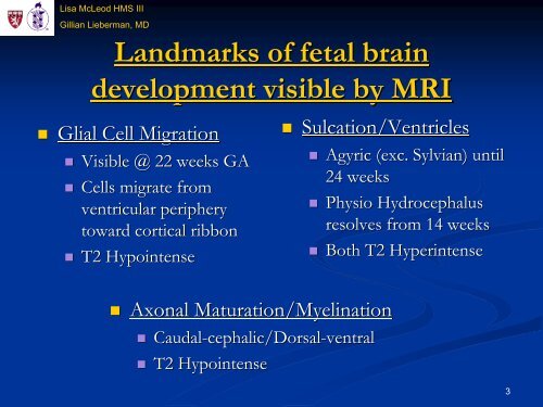
Landmarks <strong>of</strong> fetal bra<strong>in</strong><br />
development visible by <strong>MRI</strong><br />
Glial Cell Migration<br />
Visible @ 22 weeks GA<br />
Cells migrate from<br />
ventricular periphery<br />
toward cortical ribbon<br />
T2 Hypo<strong>in</strong>tense<br />
Sulcation/Ventricles<br />
Sulcation/Ventricles<br />
Axonal Maturation/Myel<strong>in</strong>ation<br />
Maturation/ Myel<strong>in</strong>ation<br />
Caudal-cephalic/Dorsal<br />
Caudal cephalic/Dorsal-ventral ventral<br />
T2 Hypo<strong>in</strong>tense<br />
Agyric (exc. Sylvian) Sylvian)<br />
until<br />
24 weeks<br />
Physio Hydrocephalus<br />
resolves from 14 weeks<br />
Both T2 Hyper<strong>in</strong>tense<br />
3


