

Use of MRI in Evaluating Fetal Ventriculomegaly
Use of MRI in Evaluating Fetal Ventriculomegaly
Use of MRI in Evaluating Fetal Ventriculomegaly

Create successful ePaper yourself
Turn your PDF publications into a flip-book with our unique Google optimized e-Paper software.
Lisa McLeod HMS III<br />
Gillian Lieberman, MD<br />
Corpus callosum<br />
BIDMC<br />
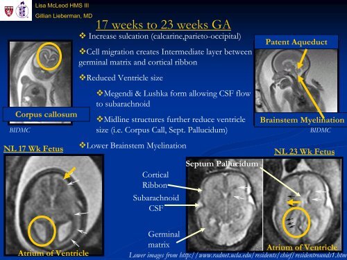
17 weeks to 23 weeks GA<br />
Increase sulcation (calcar<strong>in</strong>e,parieto-occipital)<br />
Cell migration creates Intermediate layer between<br />
germ<strong>in</strong>al matrix and cortical ribbon<br />
Reduced Ventricle size<br />
Megendi & Lushka form allow<strong>in</strong>g CSF flow<br />
to subarachnoid<br />
Midl<strong>in</strong>e structures further reduce ventricle<br />
size (i.e. Corpus Call, Sept. Pallucidum)<br />
Lower Bra<strong>in</strong>stem Myel<strong>in</strong>ation<br />
NL 17 Wk Fetus NL 23 Wk Fetus<br />
Atrium <strong>of</strong> Ventricle<br />
Cortical<br />
Ribbon<br />
Subarachnoid<br />
CSF<br />
Septum Pallucidum<br />
Patent Aqueduct<br />
Bra<strong>in</strong>stem Myel<strong>in</strong>ation<br />
BIDMC<br />
Germ<strong>in</strong>al<br />
matrix Atrium <strong>of</strong> Ventricle<br />
Lower images from http://www.radnet.ucla.edu/residents/chief/residentrounds1.htm


