

Use of MRI in Evaluating Fetal Ventriculomegaly
Use of MRI in Evaluating Fetal Ventriculomegaly
Use of MRI in Evaluating Fetal Ventriculomegaly

You also want an ePaper? Increase the reach of your titles
YUMPU automatically turns print PDFs into web optimized ePapers that Google loves.
BIDMC<br />
Lisa McLeod HMS III<br />
Gillian Lieberman, MD<br />
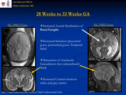
28 Weeks to 33 Weeks GA<br />
NL 28Wk Fetus NL 33Wk Fetus<br />
Increased Axonal Myel<strong>in</strong>ation <strong>of</strong><br />
Basal Ganglia<br />
Increased Sulcation (precentral<br />
gyrus, postcentral gyrus, Temporal<br />
Sulci)<br />
Maturation <strong>of</strong> Arachnoid<br />
Granulations (less subarachnoid<br />
fluid)<br />
Increased Contrast between<br />
white and grey matter<br />
http://www.radnet.ucla.edu/residents/chief/residentrounds1.htm<br />
BIDMC<br />
BIDMC


