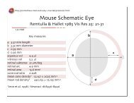
this download - University of Utah
this download - University of Utah
this download - University of Utah

Create successful ePaper yourself
Turn your PDF publications into a flip-book with our unique Google optimized e-Paper software.
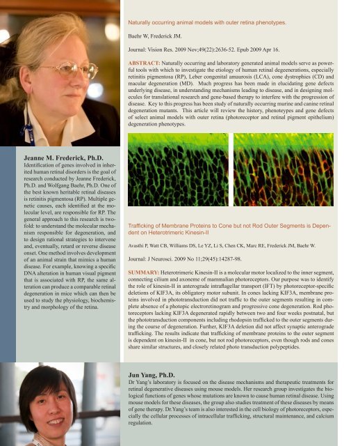
Jeanne M. Frederick, Ph.D.<br />
Identification <strong>of</strong> genes involved in inherited<br />
human retinal disorders is the goal <strong>of</strong><br />
research conducted by Jeanne Frederick,<br />
Ph.D. and Wolfgang Baehr, Ph.D. One <strong>of</strong><br />
the best known heritable retinal diseases<br />
is retinitis pigmentosa (RP). Multiple genetic<br />
causes, each identified at the molecular<br />
level, are responsible for RP. The<br />
general approach to <strong>this</strong> research is tw<strong>of</strong>old:<br />
to understand the molecular mechanism<br />
responsible for degeneration, and<br />
to design rational strategies to intervene<br />
and, eventually, retard or reverse disease<br />
onset. One method involves development<br />
<strong>of</strong> an animal strain that mimics a human<br />
disease. For example, knowing a specific<br />
DNA alteration in human visual pigment<br />
that is associated with RP, the same alteration<br />
can produce a comparable retinal<br />
degeneration in mice which can then be<br />
used to study the physiology, biochemistry<br />
and morphology <strong>of</strong> the retina.<br />
Naturally occurring animal models with outer retina phenotypes.<br />
Baehr W, Frederick JM.<br />
Journal: Vision Res. 2009 Nov;49(22):2636-52. Epub 2009 Apr 16.<br />
ABSTRACT: Naturally occurring and laboratory generated animal models serve as powerful<br />
tools with which to investigate the etiology <strong>of</strong> human retinal degeenerations, especially<br />
retinitis pigmentosa (RP), Leber congenital amaurosis (LCA), cone dystrophies (CD) and<br />
macular degeneration (MD). Much progress has been made in elucidating gene defects<br />
underlying disease, in understanding mechanisms leading to disease, and in designing molecules<br />
for translational research and gene-based therapy to interfere with the progression <strong>of</strong><br />
disease. Key to <strong>this</strong> progress has been study <strong>of</strong> naturally occurring murine and canine retinal<br />
degeneration mutants. This article will review the history, phenoteypes and gene defects<br />
<strong>of</strong> select animal models with outer retina (photoreceptor and retinal pigment epithelium)<br />
degeneration phenotypes.<br />
Trafficking <strong>of</strong> Membrane Proteins to Cone but not Rod Outer Segments is Dependent<br />
on Heterotrimeric Kinesin-II<br />
Avasthi P, Watt CB, Williams DS, Le YZ, Li S, Chen CK, Marc RE, Frederick JM, Baehr W.<br />
Journal: J Neurosci. 2009 No 11;29(45):14287-98.<br />
SUMMARY: Heterotrimeric Kinesin-II is a molecular motor localized to the inner segment,<br />
connecting cilium and axoneme <strong>of</strong> mammalian photoreceptors. Our purpose was to identify<br />
the role <strong>of</strong> kinesin-II in anterograde intraflagellar transport (IFT) by photoreceptor-specific<br />
deletions <strong>of</strong> KIF3A, its obligatory motor subunit. In cones lacking KIF3A, membrane proteins<br />
involved in phototransduction did not traffic to the outer segments resulting in complete<br />
absence <strong>of</strong> a photopic electroretinogram and progressive cone degeneration. Rod photoreceptors<br />
lacking KIF3A degenerated rapidly between two and four weeks postnatal, but<br />
the phototransduction components including rhodopsin trafficked to the outer segments during<br />
the course <strong>of</strong> degeneration. Further, KIF3A deletion did not affect synaptic anterograde<br />
trafficking. The results indicate that trafficking <strong>of</strong> membrane proteins to the outer segment<br />
is dependent on kinesin-II in cone, but not rod photoreceptors, even though rods and cones<br />
share similar structures, and closely related photo transduction polypeptides.<br />
Jun Yang, Ph.D.<br />
Dr Yang’s laboratory is focused on the disease mechanisms and therapeutic treatments for<br />
retinal degenerative diseases using mouse models. Her research group investigates the biological<br />
functions <strong>of</strong> genes whose mutations are known to cause human retinal disease. Using<br />
mouse models for these diseases, the group also studies treatment <strong>of</strong> these diseases by means<br />
<strong>of</strong> gene therapy. Dr.Yang’s team is also interested in the cell biology <strong>of</strong> photoreceptors, especially<br />
the cellular processes <strong>of</strong> intracellular trafficking, structural maintenance, and calcium<br />
regulation.