The John A. Moran Eye Center Research Profile - University of Utah
The John A. Moran Eye Center Research Profile - University of Utah
The John A. Moran Eye Center Research Profile - University of Utah

Create successful ePaper yourself
Turn your PDF publications into a flip-book with our unique Google optimized e-Paper software.
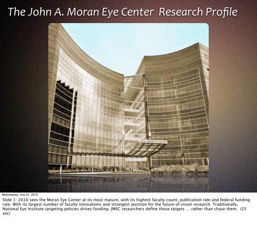
<strong>The</strong> <strong>John</strong> A. <strong>Moran</strong> <strong>Eye</strong> <strong>Center</strong> <strong>Research</strong> <strong>Pr<strong>of</strong>ile</strong><br />
Wednesday, July 21, 2010<br />
Slide 1: 2010 sees the <strong>Moran</strong> <strong>Eye</strong> <strong>Center</strong> at its most mature, with its highest faculty count, publication rate and federal funding<br />
rate. With its largest number <strong>of</strong> faculty innovations and strongest position for the future <strong>of</strong> vision research. Traditionally,<br />
National <strong>Eye</strong> Institute targeting policies drives funding. JMEC researchers define those targets … rather than chase them. (25<br />
sec)
CNS<br />
retina-‐RPE<br />
cornea<br />
Wednesday, July 21, 2010<br />
Our research operations address the fundamental mechanisms <strong>of</strong> ocular disease.<br />
<strong>The</strong> most challenging forms <strong>of</strong> blindness attack three systems in the signal transmission train:<br />
(1) the primary and irreplaceable optic element <strong>of</strong> the eye ... the cornea; (2) the most complex neural tissue known … the retina<br />
… and its obligate partner the retinal pigmented epithelium; (3) and the optic projections to the CNS. Corneal disease,<br />
vasculopathies,<br />
RP, AMD, glaucoma, and other pathologies are a spectrum <strong>of</strong> complex disorders. JMEC research touches on all these areas. (35<br />
sec)
Balamurali Krishna Ambati<br />
Ocular Angiogenesis & Corneal Disease<br />
Wednesday, July 21, 2010<br />
We have 16 major funded laboratories. Associate Pr<strong>of</strong>essor Dr. Bala Ambati is the world’s leading expert on the control <strong>of</strong><br />
vascular growth, especically but not exclusively in cornea, and is developing both molecular tools to regulate vascular<br />
pathologies and new drug delivery systems for the eye. He holds two NIH grants. (20 sec)
Wolfgang Baehr<br />
Photoreceptors, retinitis pigmentosa, & gene therapy<br />
Wednesday, July 21, 2010<br />
Wolfgang Baehr is the Ralph and Mary Tuck Pr<strong>of</strong>essor <strong>of</strong> Ophthalmology and one <strong>of</strong> the world’s most renowned molecular<br />
biologists. He is exploring the mechanisms <strong>of</strong> protein trafficking in photoreceptor cells. He is our most prolific mentor, having<br />
produced 3 stellar PhD students in two years, and our most prolific scientific author. He holds 2 NIH grants and co-directs the<br />
Biochemistry module <strong>of</strong> our vision core grant.<br />
In collaboration with my lab and Bryan Jones, we are preparing a new gene therapy for autosomal dominant RP. (40 sec)
Jun Yang<br />
Retinal degenerations, Usher Syndrome<br />
Wednesday, July 21, 2010<br />
Usher syndrome is one <strong>of</strong> the most devastating forms <strong>of</strong> RP. Assistant Pr<strong>of</strong>essor Jun Yang is one <strong>of</strong> our future hopes. She<br />
developed the most complete library <strong>of</strong> animal models for Usher Syndrome where genes interact to corrupt protein trafficking.<br />
She is designing viral vectors to reverse the defect.<br />
(20 sec)
Bryan William Jones<br />
Retinal degenerations, retinal remodeling, AMD<br />
Wednesday, July 21, 2010<br />
<strong>Research</strong> Assistant Pr<strong>of</strong>essor Bryan W. Jones is another emerging talent, with funding from RPB and the Thome foundation to<br />
study the mechanisms <strong>of</strong> retinal remodeling in RP and AMD. His collaboration with Mineo Kondo <strong>of</strong> the <strong>University</strong> <strong>of</strong> Nagoya<br />
brought the world’s only rabbit model <strong>of</strong> adRP to the US. <strong>The</strong> <strong>Moran</strong> <strong>Eye</strong> <strong>Center</strong> is the only breeding facility for it outside Japan.<br />
He and Dr. Baehr are collaborating on an RNA-based gene therapy for adRP.<br />
(30 sec)
Paul Bernstein<br />
Retinoid / Carotenoid Chemistry and AMD<br />
Wednesday, July 21, 2010<br />
Mary Boesche Pr<strong>of</strong>essor <strong>of</strong> Ophthalmology Paul Bernstein is a retinal surgeon and world class biochemist, focusing on the<br />
mechanisms <strong>of</strong> carotenoids in nutritional protection against oxidative stress leading to AMD. (15 sec)
Yingbin Fu<br />
Ocular proteases and AMD<br />
Wednesday, July 21, 2010<br />
Yingbin Fu is an Assistant Pr<strong>of</strong>essor who has been developing mouse models <strong>of</strong> AMD. His most recent successes demonstrates a<br />
disease phenotype in mouse more like neovascular AMD than any other group has achieved. (15 sec)
Edward Levine<br />
Retinal neurogenesis and retinal rescue<br />
Wednesday, July 21, 2010<br />
Associate Pr<strong>of</strong>essor Edward Levine’s NIH funded program is designed to discover how neural stem cells are programmed to<br />
become neurons. His most recent exciting findings include changing the proportions <strong>of</strong> different neuronal types with different<br />
combinations <strong>of</strong> genetic mutations. His goal is to find progenitor populations and molecular rules that can build a true retina.<br />
(25 sec)
Sabine Fuhrmann<br />
Ocular & RPE histogenesis<br />
Wednesday, July 21, 2010<br />
Assistant Pr<strong>of</strong>essor Sabine Fuhrmann just received one <strong>of</strong> the NIH top scores ever for her program to understand the genetic and<br />
molecular rules controlling how the RPE develops and is maintained. This is a critical step in the ultimate translational goal <strong>of</strong><br />
being able to replace the RPE in retinal degenerations such as AMD and some forms <strong>of</strong> arRP. (25 sec)
Margaret DeAngelis<br />
AMD Genetics<br />
Wednesday, July 21, 2010<br />
Associate Pr<strong>of</strong>essor Meg DeAngelis represents a new, and more mathematically robust way <strong>of</strong> mapping the interacting genes in<br />
various forms <strong>of</strong> AMD. Her recent work implicates an previously poorly understood retinoic acid signaling pathway in the cellular<br />
deconstructions that accompany AMD.<br />
(20 sec)
Gregory Hageman<br />
Immune disease and AMD<br />
Wednesday, July 21, 2010<br />
<strong>John</strong> <strong>Moran</strong> Presidential Pr<strong>of</strong>essor Gregory Hageman transformed the field <strong>of</strong> AMD research with his career <strong>of</strong> detailed and<br />
insightful anatomic, molecular and genetic analysis. He established that AMD is an inflammatory disease related to a large array<br />
<strong>of</strong> systemic disorders and will lead our <strong>Center</strong> for Translational <strong>Research</strong> on a mission to discover real therapeutics for AMD. (25<br />
sec)
Mary Elizabeth Hartnett<br />
Retinopathy <strong>of</strong> prematurity, AMD<br />
Wednesday, July 21, 2010<br />
Pr<strong>of</strong>essor Mary Elizabeth Hartnett is another one <strong>of</strong> our new researchers focussing on diseases resulting from disordered<br />
vascular development, such as retinopathy <strong>of</strong> prematurity and neovascular macular disease. She brings both expertise as an NIH<br />
investigator (with two grants) and as a pediatric retinal surgeon. (20 sec)
David Krizaj<br />
Retinal calcium regulation and glaucoma<br />
Wednesday, July 21, 2010<br />
Associate Pr<strong>of</strong>essor David Krizaj has discovered most <strong>of</strong> the molecular machinery associated with calcium signaling. Calcium<br />
fluxes are ionic events by which all cellular signaling is encoded. Calcium is also the key death molecules <strong>of</strong> cells. And calcium<br />
permeant channels, as shown by Dr. Krizaj, are clearly part <strong>of</strong> a multicellular mechanism that creates ganglion cell axon death in<br />
glaucoma. (30 sec)
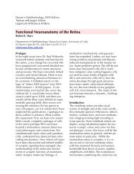
Robert Marc<br />
Retinal wiring and disease interactions<br />
Wednesday, July 21, 2010<br />
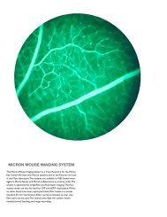
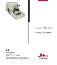
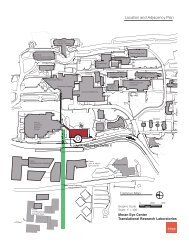
My lab is all about how retinas are wired and get unwired in disease. Using NIH funding and a generous gift <strong>of</strong> a state-<strong>of</strong>-the-art<br />
transmission electron microscope from Marth Ann Healy, as well as extensive collaborations with the <strong>University</strong> <strong>of</strong> <strong>Utah</strong> Scientific<br />
Computing and Imaging institute, we have built the world’s first retinal connectome ... a volume <strong>of</strong> tissue containing images <strong>of</strong><br />
all the retina’s connections. (30 sec)
Ning Tian<br />
Retinal developmental plasticity<br />
Wednesday, July 21, 2010<br />
Associate Pr<strong>of</strong>essor Ning Tian was the first to show that retinal neurons wire and rewire after birth; after the retina has<br />
“developed”. He has recently shown that this rewiring depends on immune signaling pathways very similar to those discovered<br />
by Dr. Hageman. This has pr<strong>of</strong>ound implications for being able to regenerate retinal networks after retinal degenerations. (25<br />
sec)
Alessandra Angelucci<br />
Cortical Visual Processing<br />
Wednesday, July 21, 2010<br />
With the work <strong>of</strong> Dr. Alessandra Angelucci, supported by NSF and NIH, we move to the challenging area <strong>of</strong> the organization <strong>of</strong><br />
the visual cortex, where flows <strong>of</strong> infomation are multiplexed into different processing geometries: a true 3D computational<br />
system. She and her students have systematically changed the models <strong>of</strong> how cortex extracts“salient” information. (25 sec)
Richard Normann<br />
Cortical neuroprosthetics<br />
Wednesday, July 21, 2010<br />
For many years Dr. Richard Normann has championed the idea that direct drive <strong>of</strong> cerebral cortex can lead to restoration <strong>of</strong><br />
sensory and motor function. With his colleage Bradley Greger, they have continually refined the tools with which brain neurons<br />
can be both monitored and activated. While true vision is some ways <strong>of</strong>f, it remains one <strong>of</strong> the biggest hopes for the pr<strong>of</strong>oundly<br />
blind for whom gene therapy cannot be considered. (30 sec)
Training<br />
Graduate Students<br />
Postdoctoral Fellows<br />
Peter Westenskow James Anderson<br />
Felix Vasquez-‐Chona Yanhua Lin<br />
Wednesday, July 21, 2010<br />
No research program can function without students - the lead scientists <strong>of</strong> our near ... not distant future. In the past two years,<br />
the Baehr, Marc, Fuhrmann and Angelucci laboratories have produced highly talented PhD students. Peter Westenskow led the<br />
research discovering the control <strong>of</strong> RPE development in Sabine Fuhrmann’s lab. James Anderson in the Marc lab supervised the<br />
construction <strong>of</strong> the first retinal connectome and single-handedly developed a Google Earth-like tool called Viking to explore it.<br />
Postdoctoral fellows take on even more advanced projects and <strong>Moran</strong> <strong>Eye</strong> <strong>Center</strong> fellows have won numerous awards from Fight<br />
for Sight and Knights Templar, like Felix and Yanhua (who has 2 PhDs). (60 sec)
<strong>The</strong> <strong>Utah</strong> Retinal<br />
Connectome RC1<br />
16.5 TB Raw, 50 TB total<br />
<strong>The</strong> first 3D retinal volume<br />
400,000 TEM images @ 5000/day<br />
371 automosaics <strong>of</strong> > 1000 tiles each<br />
Archetype for analysis <strong>of</strong> epilepsy, Alzheimer’s disease, RP & AMD<br />
Wednesday, July 21, 2010<br />
In competition with major labs at Harvard, Janelia Farm, Stanford and Max Planck in Germany, we achieved the first functional<br />
connectome assembly by teamwork, cooperation, open standards, good design and persistence. <strong>The</strong> 50 TB storage requirement<br />
is equal to a stack <strong>of</strong> 200 high-end laptops worth > $0.5 M. <strong>The</strong> volume contains <strong>of</strong> 400,000 images - 20 lifetimes worth <strong>of</strong><br />
work for an electron microscopist. (30 sec)
1985<br />
Hard Drives<br />
$983,000,000<br />
Display Boards<br />
$546,000,000<br />
Total<br />
$1,529,000,000<br />
Wednesday, July 21, 2010<br />
This is the first time in history that such a dataset could be built. In 1985, when I started my computational lab, the dataset<br />
would have cost over $1.5B to store and display. And, at imaging rates prior to our new transmission electron microscope, the<br />
imaging work would have taken decades (24-7) and the analysis centuries. (20 sec)
Bidirectional sign-conserving sign-inverting<br />
Wednesday, July 21, 2010<br />
Viking produces high density coded maps <strong>of</strong> all the real connections. This image displays a set <strong>of</strong> images that actually changes<br />
all <strong>of</strong> the existing models <strong>of</strong> how night vision systems operate. (15 sec)
Wednesday, July 21, 2010<br />
Viking movie - narrated 3 min
Retinal Connectome RC1 :: <strong>The</strong> AII Amacrine Cell<br />
AI<br />
A<br />
B<br />
AII<br />
B<br />
G<br />
ON<br />
OFF<br />
R<br />
C<br />
A<br />
G<br />
B<br />
invert<br />
conserve<br />
couple<br />
Wednesday, July 21, 2010<br />
<strong>The</strong> <strong>Utah</strong> retinal connectome shows how incomplete our existing ideas have been. One <strong>of</strong> the best mapped networks is the socalled<br />
AII cell system. This is its accepted wiring diagram. (15 sec)<br />
NF<br />
C
Retinal Connectome RC1 :: <strong>The</strong> AII Amacrine Cell<br />
WF<br />
NF<br />
A B C A B C<br />
AI AI<br />
Ax<br />
A<br />
G<br />
B<br />
AII<br />
B<br />
G<br />
ON<br />
OFF<br />
R<br />
C<br />
G<br />
invert<br />
conserve conserve<br />
couple couple<br />
Wednesday, July 21, 2010<br />
This is its correct wiring diagram. <strong>The</strong> original model was so incomplete as to be meaningless.<br />
Connectomics will change all analysis <strong>of</strong> how retinas and brains work. We are now collaborating with the Capecchi laboratory to<br />
understand the wiring mechanisms <strong>of</strong> “obsessive-compulsive disorder” pathways in mouse brains. (15 sec)<br />
Ax<br />
TH
Wednesday, July 21, 2010<br />
Why is this level <strong>of</strong> detail important. This is an Intel I7 processor chip - with over 700,000,000 transistors. If 0.5% didn’t work,<br />
the system would fail. A 50% error in biological wiring models is worse than useless -- it is dangerous.
<strong>The</strong> <strong>John</strong> A. <strong>Moran</strong> <strong>Eye</strong> <strong>Center</strong> <strong>Research</strong> <strong>Pr<strong>of</strong>ile</strong><br />
therapeutics<br />
connectomics<br />
translational science<br />
Wednesday, July 21, 2010<br />
<strong>The</strong> new <strong>John</strong> A. <strong>Moran</strong> <strong>Eye</strong> <strong>Center</strong> houses research programs directed to discovering gene-based, molecular, and cellular<br />
therapeutics for blinding diseases. It houses new research tools that will revolutionize neuroscience - like connectomics. And it<br />
is poised, by combining these skills with new ventures in translational medicine, led by Dr. Hageman, to identify new ways to<br />
prevent, ameliorate or correct debilitating diseases like AMD, heart disease and more. (30 sec)