who-eortc classification for cutaneous lymphomas - Dermatology
who-eortc classification for cutaneous lymphomas - Dermatology
who-eortc classification for cutaneous lymphomas - Dermatology
You also want an ePaper? Increase the reach of your titles
YUMPU automatically turns print PDFs into web optimized ePapers that Google loves.
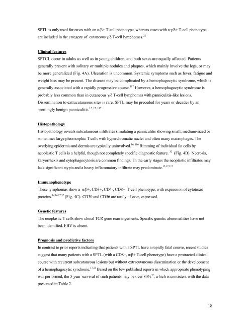
SPTL is only used <strong>for</strong> cases with an α/β+ T-cell phenotype, whereas cases with a γ/δ+ T-cell phenotype<br />
are included in the category of <strong>cutaneous</strong> γ/δ T-cell <strong>lymphomas</strong>. 32<br />
Clinical features<br />
SPTCL occur in adults as well as in young children, and both sexes are equally affected. Patients<br />
generally present with solitary or multiple nodules and plaques, which mainly involve the legs, or may<br />
be more generalized (Fig. 4A). Ulceration is uncommon. Systemic symptoms such as fever, fatigue and<br />
weight loss may be present. The disease may be complicated by a hemophagocytic syndrome, which is<br />
generally associated with a rapidly progressive course. 117 However, a hemophagocytic syndrome is<br />
probably less common than in <strong>cutaneous</strong> γ/δ T-cell <strong>lymphomas</strong> with panniculitis-like lesions.<br />
Dissemination to extra<strong>cutaneous</strong> sites is rare. SPTL may be preceded <strong>for</strong> years or decades by an<br />
15, 17, 117<br />
seemingly benign panniculitis.<br />
Histopathology<br />
Histopathology reveals sub<strong>cutaneous</strong> infiltrates simulating a panniculitis showing small, medium-sized or<br />
sometimes large pleomorphic T cells with hyperchromatic nuclei and often many macrophages. The<br />
overlying epidermis and dermis are typically uninvolved. 16, 116 Rimming of individual fat cells by<br />
neoplastic T cells is a helpful, though not completely specific diagnostic feature. 32 (Fig. 4B). Necrosis,<br />
karyorrhexis and cytophagocytosis are common findings. In the early stages the neoplastic infiltrates may<br />
lack significant atypia and a heavy inflammatory infiltrate may predominate. 15,17,117<br />
Immunophenotype<br />
These <strong>lymphomas</strong> show a α/β+, CD3+, CD4-, CD8+ T-cell phenotype, with expression of cytotoxic<br />
proteins. 14,16,17,32 (Fig. 4C). CD30 and CD56 are rarely, if ever, expressed.<br />
Genetic features<br />
The neoplastic T cells show clonal TCR gene rearrangements. Specific genetic abnormalities have not<br />
been identified. EBV is absent.<br />
Prognosis and predictive factors<br />
In contrast to prior reports indicating that patients with a SPTL have a rapidly fatal course, recent studies<br />
suggest that many patients with a SPTL (with a CD8+, α/β+ T-cell phenotype) have a protracted clinical<br />
course with recurrent sub<strong>cutaneous</strong> lesions but without extra<strong>cutaneous</strong> dissemination or the development<br />
of a hemophagocytic syndrome. 17,32 Based on the few published reports in which appropriate phenotyping<br />
was per<strong>for</strong>med, the 5-year-survival of such patients may be over 80% 32 , which is consistent with the data<br />
presented in Table 2.<br />
18


