胸腔外科常見疾病
胸腔外科常見疾病
胸腔外科常見疾病
Create successful ePaper yourself
Turn your PDF publications into a flip-book with our unique Google optimized e-Paper software.
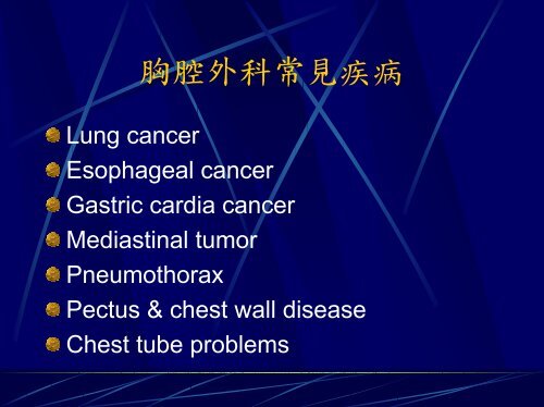
Lung cancer<br />
<strong>胸腔外科常見疾病</strong><br />
Esophageal cancer<br />
Gastric cardia cancer<br />
Mediastinal tumor<br />
Pneumothorax<br />
Pectus & chest wall disease<br />
Chest tube problems
Lung Cancer<br />
Symptoms and signs<br />
Basic Examinations<br />
Diagnosis<br />
Clinical Staging<br />
Pre-operative evaluation<br />
Surgical treatment<br />
Post-operative complications<br />
Prognosis and follow-up
Symptoms and Signs<br />
Cough<br />
Hemoptysis<br />
Chest pain<br />
Dyspnea and short of breathing<br />
Body weight loss<br />
….
Basic Examinations<br />
Chest X-ray examination<br />
Chest CT scan<br />
Compare previous CXR
Direct<br />
Sputum cytology<br />
Bronchoscope<br />
Diagnosis<br />
Percutaneous needle biopsy<br />
Indirect<br />
Pleural biopsy or cytology of pleural effusion<br />
Lymph nodes biopsy or aspiration cytology<br />
Mediastinoscope
Pathologic<br />
Classification of Lung<br />
Cancer<br />
Squamous cell carcinoma<br />
Adenocarcinoma<br />
Large cell carcinoma<br />
Small cell carcinoma<br />
…..
Clinical Staging<br />
CT scan of chest<br />
Whole body bone scan<br />
Whole abdomen sonography<br />
Brain scan or CT scan of brain
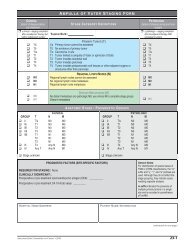
Staging of Lung Cancer<br />
Stage I T1-2 N0 M0<br />
Stage II T1-2 N1 M0<br />
T3 N0 M0<br />
Stage IIIa T3 N1 M0<br />
T1-3 N2 M0<br />
Stage IIIb T4 Nx M0<br />
Tx N3 M0<br />
Stage IV Tx Nx M1
Pre-operative Evaluation<br />
Pulmonary function tests<br />
ECG and cardiac function examiantion<br />
Pulmonary training and rehabilitation
Contraindicaions for<br />
Surgical Treatment<br />
Poor cardiopulmonary function<br />
Small cell lung cancer<br />
Extrathoracic metastasis (M1 lesion)<br />
T4 or N3 lesions<br />
Malignant pleural effusion<br />
Involved vital organs (heart, esophagus, spine)<br />
Contralateral mediastinal lymphadenopathy
Surgical Treatment<br />
Wedge resection<br />
Lobectomy/bilobectomy<br />
Pneumonectomy<br />
En-bloc resection<br />
Lymph nodes dissection
Post-Operative Adjuvent<br />
Radiotherapy<br />
Chemotherapy<br />
Therapy
Post-operative<br />
Complications<br />
Pulmonary atelectasis and pneumonia<br />
Bronchiopleural fistula (air leakage)<br />
Empyema<br />
Chylothorax<br />
Hemothorax<br />
Respiratory failure<br />
Arrythemia<br />
Stroke<br />
…..
Prognosis and Follow-up<br />
Prognosis<br />
Stage I 60-70%<br />
Stage II 40-55%<br />
Stage IIIa 20-30%<br />
Follow-up<br />
As schedule<br />
Triple scans—CXR, Brain Scan, bone scan<br />
and abdomen sonography
Recurrence and<br />
Metastasis<br />
Most within 3 years post-operation<br />
Most mortality within 2 years after<br />
metastasis diagnosis<br />
Distal metastasis more than local<br />
recurrence<br />
Most metastatic organs—brain, bone,<br />
liver and contralateral lung
Esophageal Cancer<br />
Etiology and pathogenesis<br />
Symptoms and signs<br />
Basic Examinations<br />
Diagnosis<br />
Clinical Staging<br />
Pre-operative evaluation<br />
Surgical treatment<br />
Post-operative complications<br />
Prognosis and follow-up
Epidemiology of<br />
Esophageal Cancer<br />
Incidence<br />
High in Iran, South Africa, China, Russia<br />
Age—60-70 years (average 62 years)<br />
Sex—M>F (2:1-20:1)<br />
Race—Black>White<br />
Site<br />
Tm>Tl>Tu>Ce
Surgical Anatomy of<br />
Cervical (Ce)<br />
Sternal notch<br />
Thoracic (Te)<br />
Tu<br />
Esophagus<br />
Trachea bifurcation,<br />
Tm & Tl<br />
Diaphragm<br />
Abdominal (Ae)
Pathogenesis of<br />
Esophageal Cancer<br />
Irritations to esophageal mucosachronic<br />
esophagitis<br />
Food stasis<br />
Thermal insult of food<br />
Chronic trauma from diet<br />
Overuse of alcohol & tabacco<br />
Nutritional deficienciescarcinogen—<br />
nitroamine<br />
Zinc deprivation<br />
Absence of Molybdenum<br />
Vitamin depletion—A, B, C
Etiology of Esophageal<br />
Cancer<br />
Environmental factors<br />
Geographic clustering<br />
Diet & nutritional differences<br />
Smoking & alcohol<br />
Genetic factors<br />
Predisposing factors<br />
Caustic injury<br />
Achalasia<br />
GER with Barrett’s esophagus<br />
Diverticula<br />
Previous gastric surgery<br />
Leucoplakia, Tylosis etc.
Symptoms of<br />
Esophageal Cancer (I)<br />
Early<br />
Retrosternal pain or burning<br />
sensationesophagitis<br />
Swallowing disturbancediffuse esophageal<br />
spasm<br />
Pharyngeal foreign body sensationChronic<br />
pharyngitis<br />
Substernal or epigastric pain, post-prandial<br />
fullnessgastritis<br />
Symptoms may relief after medical treatment
Symptoms of<br />
Esophageal Cancer (II)<br />
Classical symptoms<br />
Progressive dysphagia with/without<br />
retrosternal painful sensation<br />
Symptoms may persistent 6 months<br />
Onset of symptoms—75% lumen<br />
obstruction<br />
Late<br />
According to the structure or organs that<br />
being invasion or metastasis
Clinical Manifestations<br />
of Esophageal Cancer<br />
Symptoms<br />
Dysphagia 84%<br />
Weight loss 35%<br />
Pain—unspecific 22%<br />
Regurgitation 18%<br />
Cough 6%<br />
Vomiting 5%<br />
Hoarseness 4%<br />
Hematemesis 3%<br />
Choking 2%<br />
Neck pain 2%<br />
Signs<br />
Cachexia<br />
Neck mass<br />
Skin nodule<br />
SVC syndrome
Nomogram for Clinical<br />
Staging of Esophageal<br />
Cancer<br />
Suspected esophageal cancer<br />
History, physical examination<br />
Chest X-ray and chemistries<br />
Esophagogram<br />
Esophagoscope with biopsy<br />
Bronchoscope for cervical and Tu lesions<br />
CT scan of chest and upper abdomen<br />
Nuclear examination for staging
Malignant Tumors of the<br />
Epithelial<br />
Esophagus<br />
Squamous cell carcinoma (>90%)<br />
Spindle cell carcinoma<br />
Carcinosarcoma<br />
Pseudosarcoma<br />
Adenocarcinoma<br />
Adenoacanthoma<br />
Adenosquamous carcinoma<br />
….<br />
Non-epithelial<br />
Leiomyosarcoma<br />
….
UICC TNM Classification<br />
for Esophageal Tumors<br />
(I)<br />
T—primary tumor<br />
TX—primary tumor cannot be assessed<br />
T0—no evidence of primary tumor<br />
Tis—carcinoma in situ<br />
T1—tumor invaded mucosa or submucosa<br />
T2—tumor invaded muscularis propria<br />
T3—tumor invaded adventitia<br />
T4—tumor invaded adjacent structures
UICC TNM Classification<br />
for Esophageal Tumors<br />
(II)<br />
N—regional lymph nodes<br />
NX—regional lymph nodes cannot be assessed<br />
N0—no regional lymph nodes metastasis<br />
N1—regional lymph nodes metastasis<br />
M—distant metastasis<br />
MX—distant metastasis cannot be assessed<br />
M0—no distant metastasis<br />
M1—distant metastasis (including celiac nodes)
Post-Surgical Stage for<br />
Esophageal Cancer<br />
Stage I T1 N0 M0<br />
Stage IIa T2-3 N0 M0<br />
Stage IIb T1-2 N1 M0<br />
Stage III T3 N1 M0<br />
T4 N0-1 M0<br />
Stage IV anyT anyN M1
Pre-Operative Care<br />
Correct malnutrition<br />
Treat the underlying CV diseases<br />
Treat the pulmonary complications<br />
No smoking, bronchodilator, physical therapy<br />
Treat other organ disease<br />
Assessment of operation<br />
Curative v.s. palliative resection<br />
Availability of organs for reconstruction
Contraindications for<br />
Tumor Resection<br />
Poor general condition<br />
Wide spread and infiltration of adjacent<br />
organs<br />
Involvement of recurrent laryngeal nerve, phrenic<br />
nerve, sympathetic nerve<br />
Involvement of tracheobronchial trees with fistula<br />
formation<br />
Major vessels and heart involved<br />
Diffuse pleural or peritoneal carcinomatosis<br />
Malignant pleural effusion or ascites<br />
Distant organ metastasis<br />
Celiac trunk lymph nodes metastasis
Invasion and Metastasis<br />
Liver<br />
Trachea & bronchus<br />
Lung and pleura<br />
Kidney<br />
Bone<br />
Adrenal gland<br />
Brain
Management of<br />
Resectable Esophgeal<br />
Resection<br />
Cancer (I)<br />
Transthoracic esophagectomy<br />
Conventional, radical or en-bloc<br />
Transhiatal esophagectomy<br />
Lymph nodes dissection<br />
2 fields—thoraco-abdominal<br />
3 fields—cervico-thoraco-abdominal
Management of<br />
Resectable Esophageal<br />
Cancer (II)<br />
Reconstruction<br />
Organ—stomach, colon, jejunum<br />
Route—retrosternal, post. mediastinal,<br />
subcutaneous, transpleural<br />
Anastomosis site—cervical, intrathoracic<br />
Post-operative adjuvant therapy<br />
Radiotherapy<br />
Chemotherapy<br />
Radiotherapy+chemotherapy
Post-operative<br />
Complications for<br />
Esophageal Cancer<br />
Anastomosis leakage—20-30%<br />
Pneumonia<br />
Coronary artery complications<br />
Respiratory failure<br />
Sepsis<br />
Hemorrhage<br />
Empyema<br />
Chylothorax<br />
Shock<br />
Infarction of stomach
Management of<br />
Unresectable<br />
Esophageal Cancer<br />
Palliative surgery<br />
Bypass procedures with colon, stomach, jejunum<br />
Endoesophageal stent intubation<br />
Enterostomy<br />
Cervical esophagostomy<br />
Laser therapy<br />
Dilatation<br />
Radiotherapy<br />
Chemotherapy<br />
Combined therapy
Survival for<br />
Unresectable<br />
Esophageal Cancer<br />
Palliative Mean survival<br />
Bypass 8.0 months<br />
Intubation 5.5 months<br />
Enterostomy 3.5 months<br />
Dilatation 3.0 months
Definition<br />
Pneumothorax<br />
Air accumulation in the pleural space with secondary lung<br />
collapse<br />
Sources<br />
Visceral pleura<br />
Ruptured esophagus<br />
Chest wall defect<br />
Gas-forming organisms<br />
Factors determining gas reabsorption<br />
Diffusion properties of the gases<br />
Pressure gradients<br />
Area of contact<br />
Permeability of pleural surface
Classification of<br />
Pneumothorax<br />
Spontaneous<br />
Primary<br />
Secondary<br />
COPD<br />
Infection<br />
Neoplasm<br />
Catamenial<br />
Miscellaneous<br />
Traumatic<br />
Blunt<br />
Penetrating<br />
Iatrogenic<br />
Inadvertent<br />
Diagnostic<br />
Therapeutic
Primary Spontaneous<br />
Pneumothorax<br />
Symptoms and signs<br />
Sudden onset chest pain<br />
Shortness of breathing<br />
Non-productive cough<br />
Physical examination<br />
Decrease in chest wall movement on inspection<br />
Chest cavity hyperesonant and tympanic on<br />
percussion<br />
Decrease breath sounds on auscultation<br />
Diagnosis—CXR<br />
Differential diagnosis<br />
Skin fold<br />
Giant bulla
Treatment Options for<br />
Primary Spontaneous<br />
Pneumothorax<br />
Observation<br />
Needle aspiration and small chest tube<br />
drainage<br />
Convention tube thoracostomy<br />
Water seal Pleur-evac type<br />
Heimlich valve<br />
Tube thoracostomy with instillation of<br />
pleural irritant—Tetracycline HCL, Talc<br />
Surgical intervention
Indications for Surgical<br />
Intervention<br />
Second episode<br />
Persistent air leakage for greater than 7-10<br />
days<br />
First episode with unexpanded, “trapped”<br />
lung<br />
History of contralateral pneumothorax<br />
Bilateral pneumothorax<br />
Occupational risk (driver, airplane pilot)<br />
Living in a remote area<br />
Large bulla<br />
Large undrained hemothorax<br />
First episode in a patient with one lung<br />
First episode in a patient with severely
Surgical Intervention for<br />
Primary Spontaneous<br />
Pneumothorax<br />
Principal<br />
Resection of the blebs or bullae<br />
Obliteration of the pleural space<br />
Approach<br />
Video-assisted thoracoscopic surgery<br />
Thoracotomy<br />
Procedures for obliteration of the pleural<br />
space<br />
Parietal pleurectomy<br />
Abration pleurodesis<br />
Laser pleurodesis
Recurrence of Primary<br />
Spontaneous<br />
Pneumothorax<br />
Therapy Recurrence (%)<br />
Expectant 30<br />
Aspiration 20-50<br />
Chest tube drainage 20-30<br />
Pleurodesis (tetracycline) 25<br />
Pleurodesis (talc) 7<br />
Surgery 2
Complication of Primary<br />
Spontaneous<br />
Pneumothorax<br />
Tension pneumothorax<br />
Re-expansion pulmonary edema<br />
Persistent air leak<br />
Hemothorax (
Indications for<br />
Tube Thoracostomy<br />
Pneumothorax--open, tension, trauma<br />
of iatrogenic<br />
Hemothorax<br />
Empyema<br />
Pleural effusion<br />
Chylothorax<br />
Post-operative drainage--thoracic or<br />
cardiac
Chest Tube Insertion<br />
Guidewire tube thoracostomy<br />
Trocar tube thoracostomy<br />
Operative tube thoracostomy
Pleural Drainage System<br />
One-way (Heimlich) valve<br />
One-bottle collection system<br />
Two-bottles collection system<br />
Three-bottles collection system<br />
Commercial drainage system
Care of Chest Tubes<br />
Is there bubbling through the water-seal<br />
bottle/chamber ?<br />
Is the tube functioning?<br />
What is the amount and type of<br />
drainage from the tube?
Complications of Chest<br />
Tubes<br />
Misplacement of chest tube<br />
Pleural infection
Other Functions for<br />
Chest Tubes<br />
Injection of Materials<br />
Auto-transfusion
Removal of Chest Tubes<br />
Indications<br />
Methods


