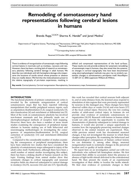
Remodeling of somotasensory hand representations following ...
Remodeling of somotasensory hand representations following ...
Remodeling of somotasensory hand representations following ...

Create successful ePaper yourself
Turn your PDF publications into a flip-book with our unique Google optimized e-Paper software.
COGNITIVE NEUROSCIENCE AND NEUROPSYCHOLOGY NEUROREPORT<br />
<strong>Remodeling</strong> <strong>of</strong> <strong>somotasensory</strong> <strong>hand</strong><br />
<strong>representations</strong> <strong>following</strong> cerebral lesions<br />
in humans<br />
Brenda Rapp, 1,2,3,CA Sharma K. Hendel 4 and Jared Medina 1<br />
Departments <strong>of</strong> 1 Cognitive Science, 2 Psychology and 3 Neuroscience, 239 Krieger Hall, Johns Hopkins University, Baltimore, MD 21218;<br />
4 Micros<strong>of</strong>t Corporation,USA<br />
There is evidence <strong>of</strong> reorganization <strong>of</strong> somatotopic maps <strong>following</strong><br />
cortical lesions in mammals such as monkeys, raccoons and rats.<br />
However, there has been a striking lack <strong>of</strong> research on somatosensory<br />
plasticity <strong>following</strong> cerebral damage in adult humans. We<br />
describe two individuals with left hemisphere damage who misperceive<br />
the locations <strong>of</strong> tactile stimuli whose presence or absence<br />
they can readily detect.We find that the mislocalizations preserve<br />
the relative topography <strong>of</strong> pre-lesion experiences, resulting in<br />
CA,1 Corresponding Author and Address<br />
Received 24 October 2001; accepted 28 November 2001<br />
shifted and compressed <strong>representations</strong> <strong>of</strong> the <strong>hand</strong> surfaces.<br />
These results not only provide evidence for systematic remodeling<br />
<strong>of</strong> somatotopic maps in humans, they also reveal that the systematic<br />
changes in cortical topography that have been documented<br />
using electrophysiological methods may give rise to similarly systematic<br />
changes in somatosensory perception itself. NeuroReport<br />
13:207^211 c 2002 Lippincott Williams & Wilkins.<br />
Key words: Cortical plasticity; Cortical reorganization; Neuroplasticity; Somatosensory maps; Somatosensory plasticity<br />
INTRODUCTION<br />
The functional plasticity <strong>of</strong> primary somatosensory cortex is<br />
revealed by the systematic reorganization <strong>of</strong> cortical<br />
somatosensory maps that has been reported <strong>following</strong><br />
manipulations that modify peripheral sensory inputs, such<br />
as amputation/deafferentation [1,2], surgical syndactyly [3],<br />
skin translocation [4] and enrichment or differential use [5].<br />
Most <strong>of</strong> the work on somatosensory plasticity has involved<br />
non-human mammals and has primarily made use <strong>of</strong><br />
electrophysiological methods <strong>of</strong> receptive field mapping.<br />
However, there have also been a number <strong>of</strong> more recent<br />
studies examining the consequences <strong>of</strong> peripheral manipulations<br />
in humans making use <strong>of</strong> both behavioral and<br />
neuroimaging methodologies in investigating the consequences<br />
<strong>of</strong> amputation/deafferentation [6–10], surgical<br />
correction <strong>of</strong> syndactyly [11] and enrichment/differential<br />
use [12,13].<br />
Compared to the considerable number <strong>of</strong> studies examining<br />
the consequences <strong>of</strong> peripheral manipulations, there<br />
have been relatively few studies that have considered the<br />
plasticity <strong>of</strong> somatosensory maps subsequent to injury <strong>of</strong> the<br />
adult cortex itself. Furthermore, although there have been a<br />
small number <strong>of</strong> animal studies investigating this subject,<br />
there has been a striking dearth <strong>of</strong> human research on this<br />
topic. Existing findings from animal studies show that<br />
systematic changes in cortical topography and neuronal<br />
receptive fields are observed subsequent to cortical damage<br />
in monkeys [14,15], raccoons [16] and rats [17]. Specifically,<br />
this work has revealed that cortical neurons both adjacent<br />
to, and at some distance from, the lesioned areas respond to<br />
stimulation <strong>of</strong> skin regions that were previously represented<br />
by neurons in the damaged area. These changes have been<br />
observed within days or weeks [14,16] and even hours [17]<br />
<strong>of</strong> the focal cortical injury. However, the very few existing<br />
studies <strong>of</strong> adult-onset cortical injury in humans do not<br />
provide clear evidence <strong>of</strong> systematic somatosensory reorganization<br />
[18,19]. Research with lesions in human adults<br />
would not only provide the opportunity to examine the<br />
characteristics <strong>of</strong> plasticity subsequent to cortical damage in<br />
humans, it would also provide a unique opportunity to<br />
examine how the topography-preserving neuronal changes<br />
observed in the animal research are experienced. Thus,<br />
while previous animal work has documented neuronal<br />
changes, it has not investigated the manner in which these<br />
changes affect perception. By examining the behavioral and<br />
phenomenological consequences <strong>of</strong> human cortical lesions<br />
we can determine if, among other things, neuronal<br />
reorganization leads to systematic changes in the perceived<br />
location and/or quality <strong>of</strong> tactile stimuli.<br />
We report here on an investigation carried out with two<br />
individuals who suffered left hemisphere strokes. We first<br />
document their ability to accurately detect the presence or<br />
absence <strong>of</strong> light tactile stimuli delivered to the contralesional<br />
<strong>hand</strong>. We then present the results <strong>of</strong> an experimental<br />
investigation <strong>of</strong> their ability to localize the tactile stimuli.<br />
We find clear evidence <strong>of</strong> systematic changes in their<br />
0959-4965 c Lippincott Williams & Wilkins Vol 13 No 2 11 February 2002 207
NEUROREPORT B. RAPP, S. K. HENDEL AND J. MEDINA<br />
perception <strong>of</strong> location. Strikingly, these changes preserve the<br />
topography <strong>of</strong> the pre-lesion sensations. The results provide,<br />
for the first time, evidence <strong>of</strong> the remodeling <strong>of</strong><br />
somatosensory representational topography subsequent to<br />
cerebral lesions in adult humans.<br />
MATERIALS AND METHODS<br />
The investigation was carried out with two right-<strong>hand</strong>ed<br />
male subjects (RSB and AKH) who were 4 years and 1.5<br />
years post-stroke, respectively. RSB was a 58-year-old<br />
toxicology researcher with a PhD and AKH was a 35-yearold<br />
social worker with a Master’s degree. No imaging data<br />
were available for AKH and it was known only that he had<br />
suffered a left hemisphere stroke to the region <strong>of</strong> the middle<br />
cerebral artery. For RSB, MRI revealed an extensive left<br />
parietal lesion extending rostrally from the post-central<br />
gyrus and caudally to the angular gyrus (Fig. 1) affecting,<br />
among other things, what is traditionally considered to be<br />
the <strong>hand</strong> area <strong>of</strong> SI as well as SII. An examination <strong>of</strong> the<br />
rostral edge <strong>of</strong> the lesion reveals that the superior rostral<br />
edge impinges upon the post-central sulcus and that the<br />
lesion advances in an anterior direction into the post-central<br />
gyrus at its inferior rostral edge. There are some signs <strong>of</strong> left<br />
frontal atrophy but all subcortical structures appear to be<br />
intact. Research with RSB and AKH was carried out in<br />
compliance with relevant laws and institutional guidelines<br />
and was approved by the Institutional Review Board <strong>of</strong> the<br />
Johns Hopkins University (Homewood Campus).<br />
Fig. 1. MRI scan for RSB. A horizontal slice from close to the inferior<br />
edge <strong>of</strong> the lesion reveals its rostral extension into the precentral gyrus<br />
and its caudal extension into the angular gyrus.The right <strong>of</strong> the image corresponds<br />
to the right hemisphere.<br />
208 Vol 13 No 2 11 February 2002<br />
Both individuals reported that they had suffered sensory<br />
loss and motoric difficulties in the period immediately<br />
<strong>following</strong> their strokes, but by the time <strong>of</strong> the investigation<br />
neither subject complained <strong>of</strong> motor difficulties or <strong>of</strong> loss <strong>of</strong><br />
somatosensation; nor did they exhibit tactile neglect or<br />
extinction. However, both did report sporadic tingling<br />
sensations in their <strong>hand</strong>s and forearms and, on occasion,<br />
stimulation presented to the ipsilesional <strong>hand</strong> produced<br />
bilateral sensations. Standard neurological testing with<br />
RSB revealed mild loss <strong>of</strong> discrimination bilaterally in<br />
<strong>hand</strong>s and feet and some difficulties in finger proprioception<br />
in the contralesional fingers. Both men experienced<br />
mild/moderate difficulties in written and spoken<br />
language production and numerical processing, but their<br />
language comprehension and other cognitive abilities were<br />
excellent.<br />
The eight neurologically intact control volunteers who<br />
participated in this investigation were all males (seven<br />
right-<strong>hand</strong>ed, one left-<strong>hand</strong>ed) with an average age <strong>of</strong> 42<br />
(range 26–61) years and education levels ranging from<br />
2 years <strong>of</strong> graduate study to PhD.<br />
First, preliminary testing was carried out to assess each<br />
subject’s ability to detect the presence or absence <strong>of</strong> a light<br />
touch to either, neither or both <strong>hand</strong>s. For this testing, the<br />
subject placed both <strong>hand</strong>s flat on the table with fingers<br />
spread at a comfortable distance from one another. The<br />
subject closed his eyes and light, brief tap was delivered<br />
either to one <strong>hand</strong>, both <strong>hand</strong>s or neither <strong>hand</strong>. The subject<br />
responded verbally with the terms right, left, both or neither<br />
(RSB: n = 216 per <strong>hand</strong>; AKH: n = 45 per <strong>hand</strong>).<br />
The primary empirical investigation consisted <strong>of</strong> the<br />
evaluation <strong>of</strong> the subjects’ perceptions <strong>of</strong> the locations <strong>of</strong><br />
the stimuli. Subjects were tested as follows. On each trial,<br />
the <strong>hand</strong> to be stimulated was placed flat on the table, with<br />
fingers spread at a comfortable distance from one another.<br />
The subject closed his eyes and a light, brief tap was<br />
delivered in pseudo-random order to one <strong>of</strong> the 22 locations<br />
on either the dorsal or ventral surface <strong>of</strong> the <strong>hand</strong>, as<br />
indicated in Fig. 2. Stimulation was presented blocked by<br />
surface. Immediately after stimulation, the subject opened<br />
his eyes and pointed to the location <strong>of</strong> sensation with the<br />
contralateral <strong>hand</strong>. The localization judgment was recorded<br />
by the experimenter on a line drawing <strong>of</strong> the subject’s <strong>hand</strong><br />
(some sessions were also videotaped to confirm the<br />
accuracy <strong>of</strong> the recording method). For each target location,<br />
the subject’s localization judgments were later encoded in x<br />
and y coordinates using a grid with the origin <strong>of</strong> the grid<br />
centered on the actual target location for each trial and with<br />
the y-axis aligned with the long axis <strong>of</strong> the finger. In this<br />
way, responses were encoded in terms <strong>of</strong> displacement<br />
distance (in mm) from the target along x and y axes. The<br />
inter-finger separation that was present at testing was<br />
removed from the displacement measurements in order to<br />
eliminate the possibility that averaged localizations would<br />
be situated in inter-finger space. Each <strong>of</strong> the 22 points was<br />
evaluated the <strong>following</strong> number <strong>of</strong> times for each subject.<br />
right dorsal: AKH 17, RSB 13; right ventral: AKH 12, RSB 16;<br />
left dorsal: AKH 11, RSB 8; left ventral: AKH 9, RSB 9. This<br />
yielded a total <strong>of</strong> 1012 data points for RSB and 1078 for<br />
AKH.<br />
These data were used to derive mean displacement values<br />
and mean localization judgments. Mean displacement
REMODELING OF SOMATOSENSORY HAND REPRESENTATIONS NEUROREPORT<br />
Fig. 2. (a) Stimulation points.The palmar surface <strong>of</strong> a right <strong>hand</strong>, marking<br />
the 22 stimulation locations used in all testing. Numbers 1^5 correspond<br />
to each <strong>of</strong> the digits; D = distal, M = medial, P = proximal,<br />
B = basal, X = palmar. Cross-hairs indicate 1 s.d. in the localization judgments<br />
<strong>of</strong> control subjects along x and y axes. (b,c) RSB’s mean localization<br />
judgments. Mean x and y values <strong>of</strong> all localization judgments are plotted<br />
for ventral and dorsal surfaces.Cross-hairs indicate1standard deviation in<br />
the judgments. To facilitate an overall evaluation <strong>of</strong> the location <strong>of</strong> the<br />
points relative to one another, outlines <strong>of</strong> <strong>hand</strong>s have been drawn around<br />
the mean localization judgment locations, approximately preserving the<br />
subject’s actual ¢nger sizes and shapes. (b) Ventral surface <strong>of</strong> RSB’s contralesional<br />
right <strong>hand</strong>, (c) dorsal surface <strong>of</strong> RSB’s contralesional right<br />
<strong>hand</strong>.<br />
values were calculated by averaging the absolute displacement<br />
values along each axis for all responses on a specific<br />
<strong>hand</strong> and surface. Mean localization judgments were<br />
determined by plotting the mean x and y values <strong>of</strong> all<br />
localization judgments for each <strong>of</strong> the 22 target location on<br />
an outline <strong>of</strong> the subjects’ <strong>hand</strong>s.<br />
Control data for localization judgments to contralesional<br />
stimuli are provided by the subjects’ responses to stimulation<br />
<strong>of</strong> their ipsilesional <strong>hand</strong>s, as well as by the responses<br />
<strong>of</strong> eight neurologically intact controls.<br />
Table 1. Mean displacement <strong>of</strong> localization judgments (absolute values<br />
in mm) along x and y axes, averaged across 22 target locations for each<br />
surface.<br />
RSB AKH Controls (range)<br />
Left dorsal<br />
x 2 3 1^3<br />
y<br />
Left ventral<br />
4 4 1^4<br />
x 2 3 0.5^2<br />
y<br />
Right dorsal<br />
4 6 1^3<br />
x 6 13 1^2<br />
y<br />
Right ventral<br />
17 31 1^5<br />
x 7 8 0^2<br />
y 15 18 1^3<br />
Left = ipsilesional, right = contralesional.<br />
RESULTS<br />
Detection accuracy: Detection accuracy was very good:<br />
RSB never failed to detect a stimulus delivered to the<br />
ipsilesional left <strong>hand</strong> and failed on only 3% <strong>of</strong> trials on the<br />
contralesional right <strong>hand</strong> AKH never failed to detect a<br />
stimulus to either <strong>hand</strong>.<br />
Localization judgments: Table 1 reports the mean absolute<br />
displacement values (averaged across the 22 stimulus<br />
locations) for dorsal and ventral surfaces <strong>of</strong> both <strong>hand</strong>s for<br />
RSB and AKH as well as for the control subjects. These data<br />
reveal that RSB and AKH’s displacements from contralesional<br />
target locations were markedly greater than the<br />
displacements <strong>of</strong> control subjects, while their displacement<br />
values for ipsilesional targets were generally within normal<br />
range. Specifically, contralesional displacement values on x<br />
and y dimensions were 12–26 s.d. from the control means<br />
for both RSB and AKH. In contrast, ipsilesional displacement<br />
values were all within the control range, except for the<br />
y-axis values on the ventral surfaces, where RSB and AKH’s<br />
mean displacement values were, respectively, 1 and 3 mm<br />
outside the range <strong>of</strong> control subjects. In addition, for both<br />
RSB and AKH, displacement values were markedly greater<br />
for contra-versus ipsilesional <strong>hand</strong>s, for both x and y-axis<br />
values, on both dorsal and ventral surfaces (the eight paired<br />
t-tests on the 22 target locations all yielded p o 0.00001).<br />
Significantly, the difference between localization accuracy<br />
for contralesional and ipsilesional <strong>hand</strong>s indicates that the<br />
localization difficulties can be specifically attributed to the<br />
neurological insult suffered by these individuals.<br />
While the results reported in Table 1 clearly indicate<br />
significant misperceptions <strong>of</strong> the locations <strong>of</strong> contralesional<br />
stimuli whose presence/absence is readily detected, they do<br />
not reveal if there is any systematicity in these misjudgments.<br />
The systematic nature <strong>of</strong> the errors can be seen in<br />
Fig. 2 and Fig. 3, which depict the mean localization<br />
judgments for each <strong>of</strong> the 22 locations on the dorsal and<br />
ventral surfaces for RSB and AKH’s contralesional <strong>hand</strong>s.<br />
Hand-shaped outlines have been drawn around the<br />
localization positions to facilitate an overall evaluation <strong>of</strong><br />
the displacements.<br />
Vol 13 No 2 11 February 2002 209
NEUROREPORT B. RAPP, S. K. HENDEL AND J. MEDINA<br />
Fig. 3. AKH’s mean localization judgments (as in Fig. 2). (a)Ventralsurface<br />
<strong>of</strong> AKH’s contralesional right <strong>hand</strong>, (b) dorsal surface <strong>of</strong> AKH’s contralesional<br />
right <strong>hand</strong>.<br />
The localization judgments clearly indicate systematic<br />
changes in the perceived locations <strong>of</strong> the stimuli. Specifically,<br />
the results reveal a striking downward shift in<br />
localization judgments; for RSB this is particularly evident<br />
for the distal and medial segments <strong>of</strong> both dorsal and<br />
ventral surfaces; for AKH all finger segments on the dorsal<br />
surface exhibit a downward shift while on the ventral<br />
surface the downward shift is seen primarily with the<br />
medial and the basal locations. RSB additionally exhibits an<br />
overall compression <strong>of</strong> the <strong>hand</strong> <strong>representations</strong> resulting<br />
from the combination <strong>of</strong> the downward shift <strong>of</strong> the distal<br />
points along with an upward shift <strong>of</strong> the palmar points and<br />
an inward shift <strong>of</strong> points along the outer edge <strong>of</strong> the <strong>hand</strong>.<br />
AKH’s judgments have considerably more error in the x<br />
dimension than do RSB’s. The x-axis error is such that<br />
stimuli delivered to dorsal digits 2 and 3 as well as to<br />
ventral digit 5 are typically experienced on adjacent digits;<br />
furthermore, stimuli delivered to the medial and proximal<br />
segments on dorsal digits 3 and 4 are virtually indistinguishable.<br />
Despite these distortions along the x-dimension,<br />
the relative positions along the y dimension are well<br />
maintained.<br />
In summary, the data clearly indicate that cerebral<br />
damage in humans may result in shifted and compressed<br />
perceptual <strong>representations</strong> that preserve the relative locations<br />
<strong>of</strong> the stimuli. In addition, the data indicate a generally<br />
similar systematic reorganization <strong>of</strong> sensory experiences for<br />
both ventral and dorsal <strong>hand</strong> surfaces.<br />
DISCUSSION<br />
This work reveals that cerebral damage in adult humans can<br />
result in a dissociation between the detection and the<br />
localization <strong>of</strong> tactile stimuli, such that stimuli that are<br />
readily detected may be significantly mislocalized. Furthermore,<br />
the data indicate that the post-lesion mislocalizations<br />
210 Vol 13 No 2 11 February 2002<br />
may preserve the relative locations <strong>of</strong> the pre-lesion<br />
topography, resulting in systematically shifted and distorted<br />
somatosensory experiences.<br />
These findings suggest that somatosensory <strong>representations</strong><br />
may have been reorganized within a reduced<br />
representational space. Inputs are not merely redirected to<br />
intact neural tissue, instead there has been a more general<br />
reorganization within the available neural substrate. This is<br />
consistent with the neurophysiological findings <strong>of</strong> animal<br />
research reviewed in the introduction indicating that the<br />
remodeling <strong>of</strong> somatosensory neural substrates subsequent<br />
to focal cortical lesions generally preserves the original<br />
neural topography. In addition, the results from AKH<br />
indicating overlapping representational surfaces for ventral<br />
digits 4 and 5 and dorsal digits 3 and 4 are consistent with<br />
the multi-digit receptive fields that have been observed<br />
subsequent to cortical lesions in animals [20]. Finally, the<br />
similarity between the results obtained for the dorsal and<br />
ventral surfaces constitutes an important contribution to the<br />
scarce literature (from either human or animal studies)<br />
regarding the plasticity <strong>of</strong> the dorsal (hairy surface) <strong>of</strong> the<br />
<strong>hand</strong>s. The degree to which the particular changes we have<br />
documented are observed in other neurologically injured<br />
individuals will, presumably, depend on specific details <strong>of</strong><br />
lesion locus and extent.<br />
Although the results provide clear evidence <strong>of</strong> systematic<br />
somatosensory remodeling, they do not elucidate the<br />
specific mechanisms underlying the observed changes and<br />
are generally consistent with various mechanisms that have<br />
been proposed: sprouting <strong>of</strong> new connections [21,22],<br />
unmasking <strong>of</strong> subthreshold or inhibited connections [23],<br />
or dynamic reweighting <strong>of</strong> connectivity [24,25]. Additionally,<br />
although RSB’s MRI scans reveal that damage is<br />
restricted specifically to cortical tissue within the parietal<br />
lobe, given the large size <strong>of</strong> the lesion it is not possible to<br />
localize the observed changes to particular areas within<br />
damaged or spared parietal cortex. Finally, we have not<br />
undertaken a systematic examination <strong>of</strong> the subjects’<br />
discrimination capacities and thresholds. Although both<br />
subjects exhibited very good detection accuracy for the<br />
stimuli we employed, one would expect that a reduction in<br />
representational space should result in the broadening <strong>of</strong><br />
receptive fields and concomitant decrements in sensitivity.<br />
CONCLUSIONS<br />
The finding <strong>of</strong> highly organized mislocalizations <strong>of</strong> tactile<br />
stimuli in two adults who have suffered left hemisphere<br />
strokes constitutes evidence for the systematic reorganization<br />
<strong>of</strong> somatasensory substrates in response to cortical<br />
damage and provides information regarding the experience<br />
and perception that may accompany the neural plasticity<br />
that has been documented in both humans and other<br />
mammals. In these ways, this work adds significantly to the<br />
growing body <strong>of</strong> neurophysiological findings <strong>of</strong> significant<br />
and systematic neural plasticity in the reorganization <strong>of</strong><br />
somatosensory substrates in response to cerebral damage.<br />
REFERENCES<br />
1. Rasmusson DD. J Comp Neurol 205, 313–326 (1982).<br />
2. Merzenich MM, Kaas JH, Wall JT et al. Neuroscience 8, 3–55 (1983).
REMODELING OF SOMATOSENSORY HAND REPRESENTATIONS NEUROREPORT<br />
3. Clark SA, Allard T, Jenkins WM and Merzenich MM. Nature 332, 444–445<br />
(1988).<br />
4. Clark SA, Allard TT, Jenkins WM and Merzenich MM. Soc Neurosci Abstr<br />
12, 391 (1986).<br />
5. Jenkins WM, Merzenich MM, Ochs MT et al. J Neurophysiol 63, 82–104<br />
(1990).<br />
6. Ramac<strong>hand</strong>ran VS, Rogers-Ramac<strong>hand</strong>ran DC and Stewart M. Science<br />
258, 1159–1160 (1992).<br />
7. Algioti S, Cortese F and Franchini C. Neuroreport 5, 473–476 (1994).<br />
8. Elbert T, Flor H, Birbaumer N et al. Neuroreport 5, 2593–2597 (1994).<br />
9. Halligan PW, Marshall JC and Wade DT. Neuroreport 5, 1341–1345 (1994).<br />
10. Yang TT, Gallen CC, Ramac<strong>hand</strong>ran VS et al. Neuroreport 5, 701–704<br />
(1994).<br />
11. Mogilner A, Grossman JAI, Ribary U et al. Proc Natl Acad Sci USA 90,<br />
3593–3597 (1993).<br />
12. Pascual-Leone A and Torres F. Brain 116, 39–52 (1993).<br />
13. Elbert T, Pantev C, Wienbruch C et al. Science 270, 305–307 (1995).<br />
14. Jenkins WM and Merzenich MM. Prog Brain Res 71, 246–266 (1987).<br />
15. Pons TP, Garraghty PE and Mishkin M. Proc Natl Acad Sci USA 85,<br />
5279–5281 (1988).<br />
16. Doetsch GS, Johnston KW and Hannan CJ. Exp Neurol 108, 162–175<br />
(1990).<br />
17. Coq JO and Xerri C. Eur J Neurosci 11, 2597–2608 (1999).<br />
18. Wikstrom H, Raine RO, Aronen HJ et al. Ann Neurol 47, 353–360 (2000).<br />
19. Aglioti SM, Beltramello A, Peru A et al. Neurocase 5, 285–292 (1999).<br />
20. Xerri C, Merzenich MM, Peterson BE and Jenkins W. J Neurophysiol 79,<br />
2119–2148 (1998).<br />
21. Darian-Smith C and Gilbert CD. Nature 368, 737–740 (1994).<br />
22. Florence SL, Taub HB and Kaas JH. Science 282, 1117–1121 (1998).<br />
23. Calford MB and Tweedale R. Nature 332, 446–448 (1988).<br />
24. Edelman GM and Finkel LH. Neuronal group selection in the cerebral<br />
cortex. In: Edelman GM, Gall WE and Cowan WM, eds. Dynamic Aspects<br />
<strong>of</strong> Neocortical Function New York: Wiley; 1984, pp. 653–695.<br />
25. Merzenich MM. Dynamic neocortical processes and the origins <strong>of</strong> higher<br />
brain functions. In: Changeux J-P and Konishi M, eds. The Neural and<br />
Molecular Bases <strong>of</strong> Learning New York: Wiley; 1987, pp. 337–358.<br />
Acknowledgements: We are extremely grateful to: Donna Gotsch for referring AKH, Shveta Mittal and Chen Zhao for dedicated<br />
data entry, Steven Small for the MRI images, John Hart, Michael Kraut, Matt Goldrick and Michael Pokorny for help in image<br />
reconstruction and interpretation, John Hart for neurological testing, Steven Hsiao and FranciscoVega-Bermudez for helpful<br />
input and Howard Egeth, Michael McCloskey and StevenYantis for comments on earlier drafts.This research was made possible by<br />
NIH grant R01NS34073.<br />
Vol 13 No 2 11 February 2002 211