Lower Lip Reconstruction with Fujimori Gate Flaps - ESPRS
Lower Lip Reconstruction with Fujimori Gate Flaps - ESPRS
Lower Lip Reconstruction with Fujimori Gate Flaps - ESPRS

Create successful ePaper yourself
Turn your PDF publications into a flip-book with our unique Google optimized e-Paper software.
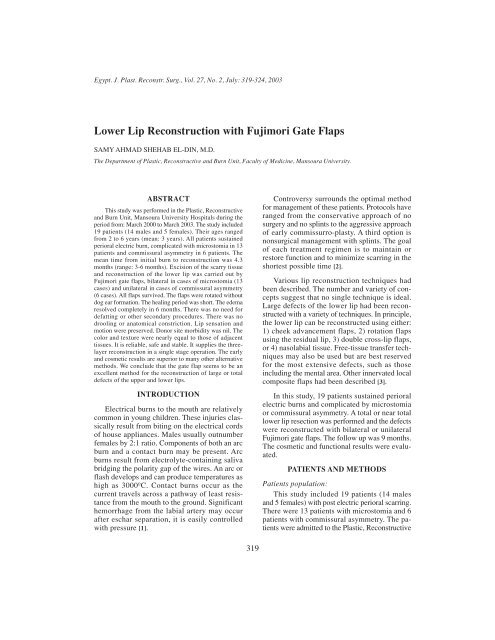
Egypt. J. Plast. Reconstr. Surg., Vol. 27, No. 2, July: 319-324, 2003<br />
<strong>Lower</strong> <strong>Lip</strong> <strong>Reconstruction</strong> <strong>with</strong> <strong>Fujimori</strong> <strong>Gate</strong> <strong>Flaps</strong><br />
SAMY AHMAD SHEHAB EL-DIN, M.D.<br />
The Department of Plastic, Reconstructive and Burn Unit, Faculty of Medicine, Mansoura University.<br />
ABSTRACT<br />
This study was performed in the Plastic, Reconstructive<br />
and Burn Unit, Mansoura University Hospitals during the<br />
period from: March 2000 to March 2003. The study included<br />
19 patients (14 males and 5 females). Their ages ranged<br />
from 2 to 6 years (mean: 3 years). All patients sustained<br />
perioral electric burn, complicated <strong>with</strong> microstomia in 13<br />
patients and commissural asymmetry in 6 patients. The<br />
mean time from initial burn to reconstruction was 4.3<br />
months (range: 3-6 months). Excision of the scarry tissue<br />
and reconstruction of the lower lip was carried out by<br />
<strong>Fujimori</strong> gate flaps, bilateral in cases of microstomia (13<br />
cases) and unilateral in cases of commissural asymmetry<br />
(6 cases). All flaps survived. The flaps were rotated <strong>with</strong>out<br />
dog ear formation. The healing period was short. The edema<br />
resolved completely in 6 months. There was no need for<br />
defatting or other secondary procedures. There was no<br />
drooling or anatomical constriction. <strong>Lip</strong> sensation and<br />
motion were preserved. Donor site morbidity was nil. The<br />
color and texture were nearly equal to those of adjacent<br />
tissues. It is reliable, safe and stable. It supplies the threelayer<br />
reconstruction in a single stage operation. The early<br />
and cosmetic results are superior to many other alternative<br />
methods. We conclude that the gate flap seems to be an<br />
excellent method for the reconstruction of large or total<br />
defects of the upper and lower lips.<br />
INTRODUCTION<br />
Electrical burns to the mouth are relatively<br />
common in young children. These injuries classically<br />
result from biting on the electrical cords<br />
of house appliances. Males usually outnumber<br />
females by 2:1 ratio. Components of both an arc<br />
burn and a contact burn may be present. Arc<br />
burns result from electrolyte-containing saliva<br />
bridging the polarity gap of the wires. An arc or<br />
flash develops and can produce temperatures as<br />
high as 3000ºC. Contact burns occur as the<br />
current travels across a pathway of least resistance<br />
from the mouth to the ground. Significant<br />
hemorrhage from the labial artery may occur<br />
after eschar separation, it is easily controlled<br />
<strong>with</strong> pressure [1].<br />
319<br />
Controversy surrounds the optimal method<br />
for management of these patients. Protocols have<br />
ranged from the conservative approach of no<br />
surgery and no splints to the aggressive approach<br />
of early commissurro-plasty. A third option is<br />
nonsurgical management <strong>with</strong> splints. The goal<br />
of each treatment regimen is to maintain or<br />
restore function and to minimize scarring in the<br />
shortest possible time [2].<br />
Various lip reconstruction techniques had<br />
been described. The number and variety of concepts<br />
suggest that no single technique is ideal.<br />
Large defects of the lower lip had been reconstructed<br />
<strong>with</strong> a variety of techniques. In principle,<br />
the lower lip can be reconstructed using either:<br />
1) cheek advancement flaps, 2) rotation flaps<br />
using the residual lip, 3) double cross-lip flaps,<br />
or 4) nasolabial tissue. Free-tissue transfer techniques<br />
may also be used but are best reserved<br />
for the most extensive defects, such as those<br />
including the mental area. Other innervated local<br />
composite flaps had been described [3].<br />
In this study, 19 patients sustained perioral<br />
electric burns and complicated by microstomia<br />
or commissural asymmetry. A total or near total<br />
lower lip resection was performed and the defects<br />
were reconstructed <strong>with</strong> bilateral or unilateral<br />
<strong>Fujimori</strong> gate flaps. The follow up was 9 months.<br />
The cosmetic and functional results were evaluated.<br />
PATIENTS AND METHODS<br />
Patients population:<br />
This study included 19 patients (14 males<br />
and 5 females) <strong>with</strong> post electric perioral scarring.<br />
There were 13 patients <strong>with</strong> microstomia and 6<br />
patients <strong>with</strong> commissural asymmetry. The patients<br />
were admitted to the Plastic, Reconstructive
320 Vol. 27, No. 2 / <strong>Lower</strong> <strong>Lip</strong> <strong>Reconstruction</strong> <strong>with</strong> <strong>Fujimori</strong> <strong>Gate</strong> <strong>Flaps</strong><br />
and Burn Unit, Mansoura University Hospitals,<br />
Egypt, during the period from March 2000<br />
through March 2003. The mean age was 3 years<br />
<strong>with</strong> a range of 2-6 years. The mean time from<br />
initial burn to reconstruction was 4.3 months (a<br />
range 3-6 months). The follow up was 9 months.<br />
Operative technique [4&5]:<br />
The whole of the affected lower lip is excised<br />
as a rectangle (Fig. 1A, BB’ DD’). The inferior<br />
margin of resection (DD’) follows the mentolabial<br />
groove. Whenever possible, a 3-to 4-mmwide<br />
strip of labial mucous membrane is left<br />
attached near the labioalveolar sulcus. The lateral<br />
margin of resection (BD and B’D’) is usually<br />
placed 0.5 to 1.0 cm laterally to perpendiculars<br />
dropped from the labial commissures (OO’), but<br />
it may extend 2 cm laterally in older patients<br />
<strong>with</strong> wrinkled faces. The width of the nasolabial<br />
skin-muscle-mucosal flaps (BC = DE, B’C’ =<br />
D’E’) is usually 3 cm and the suture lines of the<br />
flap donor sites (AED, A’E’D’) should follow<br />
the natural nasolabial fold. The dotted lines<br />
(AIB, AmC, A’T’B’, A’m’C’) represent incisions<br />
made through the mucous membrane, which<br />
should be about 1 cm wider than the flap itself,<br />
so excess mucous membrane can be available<br />
for reconstruction of the new red lip.<br />
When making flap incisions CED and C’E’D’<br />
below the line CC’ that connects both labial<br />
commissures, only skin and subcutaneous tissue<br />
should be cut, keeping the muscle and mucous<br />
membrane intact in the flap pedicle. Further<br />
undermining of the flap pedicle skin is required<br />
to allow flap rotation. <strong>Flaps</strong> prepared in this<br />
fashion contain innervated muscles: orbicularis<br />
oris, caninus, zygomaticus major or minor, riso-<br />
rius, triangularis and buccinator. As soon as the<br />
flaps have been mobilized, incisions should be<br />
closed in the four layers of mucous membrane,<br />
muscle layer, subcutaneous tissue and skin. The<br />
excess mucous membrane over the upper border<br />
of the transposed flap is used to replace vermilion.<br />
This new red lip will be thinner than the<br />
normal lip. Because of cicatricial contraction,<br />
the newly reconstructed lower lip tends to become<br />
puffy and rounded. This distortion can be<br />
effectively corrected by keeping continuous<br />
pressure on the lower lip by the sponge fixation<br />
method for 3 months postoperatively.<br />
RESULTS<br />
Nineteen procedures had been performed to<br />
reconstruct the lower lip in 19 patients: Bilateral<br />
<strong>Fujimori</strong> <strong>Gate</strong> <strong>Flaps</strong> were used in 13 patients<br />
<strong>with</strong> microstomia (Fig. 2) and Unilateral <strong>Fujimori</strong><br />
<strong>Gate</strong> <strong>Flaps</strong> were applied in 6 patients <strong>with</strong> commissural<br />
asymmetry (Fig. 3).<br />
All flaps survived. The flaps were rotated<br />
well <strong>with</strong>out dog ears. The perfusion was reliable.<br />
The healing period was short. The edema resolved<br />
completely in 6 months and the results<br />
were quite satisfactory. There was no need for<br />
defatting or other secondary procedures. There<br />
were no difficulties in maintaining oral competence<br />
or <strong>with</strong> drooling in any patient. Patients<br />
are provided <strong>with</strong> a generous and functional oral<br />
aperture that allowed oral hygiene and oral<br />
feeding <strong>with</strong>out anatomical constriction. <strong>Lip</strong><br />
sensation and motion were entirely preserved.<br />
Donor site morbidity was nil. The color and<br />
texture are nearly equal to those of adjacent<br />
tissues. The follow up period was 9 months.<br />
Fig. (1): <strong>Gate</strong> flap. A: Distance OB = O’B’ = 0.5 to 1.0 cm in normal patients, but it can be extended to 2 cm in the case of older patients<br />
<strong>with</strong> wrinkled faces. Distance BC = B’C’ = 3 cm. Skin incision along AB and AC should be made through-and-through into the<br />
mouth. Skin incision along CE and ED should be made only through subcutaneous tissue. Incision in the mucous membrane is<br />
performed along the dotted lines (A/B, AmC, A’I’B’, A’m’C’) that run laterally to the skin incision lines (AB, AC). Dotted area is<br />
undermined subcutaneously. B: Suture lines run along the nasolabial fold and mentolabial groove centrally (From <strong>Fujimori</strong>, ref. [5]).
Egypt, J. Plast. Reconstr. Surg., July 2003 321<br />
Fig. (2): Child aged 2 years sustained perioral electric burn resulting in microstomia.<br />
Fig. (2-A): Preoperative view, <strong>with</strong> tracheostomy. Fig. (2-B): Design of bilateral gate flaps.<br />
Fig. (2-C): Immediate postoperative view. Fig. (2-D): One week postoperative view.<br />
Fig. (2-E): 6 months postoperative view: mouth opened. Fig. (2-F): 6 months postoperative view: mouth closed.
322 Vol. 27, No. 2 / <strong>Lower</strong> <strong>Lip</strong> <strong>Reconstruction</strong> <strong>with</strong> <strong>Fujimori</strong> <strong>Gate</strong> <strong>Flaps</strong><br />
Fig. (3): Child aged 2 years sustained perioral electric burn resulting in commissural asymmetry.<br />
Fig. (3-A): Preoperative view.<br />
DISCUSSION<br />
Although burn-related injury is the leading<br />
cause of accidental death in the home for children<br />
under 14 years of age, low voltage electrical<br />
burns represent less than 4% of all burn injuries<br />
in children [6]. Gifford et al. (1971) reported that<br />
most low-voltage electrical injuries in children<br />
are around the mouth. These injuries occur most<br />
often from biting on an electrical cord or sucking<br />
on a wall socket. The most frequently affected<br />
site is the upper and lower lip <strong>with</strong> the connecting<br />
commissure followed by the tongue and the<br />
alveolus. The wounds can be extensive <strong>with</strong><br />
thermal injury to the surrounding cheek regions<br />
[7].<br />
Canady et al. retrospectively evaluated 24<br />
patients <strong>with</strong> oral commissure burns who were<br />
treated conservatively <strong>with</strong>out splinting or early<br />
surgery at the university of Iowa. Early surgical<br />
intervention was not indicated because the extent<br />
of soft tissue injury could not be precisely defined.<br />
Patients were allowed to heal their wounds<br />
<strong>with</strong>out splinting of the commissure. To soften<br />
the resulting scars, massage, triamcinolone cream<br />
and vitamin E were used. Prolonged use of splints<br />
was thought to cause excessive scarring, which<br />
could complicate later reconstruction. They<br />
concluded that conservative surgical management<br />
after scar maturation resulted in successful functional<br />
and aesthetic outcomes [1].<br />
Barone et al. retrospectively evaluated 29<br />
patients <strong>with</strong> perioral electric burns. Patients<br />
Fig. (3-B): Immediate postoperative view.<br />
were divided into three groups based on treatment<br />
modalities and were evaluated for aesthetic and<br />
functional outcome. The parameters judged were:<br />
lip length, scar, vermilion quality and lip roll.<br />
Group 1 patients were managed <strong>with</strong>out surgery<br />
and <strong>with</strong>out splints. Group 2 patients were treated<br />
<strong>with</strong> extra-oral splints only. Group 3 patients<br />
underwent commissuroplasty. Although the timing<br />
of commissuroplasty was not clearly defined,<br />
those authors believed that splinting alone provided<br />
the best overall results [8].<br />
Neale et al. reviewed 116 children <strong>with</strong> perioral<br />
electrical burns and also advocated the use<br />
of splints until scar maturation occurred. Once<br />
wounds were completely healed, commissuroplasty<br />
was considered [9].<br />
Earlier studies by de La Plaza et al. of 58<br />
children who sustained electrical burns to the<br />
mouth revealed better overall results <strong>with</strong> aggressive<br />
early surgical management. They concluded<br />
that surgery allows early resolution of<br />
the problem by excision of the devitalized tissues,<br />
<strong>with</strong> shorter hospital stays and fewer operations<br />
to achieve a satisfactory final result [10].<br />
The use of dynamic splinting to prevent<br />
microstomia has gained in popularity. Most<br />
authors believe that better functional restoration<br />
and overall aesthetic results are obtained through<br />
nonsurgical management of these patients using<br />
intraoral splints. The primary drawback of this<br />
type of treatment is noncompliance [2,11].
Egypt, J. Plast. Reconstr. Surg., July 2003 323<br />
Once contraction of the commissure occurs,<br />
surgical intervention is the only alternative to<br />
correct microstomia or commissural asymmetry.<br />
Careful preoperative planning for repositioning<br />
of the commissure is crucial to achieve a satisfactory<br />
outcome [2].<br />
The goal of reconstruction of the acquired<br />
lip defect or deformity is to restore function and<br />
appearance as close to preinjury (pretumor)<br />
status as possible. Specific goals, in priority<br />
order, are: 1) prevent drooling, 2) allow a water<br />
tight seal of the mouth to prevent food or liquid<br />
expulsion during chewing, 3) allow oral access<br />
for dentures, eating utensils, dental work, airway<br />
access, 4) preserve or recreate symmetrical appearance<br />
at rest, 5) provide accurate manipulation<br />
for labiodental speech sounds, 6) preserve voluntary<br />
and involuntary expression of emotion,<br />
7) permit pursing of the lips for sucking or<br />
whistling and 8) preserve lip sensation for preview<br />
of hot, cold or sharp objects [12].<br />
Our protocol in lower lip reconstruction<br />
follows the algorism advised by Behmand and<br />
Rees [13]. Small defects (less than one third of<br />
the lower lip) may be closed primarily. Wedge<br />
excision of lesions may be V-shaped, W-shaped,<br />
shield excision or single or double barrel excision.<br />
The lesion is excised in full-thickness of<br />
the lip. Four operations have been described to<br />
reconstruct medium defects (one third to two<br />
thirds of the lower lip) these are the Abbé switch<br />
flap [14], Karapandzic flap [15,16], modified<br />
Bernard’s procedure [17] and the Estlander switch<br />
flap [18]. If there is sufficient lip tissue and the<br />
commissure is not involved, Abbé switch flap<br />
or Karapandzic flap is indicated. In case the<br />
commissure is involved, Karapandzic flap (first<br />
choice) or Estlander flap (second choice) is used.<br />
If there is insufficient lip tissue, Bernard-<br />
Burrow’s flap is indicated. Large defects (more<br />
than two thirds of the lower lip) can be reconstructed<br />
by Bernard-Burrow’s procedure or Karapandzic<br />
flap or composite nasolabial flaps (<strong>Fujimori</strong><br />
gate flaps [4,5]) if there is sufficient adjacent<br />
cheek tissue. If adjacent cheek tissue is insufficient,<br />
distant or free flap reconstruction is indicated<br />
[19].<br />
The Karapandzic flap can provide a sensate<br />
lip <strong>with</strong> near-normal function; however, but if<br />
the defect is large, it inevitably results in microstomia<br />
and commissural distortion. Secondary<br />
commissuroplasty is often required because of<br />
the finite amount of remaining lip tissue. Oral<br />
feeding, hygiene and denture placement may be<br />
compromised, leaving some patients as oral<br />
cripples <strong>with</strong> excessively prominent perioral<br />
scarring [13].<br />
Modified Bernard procedures can produce<br />
excellent results but sacrifice extremely large<br />
amounts of skin and subcutaneous tissue at the<br />
labiomental and nasolabial folds. This invariably<br />
results in a tight lower lip and significant perioral<br />
scarring and contour deformity. The extent of<br />
mobilization required restricts the use of this<br />
technique to patients <strong>with</strong> substantial cheek<br />
laxity [3].<br />
In distant and free flaps, a functional oral<br />
sphincter is hard to create and the flaps lack the<br />
harmony of the face. Patients may have some<br />
donor site morbidity and the operation time is<br />
longer. Patients need microvascular anastomosis<br />
and require a longer healing period and hospitalization<br />
time [3].<br />
The gate flap, which uses a flap from each<br />
nasolabial fold, is an improvement over previous<br />
procedures. The flaps were rotated well and did<br />
not create dog-ear. The perfusion was reliable<br />
because of the angular artery. The healing period<br />
was short. The edema resolved completely in 6<br />
months and the results were quite satisfactory.<br />
There was no need for defatting or other secondary<br />
procedures. The gate flap technique is reliable,<br />
safe and stable. It avoids the problems<br />
related to distant or free tissue transfers. It<br />
supplies the three-layer reconstruction in a single<br />
operation and seems to be superior in both functional<br />
and cosmetic results. It can be used in<br />
upper lip reconstruction [20]. It is in harmony<br />
<strong>with</strong> adjacent tissues and the neo-oral sphincter<br />
functions well. The early and cosmetic results<br />
are superior to many other alternative methods.<br />
Our conclusion is that the gate flap seems to be<br />
an excellent method for the reconstruction of<br />
wide or total defects of the upper and lower lips.<br />
Prevention is better than cure: The facial<br />
surgeon has an inherent responsibility to participate<br />
in a proactive, ongoing educational program<br />
for his patients, fellow physicians and the community<br />
of lay persons. No one can appreciate<br />
the devastating effect of electrical, caustic and<br />
flame burns on patients than we who care for<br />
them and it is our responsibility to activate<br />
whatever measures we can to prevent these
324 Vol. 27, No. 2 / <strong>Lower</strong> <strong>Lip</strong> <strong>Reconstruction</strong> <strong>with</strong> <strong>Fujimori</strong> <strong>Gate</strong> <strong>Flaps</strong><br />
injuries. Speaking to parent-teacher associations,<br />
civic groups, industrial and business persons<br />
and writing short medical vignettes for the community<br />
newspapers can all serve to make families<br />
and other responsible parties aware of these<br />
health hazards and stimulate them to make the<br />
home, school and workplace safer [21].<br />
REFERENCES<br />
1- Canady T.W., Thompson S.A. and Bardach J.: Oral<br />
commissure burns in children. Plast. Reconstr. Surg.,<br />
97: 738, 1996.<br />
2- McCauley R.L. and Barret J.P.: Electrical injuries. In:<br />
Achauer B.M., Eriksson E., Wilkins E.G. and<br />
Vanderkam V.M. (eds.): Plastic Surgery, Indications,<br />
Operations and Outcomes. Volume 1, Chapter 25, p<br />
375, Mosby, 2000.<br />
3- Rudkin G.H., Carlsen B.T. and Miller T.A.: Nasolabial<br />
flap reconstruction of large defects of the lower lip.<br />
Plast. Reconstr. Surg., 111: 810, 2003.<br />
4- <strong>Fujimori</strong> R.: “<strong>Gate</strong> flap” for the total reconstruction<br />
of the lower lip. Br. J. Plast. Surg., 33: 340, 1981.<br />
5- <strong>Fujimori</strong> R.: Nasolabial (gate) skin-muscle-mucosal<br />
flap to lower lip. In: Strauch B., Vasconez L.O. and<br />
Hall-Findaly E.J. (eds.): Grabb’s Encyclopedia of<br />
<strong>Flaps</strong>. 2nd ed., Chapter 153, p 586, <strong>Lip</strong>pincott-Raven,<br />
Philadelphia, 1998.<br />
6- Keusch C.F., Gifford G.H. and Erikson E.: Pediatric<br />
electrical burns. In: Rees R.C., Cravalho E.G. and<br />
Burke J.F. (eds.): Electrical trauma: Pathophysiology,<br />
Manifestations and Clinical Management. New York,<br />
Cambridge University Press, 1992.<br />
7- Gifford G.H., Marty A.T. and Collum M.A.: The<br />
management of electrical mouth burns in children.<br />
Pediatrics, 47: 113, 1971.<br />
8- Barone C.M., Hulnick S.J., DeLinde L.G., et al.:<br />
Evaluation of treatment modalities in perioral electrical<br />
burns. J. Burn Care Rehabil, 15: 335, 1994.<br />
9- Neale H.W., Billmire D.A. and Gregory R.O.: Management<br />
of perioral burn scarring in the child and<br />
adolescent. Ann. Plast. Surg., 15: 212, 1995.<br />
10- De La Plaza R., Quetglas A. and Rodriguez E.: Treatment<br />
of electrical burns of the mouth. Burns, 10: 49,<br />
1983.<br />
11- Leake J.E. and Curtin J.W.: Electrical burns of the<br />
mouth in children. Clin. Plast. Surg., 11: 669, 1994.<br />
12- Calhoun K.H.: Introduction to reconstruction. In:<br />
Calhoun K.H., Stienberg C.M., Bailey B.J. and Holt<br />
G.R. (eds.): Surgery of the <strong>Lip</strong>. Chapter 3, p22, Geirg<br />
Thieme Verlag, Stuttgart-New York, 1992.<br />
13- Behmand R.A. and Rees R.S.: Reconstructive lip<br />
surgery. In: Achauer B.M., Eriksson E., Wilkins E.G.<br />
and VanderKam V.M. (eds.): Plastic Surgery, Indications,<br />
Operations and Outcomes. Volume 3, Chapter<br />
75, p 1193, Mosby, 2000.<br />
14- Millard D.R. and McLaughlin C.A.: Abbé flap on<br />
mucosal pedicle. Ann. Plast. Surg., 3: 544, 1979.<br />
15- Karapandzic M.: <strong>Reconstruction</strong> of lip defects by local<br />
arterial flaps. Br. J. Plast. Surg., 27: 93, 1974.<br />
16- Karapandzic M.: Innervated musculocutaneous lip and<br />
cheek flaps. In: Strauch B., Vasconez L.O. and Hall-<br />
Findlay E.J. (eds.): Grabb’s Encyclopedia of <strong>Flaps</strong>.<br />
2nd ed., Chapter 161, p 615, <strong>Lip</strong>pincott-Raven, Philadelphia,<br />
1998.<br />
17- Madden J.J., Erhardt W.L., Franklin J.D., et al.: <strong>Reconstruction</strong><br />
of the upper and lower lip using a modified<br />
Bernard-burrow technique. Ann. Plast. Surg., 5: 100,<br />
1980.<br />
18- Estlander J.A.: A method of reconstructing loss of<br />
substance in one lip from the other lip. Plast. Reconstr.<br />
Surg., 42: 361, 1968.<br />
19- Serletti J.M., Tavin E., Moran S.L. and Coniglio J.U.:<br />
Total lower lip reconstruction <strong>with</strong> a sensate composite<br />
radial forearm-palmaris longus free flap and a tongue<br />
flap. Plast. Reconstr. Surg., 99: 559, 1997.<br />
20- Aytekin A., Ay A. and Aytekin O.: Total upper lip<br />
reconstruction <strong>with</strong> bilateral <strong>Fujimori</strong> gate flaps. Plast.<br />
Reconstr. Surg., 111: 797, 2003.<br />
21- Holt G.R.: Burns of the lip. In: Calhoun K.H., Stiernberg<br />
C.M., Bailey B.J. and Holt G.R. (eds.): Surgery of the<br />
lip. Chapter 12, p 84, Georg Thieme Verlag, Stuttgart-<br />
New York, 1992.