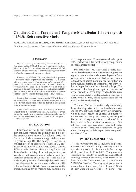
Childhood Chin Trauma and Temporo-Mandibular Joint ... - ESPRS
Childhood Chin Trauma and Temporo-Mandibular Joint ... - ESPRS
Childhood Chin Trauma and Temporo-Mandibular Joint ... - ESPRS

Create successful ePaper yourself
Turn your PDF publications into a flip-book with our unique Google optimized e-Paper software.
Egypt, J. Plast. Reconstr. Surg., Vol. 35, No. 2, July: 175-182, 2011<br />
<strong>Childhood</strong> <strong>Chin</strong> <strong>Trauma</strong> <strong>and</strong> <strong>Temporo</strong>-M<strong>and</strong>ibular <strong>Joint</strong> Ankylosis<br />
(TMJ): Retrospective Study<br />
ALMODDATHER M. EL-HADIDY, M.D.; AHMED A.M. KHALIL, M.D. <strong>and</strong> HOSSAM EL-DIN ALI, M.D.<br />
The Plastic <strong>and</strong> Reconstructive Surgery Unit, Faculty of Medicine, Mansoura University, Egypt<br />
ABSTRACT<br />
Objective: To study the relationship between the childhood<br />
chin trauma <strong>and</strong> the TMJ ankylosis <strong>and</strong> to assess our experience<br />
which is better for clinical <strong>and</strong> aesthetic outcome of TMJ<br />
ankylotic patients; the using of distraction osteogenesis before<br />
or after the resection of the ankylotic joint.<br />
Patients <strong>and</strong> Methods: This study involved 18 patients;<br />
11 males <strong>and</strong> 7 females presented long-st<strong>and</strong>ing TMJ ankylosis<br />
with a previous history of chin trauma before the age of 10<br />
years. Their ages ranged from 14 to 25 years. Distraction<br />
osteogenesis was used in all patients before or after the<br />
resection of the ankylotic mass <strong>and</strong> the joint reconstructed by<br />
costochondral graft <strong>and</strong> an interpositional temporalis myofascial<br />
flap. Follow-up period ranged from 15 to 36 months.<br />
Results: The postpond resection of the TMJ ankylosis to<br />
the second stage of surgery after distraction osteogenesis gave<br />
us the favorable results rather than the distraction osteogenesis<br />
came as the second stage.<br />
Conclusion: There is a direct relationship between the<br />
neglected childhood chin trauma <strong>and</strong> TMJ ankylosis. Furthermore,<br />
the m<strong>and</strong>ibular distraction osteogenesis before the<br />
resection the TMJ ankylosis is an effective in the management<br />
of TMJ ankylosis.<br />
INTRODUCTION<br />
<strong>Childhood</strong> injuries to chin resulting in m<strong>and</strong>ibular<br />
condylar fracture are common [1]. Falls are<br />
the most common cause of m<strong>and</strong>ibular condylar<br />
fracture with chin trauma among children below<br />
10 years of age [2]. The condylar fractures in<br />
children are often difficult to diagnose [3]. This<br />
difficulty returned to one of the following causes;<br />
difficult evaluation <strong>and</strong> history taking from child,<br />
the trouble in obtaining plane radiographs, the poor<br />
quality of radiographs <strong>and</strong> overlap of multiple<br />
anatomic structure of the small pediatric skull. In<br />
addition, the presence of other injuries in the acute<br />
stage elsewhere in the body may add to the difficulty<br />
in diagnosis [4,5,6]. Failure to recognize the<br />
presence of a condylar fracture may translate into<br />
175<br />
late complications. <strong>Temporo</strong>-m<strong>and</strong>ibular joint<br />
(TMJ) ankylosis is the most serious complication<br />
of condylar fracture [7].<br />
Patients with TMJ ankylosis usually have<br />
speech impairment, difficult mastication, poor oral<br />
hygiene, dental caries <strong>and</strong> various degrees of anatomical<br />
facial deformities including microgenia,<br />
reduced facial height, poor jaw neck definition <strong>and</strong><br />
more occlusal canting in unilateral TMJ ankylosis<br />
due to hypoplasia on the affected side [8]. The<br />
treatment of TMJ ankylosis requires restoration of<br />
proper m<strong>and</strong>ibular form, length <strong>and</strong> vertical dimension,<br />
occlusal stability <strong>and</strong> satisfactory joint movement.<br />
With children, future symmetrical growth<br />
must also be considered [9].<br />
The aim of this retrospective study was to study<br />
the relationship between the childhood chin trauma<br />
<strong>and</strong> the TMJ ankylosis <strong>and</strong> to assess our experience<br />
which is more better for clinical <strong>and</strong> aesthetic<br />
outcome of TMJ ankylotic patients; the using of<br />
distraction osteogenesis for correction of facial<br />
deformities before or after the resection of the<br />
ankylotic joint with coronoidectomy <strong>and</strong> reconstruction<br />
by the autogenous costochondral graft<br />
which is wrapped with interpositional temporalis<br />
myofascial flap.<br />
PATIENTS AND METHODS<br />
This retrospective study included 18 patients<br />
presenting with long-st<strong>and</strong>ing TMJ ankylosis with<br />
a previous history of chin trauma before the age<br />
of 10 years from 2001 to 2009. The presenting<br />
complaint was inability of mouth opening, difficult<br />
chewing, difficult breathing, sleep troubles <strong>and</strong><br />
facial deformity. There were 11 (61.1%) males <strong>and</strong><br />
7 (38.9%) females. Their ages at presentation<br />
ranging from 14 to 25 years (mean 18 years). Seven<br />
of them were unilateral <strong>and</strong> 11 bilateral. Three
176 Vol. 35, No. 2 / <strong>Childhood</strong> <strong>Chin</strong> <strong>Trauma</strong> & <strong>Temporo</strong>-M<strong>and</strong>ibular<br />
patients had recurrent ankylosis after previous<br />
failed surgery.<br />
Eleven patients were undertaking the distractor<br />
application in the 1 st surgical stage. Three months<br />
later, the 2 nd surgical stage including the resection<br />
of the ankylotic mass with coronoidectomy <strong>and</strong><br />
reconstruction by costochondral graft with intervention<br />
of an interpositional temporalis myofascial<br />
flap. While the remaining cases underwent the<br />
resection of the ankylotic mass with coronoidectomy<br />
<strong>and</strong> reconstruction by costochondral graft with<br />
intervention of an interpositional temporalis myofascial<br />
flap. After 3 months, the m<strong>and</strong>ibular distractor<br />
was applied. Intraoral distractor was used<br />
in 7 cases <strong>and</strong> an external distractor was used in<br />
the remaining patients.<br />
Preoperative evaluation:<br />
The preoperative evaluation included a patient<br />
history, physical examinations, radiological investigations<br />
<strong>and</strong> photography was taken for all patients.<br />
A patient history included patient’s age, sex, history<br />
of previous trauma, medical condition <strong>and</strong> complaint<br />
at presentation. Physical examinations involved<br />
presence or absence of any scar mark under<br />
chin, any facial asymmetry at presentation, jaw<br />
movements <strong>and</strong> measuring of maximal mouth opening.<br />
Radiological investigations included panoramic<br />
view, facial CT scan <strong>and</strong> MRI (Fig. 1).<br />
Surgical procedures:<br />
The procedures were performed under general<br />
anesthesia <strong>and</strong> intubation either nasotracheal with<br />
the aid of fibroptic laryngoscope or via elective<br />
tracheostomy.<br />
A- Distractor application (Figs. 2-6):<br />
An intraoral approach was made along the<br />
anterior border of the ramus. Subperiosteal elevation<br />
on both surfaces of ramus <strong>and</strong> corticotomy<br />
were done. The intraoral distractor device was<br />
applied through the same approach. While external<br />
distractor was applied through a small stab incision<br />
extraorally by a trocar system. Our vector is oblique<br />
over the angle to do distraction in both directions<br />
towards the ramus <strong>and</strong> the body. The distractor<br />
was fixed to the lateral surface of the m<strong>and</strong>ible<br />
with its rods <strong>and</strong> the m<strong>and</strong>ibular osteotomy was<br />
completed through green stick fracture in the ramus<br />
to preserve the inferior alveolar pundle. Then, the<br />
distractor was secured in its position. After the<br />
latent period, distraction was performed at a rate<br />
of 0.5mm daily until satisfaction results were<br />
gained. After the consolidation period (3 months),<br />
the distraction device was removed.<br />
B- Resection of the ankylotic mass with coronoidectomy<br />
<strong>and</strong> reconstruction by costochondral graft<br />
with intervention of an interpositional temporalis<br />
myofascial flap (Figs. 7-11):<br />
The TMJ was surgically explored through a<br />
st<strong>and</strong>ard preauricular approach with temporal extension.<br />
The ankylotic mass (disc, condylar head,<br />
<strong>and</strong> root of the zygomatic arch) <strong>and</strong> the coronoid<br />
process were exposed <strong>and</strong> excised using a surgical<br />
burs <strong>and</strong> osteotomies. Through the same approach,<br />
ptreygo-massetric sling was disinserted <strong>and</strong> in<br />
some patients via a separate approach extraorally<br />
(Risdon approach). Maximum mouth opening was<br />
tried with demo work on seeing of the space of<br />
excised ankylotic mass <strong>and</strong> measured the maximum<br />
interincisal distance. Then, the IMF was applied.<br />
The costochondral graft was taken from the 7 th<br />
rib <strong>and</strong> the wound was closed in layers. The harvested<br />
graft involved 3-5cm bone <strong>and</strong> 0.5cm cartilage.<br />
The graft was trimmed <strong>and</strong> multiple partial<br />
thickness bur holes were made in the graft <strong>and</strong> the<br />
recipient surface of the m<strong>and</strong>ible. Then, the graft<br />
was placed on the lateral surface of the ramus as<br />
an onlay graft.<br />
The inferiorly based temporalis myofascial flap<br />
was used as a wrap over the costochondral graft<br />
mimic normal joint. In all patients, a vacuum drain<br />
was inserted for 48 hours <strong>and</strong> the overlying tissues<br />
were closed in layers <strong>and</strong> dressed.<br />
Post-operative protocol:<br />
Caring of respiration was seriously monitoring.<br />
Prophylactic antibiotics, analgesics <strong>and</strong> antiinflammatory<br />
medications for one week were described<br />
for all patients. They were discharged 3 to<br />
5 days postoperatively. The IMF was removed after<br />
two weeks. Oral hygiene <strong>and</strong> fluids were recommended<br />
for two weeks then soft food for another<br />
four weeks.<br />
All patients underwent physical therapy including<br />
home training <strong>and</strong> physiotherapy. Home training<br />
involved frequent chewing of gums <strong>and</strong> wooden<br />
tongue depressor application three times daily for<br />
15-20 minutes for each time with increasing the<br />
number of depressors for 3 weeks to gain maximum<br />
mouth opening. The maximum mouth opening was<br />
measured after one month. All patients were seen<br />
weekly for 6 weeks. Follow-up period ranged from<br />
15 to 36 months.<br />
RESULTS<br />
The results of our study (Table 1) were depended<br />
on the clinical observations, maximal mouth<br />
opening <strong>and</strong> panoramic view.
Egypt, J. Plast. Reconstr. Surg., July 2011 177<br />
Table (1): Patients parameters.<br />
No.<br />
1<br />
2<br />
3<br />
4<br />
5<br />
6<br />
7<br />
8<br />
9<br />
10<br />
11<br />
12<br />
13<br />
14<br />
15<br />
16<br />
17<br />
18<br />
Mean<br />
Age (year)<br />
16<br />
14<br />
18<br />
25<br />
16<br />
18<br />
15<br />
23<br />
18<br />
23<br />
21<br />
14<br />
17<br />
19<br />
14<br />
14<br />
16<br />
23<br />
18<br />
Sex<br />
M<br />
F<br />
M<br />
M<br />
F<br />
M<br />
F<br />
M<br />
M<br />
M<br />
M<br />
F<br />
F<br />
M<br />
F<br />
F<br />
M<br />
M<br />
Ankylotic<br />
side<br />
Uniateral [L]<br />
Uniateral [L]<br />
Bilateral<br />
Bilateral<br />
Uniateral [R]<br />
Bilateral<br />
Uniateral [L]<br />
Bilateral<br />
Bilateral<br />
Bilateral<br />
Bilateral<br />
Uniateral [R]<br />
Bilateral<br />
Bilateral<br />
Uniateral [L]<br />
Unilateral [L]<br />
Bilateral<br />
Bilateral<br />
All patients tolerated the surgical procedures<br />
<strong>and</strong> recovered well. Anaesthetized patients with<br />
the aid of fibroptic laryngoscope were more calm,<br />
easily breath, well recover <strong>and</strong> early return to their<br />
life activity.<br />
Patients with intraoral distraction device were<br />
more satisfied than those with external distraction<br />
device because they were returned back to perform<br />
their normal activity. Two patients reported loosing<br />
the supporting screws of their external devices.<br />
The distraction osteogenesis was achieved successfully<br />
in all patients with good bone formation<br />
which documented radiologically. Three patients,<br />
who released TMJ ankylosis firstly, experienced<br />
pain in the ipsilateral TMJ during the distraction<br />
period. No wound infection was reported. Mild<br />
skin infection was found around the external distraction<br />
rods in 5 patients that healed after application<br />
of topical antibiotics.<br />
The clinical observations showed nearly symmetrical<br />
facial appearance (Figs. 12,13), horizontal<br />
occlusal plane (Fig. 14) with improvement of<br />
occlusion. In addition, breath <strong>and</strong> sleep behavior<br />
were improved as a result of upper airway improvement<br />
<strong>and</strong> the mouth room became capacious in all<br />
patients. An important finding was good joint<br />
Maximum mouth opening<br />
(mm)<br />
Latent period<br />
(day)<br />
5<br />
6<br />
7<br />
7<br />
7<br />
6<br />
7<br />
6<br />
7<br />
7<br />
7<br />
6<br />
5<br />
5<br />
7<br />
7<br />
7<br />
5<br />
6.33<br />
Preoperative<br />
7<br />
0<br />
2<br />
2<br />
0<br />
3<br />
2<br />
4<br />
2<br />
2<br />
3<br />
4<br />
5<br />
0<br />
0<br />
3<br />
3<br />
2<br />
2.44<br />
Postoperative<br />
(1 month later)<br />
41<br />
27<br />
28<br />
30<br />
24<br />
35<br />
32<br />
32<br />
29<br />
32<br />
30<br />
34<br />
29<br />
34<br />
34<br />
34<br />
35<br />
32<br />
31.78<br />
Follow-up period<br />
(month)<br />
36<br />
30<br />
24<br />
24<br />
24<br />
20<br />
20<br />
20<br />
20<br />
20<br />
18<br />
18<br />
18<br />
18<br />
18<br />
17<br />
16<br />
15<br />
20.89<br />
function with an adequate range of m<strong>and</strong>ibular<br />
movement <strong>and</strong> pain free inspite of the absence of<br />
the condyle. The mean of maximum interincisal<br />
opening was 2.44mm preoperatively <strong>and</strong> 31.78mm<br />
postoperatively (Fig. 15) after a mean follow-up<br />
period of 20.89 months. Re-ankylosis was reported<br />
in 2 patients who underwent the release of TMJ<br />
ankylosis firstly before the distraction process.<br />
Facial scar was an unsatisfactory result in 7 patients.<br />
Fig. (1): Facial CT scan showed left TMJ ankylosis.
178 Vol. 35, No. 2 / <strong>Childhood</strong> <strong>Chin</strong> <strong>Trauma</strong> & <strong>Temporo</strong>-M<strong>and</strong>ibular<br />
Fig. (2): Intraoral approach <strong>and</strong> corticotomy. Fig. (3): Intraoral distractor application.<br />
Fig. (4): Extraoral approach <strong>and</strong> corticotomy. Fig. (5): External distractor application.<br />
Fig. (6): Panoramic view during distraction period. Fig. (7): St<strong>and</strong>ard preauricular approach.
Egypt, J. Plast. Reconstr. Surg., July 2011 179<br />
Fig. (8): Exposure <strong>and</strong> excision of ankylotic mass. Fig. (9): Harvesting of costochondoral graft.<br />
Fig. (10): Placing the graft <strong>and</strong> wrap temporalis myofascial<br />
flap.<br />
Fig. (11): Open the mouth <strong>and</strong> measure the maximum mouth<br />
opening.<br />
Fig. (12): Pre <strong>and</strong> post-operatives facial appearance (Frontal view).
180 Vol. 35, No. 2 / <strong>Childhood</strong> <strong>Chin</strong> <strong>Trauma</strong> & <strong>Temporo</strong>-M<strong>and</strong>ibular<br />
Fig. (13): Pre <strong>and</strong> post-operatives facial appearance (Lateral view).<br />
Fig. (14): Pre <strong>and</strong> post-operatives occlusal plane.<br />
Fig. (15): Pre <strong>and</strong> post-operative maxium interincisal distance.
Egypt, J. Plast. Reconstr. Surg., July 2011 181<br />
DISCUSSION<br />
Many studies [10-13] reported that the trauma<br />
was the most common aetiology of TMJ ankylosis<br />
(13-100%). Khan et al., 2010 found that trauma<br />
was the most common cause of TMJ ankylosis <strong>and</strong><br />
was confirmed in 96.7% of patients by an obvious<br />
scar mark under their chin <strong>and</strong> a history of chin<br />
trauma before the age of 10 years [14]. This study<br />
was agreed with the previous studies as the trauma<br />
was the most common cause of TMJ ankylosis.<br />
Moreover, the neglected diagnosis <strong>and</strong> the badly<br />
management hematoma of the TMJ were progressed<br />
up to fibrosis <strong>and</strong> ultimately to ankylosis.<br />
Majority of post-traumatic TMJ ankylosis was<br />
primarily attributed to delay or non-treatment of<br />
condylar fractures due to several factors such as<br />
poor educational levels, non-availability of surgical<br />
expertise, poor economic status <strong>and</strong> prolonged<br />
immobilization of the joint due to pain after injury<br />
[15].<br />
Our objective in the management of the TMJ<br />
ankylosis was to restore mouth opening, to establish<br />
a function outcome of the joint, to correct the facial<br />
profile <strong>and</strong> to relive the upper airway obstruction<br />
with minimal complications.<br />
Reconstruction of the m<strong>and</strong>ibular condyle remains<br />
a challenge because of its unique anatomical<br />
structure. Autogenous graft is generally considered<br />
the best reconstruction material as it is less in cost<br />
<strong>and</strong> time for preparation in comparison with allograft<br />
<strong>and</strong> it heals normally with little complications<br />
[16]. Costochondral graft is the most widely<br />
accepted autogenous technique for m<strong>and</strong>ibular<br />
condyle reconstruction. The costochondral graft<br />
is readily available, possesses good mechanical<br />
properties <strong>and</strong> has the capacity for remodeling into<br />
an adaptive m<strong>and</strong>ibular condyle [17].<br />
The unpredictable growth pattern of the costochondral<br />
grafts has often been cited as a disadvantage.<br />
Aberrant growth can cause progressive dental<br />
midline shifts, occlusal changes, chin deviation<br />
<strong>and</strong> enlargement of the graft itself [18].<br />
When treating the TMJ ankylosis with costochondral<br />
graft in this study, the good healing of<br />
the costochondral graft with the m<strong>and</strong>ibular ramus<br />
was confirmed <strong>and</strong> most patients showed no reankylosis<br />
inspite of Saeed <strong>and</strong> Kent in 2003 reported<br />
re-ankylosis <strong>and</strong> limited improvement in mouth<br />
opening [13]. Our explanation regarded to use an<br />
adequate amount of myofascial temporalis muscle<br />
flap as an interpositional graft was effective in the<br />
prevention of ankylosis recurrence. The main ad-<br />
vantages of using the autogenous myofascial temporalis<br />
muscle flap are its proximity to the operative<br />
site <strong>and</strong> its good blood supply.<br />
Since McCarthy use a distraction technique for<br />
m<strong>and</strong>ibular lengthening in the patients with hemifacial<br />
microsomia [19], Distraction osteogenesis<br />
has become a widely accepted natural surgical<br />
procedure in the treatment of craniofacial deformities<br />
<strong>and</strong> defects. Several series [20,21,22] confirmed<br />
that distraction osteogenesis is a promising treatment<br />
option for patients with TMJ ankylosis. Distraction<br />
osteogenesis has become a popular surgical<br />
modality due to many advantages: minimal complexity<br />
of the procedure, minimal operative time,<br />
minimal hospital stay, low risk of complications,<br />
no donor site morbidity, no need for blood <strong>and</strong> no<br />
IMF fixation required [23].<br />
Muscular resistance, particularly from masseter<br />
<strong>and</strong> medial pterygoid muscles is one of the most<br />
crucial factors in creating resistance during distraction<br />
osteogenesis, as well as during jaw exercises<br />
after releasing the ankylosis [23]. So, in this series<br />
the ptreygo-massertic sling was disinserted freely<br />
that resulting in maximal mouth opening.<br />
The authors regarded the reported experienced<br />
pain during the distraction period to the distraction<br />
forces pushing the remaining ramus up into the<br />
glenoid fossa.<br />
We observed that the postpond resection of the<br />
TMJ ankylosis to the second stage of surgery after<br />
distraction osteogenesis gave us the favorable<br />
results rather than the distraction osteogenesis<br />
came as the second stage. This emphasis that the<br />
immobile joint represented a fixed point that the<br />
distraction was pushed the m<strong>and</strong>ible downward<br />
for m<strong>and</strong>ibular lengthening with no harm effect<br />
on the non-reconstructed joint. Moreover, the<br />
enlarged bone segment in this stage appreciate the<br />
convenient osteotomy <strong>and</strong> appropriate placing of<br />
the costochondral graft in the second stage. On the<br />
other h<strong>and</strong>, the resection of TMJ ankylosis firstly<br />
<strong>and</strong> the distraction osteogenesis came later, it is<br />
going to lengthening the ramus towards the reconstructed<br />
joint destroying it <strong>and</strong> may hasted the<br />
ankylosis again.<br />
From the current study, the authors summarize<br />
<strong>and</strong> conclude that there is a direct relationship<br />
between the neglected childhood chin trauma <strong>and</strong><br />
TMJ ankylosis. So, we recommend to do<br />
ultrasonography on the TMJ with regular followup<br />
to examine the TMJ function as a routine for<br />
any patient with m<strong>and</strong>ibular trauma. Furthermore,<br />
the m<strong>and</strong>ibular distraction osteogenesis before the
182 Vol. 35, No. 2 / <strong>Childhood</strong> <strong>Chin</strong> <strong>Trauma</strong> & <strong>Temporo</strong>-M<strong>and</strong>ibular<br />
resection the TMJ ankylosis is an effective in the<br />
management of TMJ ankylosis <strong>and</strong> the advantage<br />
of the postponding resection of the ankylotic joint<br />
is prevention of the rotation <strong>and</strong> the upward movement<br />
of the m<strong>and</strong>ibular ramus during the distraction<br />
course.<br />
REFERENCES<br />
1- Holan G.: <strong>Trauma</strong>tic injuries to chin: A survey in a<br />
paediatric dental practice. International Journal of paediatric<br />
dentistry, 8: 143-48, 1998.<br />
2- Ogunlewe M.O., James O., Ladeinde L.A. <strong>and</strong> Adeyemo<br />
W.L.: Pattern of paediatric maxillofacial fractures in<br />
Lagos, Nigeria. International Journal of Paediatric Dentistry,<br />
16: 358-62, 2006.<br />
3- Tejani Z., Johnson A., Mason C. <strong>and</strong> Goodman J.: Multiple<br />
crown root fractures in primary molars <strong>and</strong> a suspected<br />
subcondylar fracture following trauma: A report of a case.<br />
Dental <strong>Trauma</strong>tology, 24: 253-56, 2008.<br />
4- Lee C.Y.S., Mc Cullon C., Blaustein D. <strong>and</strong> Mohammadi<br />
H.: Sequelae of unrecognized, untreated m<strong>and</strong>ibular<br />
condylar fractures in the pediatric patients. Ann. Dent.,<br />
52: 5-8, 1993.<br />
5- Dimitroulis G.: Condylar injuries in growing patients.<br />
Australian Dental Journal, 42 (6): 367-71, 1997.<br />
6- Oji C.: Fractures of the facial skeleton in children: A<br />
survey of patients under the age of 11 years. Journal of<br />
Cranio-Maxillofacial Surgery, 26: 322-25, 1998.<br />
7- Ferretti, Bryant R., Becker P. <strong>and</strong> Lawrence C.: <strong>Temporo</strong>m<strong>and</strong>ibular<br />
joint morphology following post-traumatic<br />
ankylosis in 26 patients. Int. J. Oral Maxillofac. Surg.,<br />
34: 376-81, 2005.<br />
8- Perrott D.H., Umeda H. <strong>and</strong> Kaban L.B.: Costochondral<br />
graft construction/reconstruction of the ramus/condyle<br />
unit: Long-term follow-up. Int. J. Oral Maxillofac. Surg.,<br />
23: 321-28, 1994.<br />
9- Kaban L.B., Perrott D.H. <strong>and</strong> fisher K.: A protocol for<br />
management of temporom<strong>and</strong>ibular joint ankylosis. J.<br />
Oral Maxillofac. Surg., 48: 1145-51, 1990.<br />
10- Demir Z., Velideglu H., Sahin U., Kurtay A. <strong>and</strong> Coskunfirat<br />
O.K.: Preserved costal cartilage homograft application<br />
for treatment of temporom<strong>and</strong>ibular joint ankylosis. Plast.<br />
Reconstr. Surg., 108: 44-51, 2001.<br />
11- El-Sheikh M.M. <strong>and</strong> Medra A.M.: Management of unilat-<br />
eral temporom<strong>and</strong>ibular joint ankylosis associated with<br />
facial asymmetry. J. Craniomaxillofac. Surg., 25: 109-<br />
15, 1997.<br />
12- Miyamoto H., Kurita K., Ogi N., Ishimaru J.I. <strong>and</strong> Goss<br />
A.N.: The effect of an intra-articular bone fragment in<br />
the genesis of the temporom<strong>and</strong>ibular joint ankylosis. Int.<br />
J. Oral Maxillofac. Surg., 29: 290-95, 2000.<br />
13- Saeed N.R. <strong>and</strong> Kent J.N.: A retrospective study of the<br />
costochondral graft in TMJ reconstruction. Int. J. Oral<br />
Maxillofac. Surg., 32: 606-9, 2003.<br />
14- Khan Z., Alam J., Khan S., Abid H. <strong>and</strong> Warraich R.A.:<br />
Correlation between childhood chin trauma, condylar<br />
fracture & TMJ ankylosis. Pakistan Oral & Dental Journal,<br />
30 (1): 47-51, 2010.<br />
15- Jain, et al.: <strong>Temporo</strong>m<strong>and</strong>ibular joint ankylosis: A review<br />
of 44 cases. Oral Maxillofac. Surg., 12: 61-66, 2008.<br />
16- Maclntosh R.B.: The use of autogenous tissues for temporom<strong>and</strong>ibular<br />
joint reconstruction. J. Oral Maxillofac.<br />
Surg., 58: 63-69, 2000.<br />
17- Matsuura H., Miyamoto H., Kurita K. <strong>and</strong> Goss A.N.:<br />
The effect of autogenous costochondral grafts on temporom<strong>and</strong>ibular<br />
joint fibrous <strong>and</strong> bony ankylosis: A<br />
preliminary experimental study. J. Oral Maxillofac. Surg.,<br />
64: 1517-25, 2006.<br />
18- Ko E.W., Huang C. <strong>and</strong> Chen Y.: <strong>Temporo</strong>m<strong>and</strong>ibular<br />
joint reconstruction in children using costochondral grafts.<br />
J. Oral Maxillofac. Surg., 57: 789-98, 1999.<br />
19- McCarthy J.G., Schreiber J. <strong>and</strong> Karp N.: Lengthening<br />
the human m<strong>and</strong>ible by gradual distraction. Plast. Reconstr.<br />
Surg., 89: 1-8, 1992.<br />
20- Stucki-McCormick S.U.: Reconstruction of the m<strong>and</strong>ibular<br />
condyle using transport distraction osteogenesis. J. Craniofac.<br />
Surg., 8: 48-52, 1997.<br />
21- Cheung L.K. <strong>and</strong> Lo J.: The long-term effect of transport<br />
distraction in the management of temporom<strong>and</strong>ibular joint<br />
ankylosis. Plast. Reconstr. Surg., 119: 1003-9, 2007.<br />
22- Schwartz H.C. <strong>and</strong> Relle R.J.: Distraction osteogenesis<br />
for temporom<strong>and</strong>ibular joint reconstruction. J. Oral Maxillofac.<br />
Surg., 66: 718-23, 2008.<br />
23- Sadakah A.A., Elgazzar R.F. <strong>and</strong> Abdelhady A.I.: Intraoral<br />
distraction osteogenesis for the correction of facial deformities<br />
following temporom<strong>and</strong>ibular joint ankylosis: A<br />
modified technique. Int. J. Oral Maxillofac. Surg., 35:<br />
399-406, 2006.