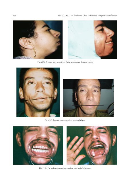
180 Vol. 35, No. 2 / <strong>Childhood</strong> <strong>Chin</strong> <strong>Trauma</strong> & <strong>Temporo</strong>-M<strong>and</strong>ibular Fig. (13): Pre <strong>and</strong> post-operatives facial appearance (Lateral view). Fig. (14): Pre <strong>and</strong> post-operatives occlusal plane. Fig. (15): Pre <strong>and</strong> post-operative maxium interincisal distance.
Egypt, J. Plast. Reconstr. Surg., July 2011 181 DISCUSSION Many studies [10-13] reported that the trauma was the most common aetiology of TMJ ankylosis (13-100%). Khan et al., 2010 found that trauma was the most common cause of TMJ ankylosis <strong>and</strong> was confirmed in 96.7% of patients by an obvious scar mark under their chin <strong>and</strong> a history of chin trauma before the age of 10 years [14]. This study was agreed with the previous studies as the trauma was the most common cause of TMJ ankylosis. Moreover, the neglected diagnosis <strong>and</strong> the badly management hematoma of the TMJ were progressed up to fibrosis <strong>and</strong> ultimately to ankylosis. Majority of post-traumatic TMJ ankylosis was primarily attributed to delay or non-treatment of condylar fractures due to several factors such as poor educational levels, non-availability of surgical expertise, poor economic status <strong>and</strong> prolonged immobilization of the joint due to pain after injury [15]. Our objective in the management of the TMJ ankylosis was to restore mouth opening, to establish a function outcome of the joint, to correct the facial profile <strong>and</strong> to relive the upper airway obstruction with minimal complications. Reconstruction of the m<strong>and</strong>ibular condyle remains a challenge because of its unique anatomical structure. Autogenous graft is generally considered the best reconstruction material as it is less in cost <strong>and</strong> time for preparation in comparison with allograft <strong>and</strong> it heals normally with little complications [16]. Costochondral graft is the most widely accepted autogenous technique for m<strong>and</strong>ibular condyle reconstruction. The costochondral graft is readily available, possesses good mechanical properties <strong>and</strong> has the capacity for remodeling into an adaptive m<strong>and</strong>ibular condyle [17]. The unpredictable growth pattern of the costochondral grafts has often been cited as a disadvantage. Aberrant growth can cause progressive dental midline shifts, occlusal changes, chin deviation <strong>and</strong> enlargement of the graft itself [18]. When treating the TMJ ankylosis with costochondral graft in this study, the good healing of the costochondral graft with the m<strong>and</strong>ibular ramus was confirmed <strong>and</strong> most patients showed no reankylosis inspite of Saeed <strong>and</strong> Kent in 2003 reported re-ankylosis <strong>and</strong> limited improvement in mouth opening [13]. Our explanation regarded to use an adequate amount of myofascial temporalis muscle flap as an interpositional graft was effective in the prevention of ankylosis recurrence. The main ad- vantages of using the autogenous myofascial temporalis muscle flap are its proximity to the operative site <strong>and</strong> its good blood supply. Since McCarthy use a distraction technique for m<strong>and</strong>ibular lengthening in the patients with hemifacial microsomia [19], Distraction osteogenesis has become a widely accepted natural surgical procedure in the treatment of craniofacial deformities <strong>and</strong> defects. Several series [20,21,22] confirmed that distraction osteogenesis is a promising treatment option for patients with TMJ ankylosis. Distraction osteogenesis has become a popular surgical modality due to many advantages: minimal complexity of the procedure, minimal operative time, minimal hospital stay, low risk of complications, no donor site morbidity, no need for blood <strong>and</strong> no IMF fixation required [23]. Muscular resistance, particularly from masseter <strong>and</strong> medial pterygoid muscles is one of the most crucial factors in creating resistance during distraction osteogenesis, as well as during jaw exercises after releasing the ankylosis [23]. So, in this series the ptreygo-massertic sling was disinserted freely that resulting in maximal mouth opening. The authors regarded the reported experienced pain during the distraction period to the distraction forces pushing the remaining ramus up into the glenoid fossa. We observed that the postpond resection of the TMJ ankylosis to the second stage of surgery after distraction osteogenesis gave us the favorable results rather than the distraction osteogenesis came as the second stage. This emphasis that the immobile joint represented a fixed point that the distraction was pushed the m<strong>and</strong>ible downward for m<strong>and</strong>ibular lengthening with no harm effect on the non-reconstructed joint. Moreover, the enlarged bone segment in this stage appreciate the convenient osteotomy <strong>and</strong> appropriate placing of the costochondral graft in the second stage. On the other h<strong>and</strong>, the resection of TMJ ankylosis firstly <strong>and</strong> the distraction osteogenesis came later, it is going to lengthening the ramus towards the reconstructed joint destroying it <strong>and</strong> may hasted the ankylosis again. From the current study, the authors summarize <strong>and</strong> conclude that there is a direct relationship between the neglected childhood chin trauma <strong>and</strong> TMJ ankylosis. So, we recommend to do ultrasonography on the TMJ with regular followup to examine the TMJ function as a routine for any patient with m<strong>and</strong>ibular trauma. Furthermore, the m<strong>and</strong>ibular distraction osteogenesis before the