Flexor Tendon Injuries: A Protocol Based on Factors that ... - ESPRS
Flexor Tendon Injuries: A Protocol Based on Factors that ... - ESPRS
Flexor Tendon Injuries: A Protocol Based on Factors that ... - ESPRS

You also want an ePaper? Increase the reach of your titles
YUMPU automatically turns print PDFs into web optimized ePapers that Google loves.
148 Vol. 33, No. 2 / <str<strong>on</strong>g>Flexor</str<strong>on</strong>g> <str<strong>on</strong>g>Tend<strong>on</strong></str<strong>on</strong>g> <str<strong>on</strong>g>Injuries</str<strong>on</strong>g><br />
of z<strong>on</strong>e III injury had either grade A or B functi<strong>on</strong>al<br />
outcome. Minor complicati<strong>on</strong>s related to the skin<br />
wound and <strong>that</strong> did not affect the final outcome<br />
occurred in 12 patients (5.6%), <strong>that</strong>’s including<br />
mild wound infecti<strong>on</strong> <strong>that</strong> was self-c<strong>on</strong>trolled,<br />
hematoma <strong>that</strong> may have required aspirati<strong>on</strong>,<br />
hypertrophic scar in which silic<strong>on</strong> patch was applied<br />
and an adherent scar occurred in single patient.<br />
Total failure of the repair occurred <strong>on</strong>ly in 3 patients,<br />
who experienced tend<strong>on</strong> rupture (1.4%) and<br />
require re-suturing (two cases in z<strong>on</strong>e II and <strong>on</strong>e<br />
case in z<strong>on</strong>e I and final outcome of such cases was<br />
added to the previous results).<br />
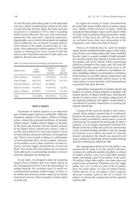
Table (3): Final outcome according to the injured z<strong>on</strong>e.<br />
Injured z<strong>on</strong>e<br />
Z<strong>on</strong>e I (Green)<br />
Z<strong>on</strong>e II (Red)<br />
Z<strong>on</strong>e III (Yellow)<br />
Z<strong>on</strong>e IV (Yellow)<br />
Z<strong>on</strong>e V (Yellow)<br />
Total<br />
Total<br />
number<br />
33 (14%)<br />
48 (23%)<br />
36 (17%)<br />
38 (18%)<br />
59 (28%)<br />
214 (100%)<br />
DISCUSSION<br />
Excellent-<br />
Good<br />
outcome<br />
31 (93.9%)<br />
44 (92.7%)<br />
36 (100%)<br />
37 (97.4%)<br />
57 (96.6%)<br />
205 (95.8%)<br />
Fairpoor<br />
outcome<br />
2 (6.1%)<br />
4 (8.3%)<br />
–<br />
1 (2.6%)<br />
2 (3.4%)<br />
9 (4.2%)<br />
Treatment of tend<strong>on</strong> injuries is an important<br />
part of hand surgery practice worldwide. Adhesi<strong>on</strong><br />
formati<strong>on</strong>, rupture of the repairs, stiffness of finger<br />
joints, remain the principal problems of primary<br />
tend<strong>on</strong> repairs. <str<strong>on</strong>g>Tend<strong>on</strong></str<strong>on</strong>g> injuries happen in all parts<br />
of the hand and forearm, but the tend<strong>on</strong> injuries<br />
in the digital flexor sheath area (z<strong>on</strong>es 1 and 2)<br />
are the most difficult to treat and remain a focus<br />
of both clinical attenti<strong>on</strong> and basic investigati<strong>on</strong>s<br />
[12]. There is now ample evidence to substantiate<br />
several important facts. As an example, intrasynovial<br />
tend<strong>on</strong>s receive their nutriti<strong>on</strong> via both intrinsic<br />
vascular supply and perfusi<strong>on</strong> of synovial<br />
fluid. This means <strong>that</strong> the tend<strong>on</strong>s do not need to<br />
form adhesi<strong>on</strong>s to surrounding tend<strong>on</strong>s to receive<br />
nutriti<strong>on</strong> adequate for healing [1].<br />
In our study, we designed a plan for repairing<br />
injured flexor tend<strong>on</strong>s <strong>that</strong> was totally based <strong>on</strong><br />
the background known from the physiology of<br />
tend<strong>on</strong> healing. We included cases in which we<br />
could perform primary tend<strong>on</strong> repair, as there is<br />
no doubt <strong>that</strong> primary tend<strong>on</strong> repair gives better<br />
functi<strong>on</strong>al recovery than sec<strong>on</strong>dary tend<strong>on</strong> repair<br />
or graft [13].<br />
In regard the timing of repair, Swi<strong>on</strong>tkowski<br />
[6] stated <strong>that</strong> acute tend<strong>on</strong> injuries require urgent<br />
care, ideally within 24 hours of injury. Zidel [4]<br />
c<strong>on</strong>sidered <strong>that</strong> primary repair can be d<strong>on</strong>e within<br />
24 hours and c<strong>on</strong>sidered delayed primary repair<br />
with the 1 st day up to the 14 th day. In our study,<br />
we included cases <strong>that</strong> were presenting to the<br />
emergency unit within less than 24 hours.<br />
Variety of methods may be used for tend<strong>on</strong><br />
repair, but the modified Kessler repair is still widely<br />
used for the core tend<strong>on</strong> suture [14]. Also, modified<br />
Kessler repair is a good example of high-strength,<br />
low-fricti<strong>on</strong> repairs <strong>that</strong> minimize fricti<strong>on</strong> between<br />
the tend<strong>on</strong> and flexor sheath while maintaining<br />
sufficient strength to the repair [15]. We used the<br />
modified Kessler repair in all of our cases as the<br />
standard core suture in additi<strong>on</strong> to peripheral sutures.<br />
Handling tend<strong>on</strong>s was atraumatic to minimize<br />
mobilizati<strong>on</strong> as possible during preparati<strong>on</strong> and<br />
sutures were preferentially placed nearer to the<br />
volar surface to least interfere with intratendinous<br />
circulati<strong>on</strong> <strong>that</strong> enter dorsally.<br />
Appropriate management of tend<strong>on</strong> sheath and<br />
pulleys is c<strong>on</strong>cern of hand surge<strong>on</strong>s in dealing with<br />
tend<strong>on</strong> injuries in digital sheath area. Suturing the<br />
sheath is c<strong>on</strong>troversial. Avoiding compressi<strong>on</strong> of<br />
the repaired tend<strong>on</strong> by the tightly closed sheath is<br />
c<strong>on</strong>sidered of primary importance in treating the<br />
injured sheath [16].<br />
Closure of the synovial sheath is still c<strong>on</strong>troversial.<br />
Some authors menti<strong>on</strong> <strong>that</strong> it is indicated,<br />
based <strong>on</strong> the fact <strong>that</strong> since intrinsic tend<strong>on</strong> vasculature<br />
is easily occluded by sutures and so, synovial<br />
nutriti<strong>on</strong> may be required for healing [8]. In other’s<br />
opini<strong>on</strong>, it is no l<strong>on</strong>ger c<strong>on</strong>sidered essential [17].<br />
<str<strong>on</strong>g>Based</str<strong>on</strong>g> <strong>on</strong> the fact of <strong>that</strong> the synovial nutriti<strong>on</strong> has<br />
a role in tend<strong>on</strong> healing and <strong>that</strong> it may be enough<br />
for healing even without the need of intrinsic<br />
tend<strong>on</strong> vasculature, the sheath was sutured in all<br />
cases, aiming for enhancing intrinsic tend<strong>on</strong> healing<br />
and thus minimizing adhesi<strong>on</strong>s [18].<br />
Our management protocol for the pulleys was<br />
as prescribe by Tang, et al. [19], which is the preservati<strong>on</strong><br />
of a sufficient number of pulleys is critical<br />
to tend<strong>on</strong> moti<strong>on</strong>. Loss of an individual annular<br />
pulley (including a part of A2 pulley or the entire<br />
A4 pulley) when other pulleys are intact does not<br />
result in loss of functi<strong>on</strong>. Therefore, loss of a single<br />
pulley (A1, A3, or A4) or a part of the A2 pulley<br />
does not need repair. In case of tend<strong>on</strong> repairs<br />
within narrow A2 or A4 pulleys, some surge<strong>on</strong>s<br />
advocate venting a part of the A2 or entire A4<br />
pulleys to release the compressi<strong>on</strong> of the repaired<br />
tend<strong>on</strong>s [20].