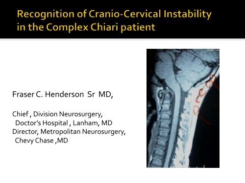
Recognition of Cranio-Cervical Instability in the Complex Chiari ...
Recognition of Cranio-Cervical Instability in the Complex Chiari ...
Recognition of Cranio-Cervical Instability in the Complex Chiari ...

You also want an ePaper? Increase the reach of your titles
YUMPU automatically turns print PDFs into web optimized ePapers that Google loves.
Fraser C. Henderson Sr MD,<br />
Chief , Division Neurosurgery,<br />
Doctor’s Hospital , Lanham, MD<br />
Director, Metropolitan Neurosurgery,<br />
Chevy Chase ,MD
¡ Double vision<br />
¡ Memory loss<br />
¡ Speech difficulties<br />
¡ Dizz<strong>in</strong>ess<br />
¡ Vertigo, T<strong>in</strong>nitus<br />
¡ Difficulty swallow<strong>in</strong>g<br />
¡ Disorder <strong>of</strong> breath<strong>in</strong>g<br />
¡ Chok<strong>in</strong>g<br />
¡ Dysautonomia<br />
¡ Numbness,Weakness<br />
¡ Rapid fatigue<br />
¡ Unsteady gait<br />
¡ Ur<strong>in</strong>ary frequency<br />
¡ Irritable bowel disease
§ A significant number <strong>of</strong> patients worsened<br />
after suboccipital decompression<br />
Dyst ,1988; B<strong>in</strong>dal ,1995; Dauser, 1988<br />
§ 50% pediatric group have “Ventral<br />
Bra<strong>in</strong>stem Flatten<strong>in</strong>g”<br />
Grabb,1999<br />
§ 40% have basilar impression<br />
Cahan L, J NSGY,1982<br />
§ Angulation over <strong>the</strong> odontoid process<br />
Menezes,1988
Bra<strong>in</strong>stem herniation (<strong>Chiari</strong><br />
1.5)<br />
Medullary k<strong>in</strong>k / retr<strong>of</strong>lexed<br />
odontoid<br />
Abnormal clivoaxial angle/<br />
basilar <strong>in</strong>vag<strong>in</strong>ation<br />
to treat <strong>the</strong> deformative stress<br />
imparted by <strong>the</strong>se<br />
anomalies,<br />
56% required occipito-‐cervical<br />
fusion<br />
22% transoral odontoidectomy
¡ Discuss How deformative stress affects <strong>the</strong> nervous<br />
system<br />
¡ Identify established metrics <strong>of</strong> anatomic and dynamic<br />
deformative stress
Stress results from stra<strong>in</strong> Ɛ<br />
Ɛ = d L / Lo<br />
Normal human neuraxis develops a stra<strong>in</strong><br />
Ɛ = .17 on full flexion<br />
Giant squid axon loses function at<br />
Ɛ = .2
Kitihara, Neurol Med Chir<br />
(Tokyo)1995<br />
Margolies, IRCOB Conference,<br />
1992<br />
Tunturi, J Neurosurg,1978<br />
Breig A: Overstretch<strong>in</strong>g <strong>of</strong> <strong>the</strong><br />
sp<strong>in</strong>al cord-‐-‐a basic cause <strong>of</strong><br />
symptoms <strong>in</strong> cord disorders. J<br />
Biomech 3:7-‐9, 1970<br />
neutral<br />
flexion
¡ = “out <strong>of</strong> plane” load<strong>in</strong>g from a<br />
retr<strong>of</strong>lexed odontoid<br />
¡ Causes local histo-‐pathological<br />
changes and also <strong>in</strong>creases<br />
tensile stress<br />
Breig, “Adverse Mechanical Tension <strong>in</strong> <strong>the</strong><br />
CNS”, 1978<br />
Sp<strong>in</strong>al Cord
¡ Clump<strong>in</strong>g and loss <strong>of</strong><br />
neur<strong>of</strong>ilaments and<br />
microtubules<br />
Povlishok, Bra<strong>in</strong> Path,1995<br />
Maxwell, J Neurotrama,2002<br />
Jafari J Neurocytol,1997
Giant squid axon becomes non functional at Ɛ= .2<br />
Stretch <strong>in</strong>jury → Accumulation <strong>of</strong> Neur<strong>of</strong>ilament <strong>in</strong> Axons with<strong>in</strong> 2 hrs.<br />
Confocal microscopy <strong>of</strong> immuno- sta<strong>in</strong>ed to reveal neur<strong>of</strong>ilament prote<strong>in</strong><br />
12
Mouse optic nerve stretched<br />
2mm (ε = .2)<br />
Saatman KE J Cereb Blood Flow Metab. 23(1):34-42, 2003<br />
13<br />
50 um
Distortion <strong>of</strong><br />
Na+<br />
channels<br />
Calcium <strong>in</strong>flux after stretch <strong>in</strong>jury<br />
Reversal Na<br />
+ Ca++<br />
gradient<br />
Depolarizat<br />
ion voltage-<br />
gated Ca+<br />
+ channels<br />
Wolf et al, J Neurosci 2001
Calcium <strong>in</strong>flux after stretch <strong>in</strong>jury, blocked by TTX<br />
Pre stretch Post-stretch<br />
Wolf, Sties, Lizard, Smith, J<br />
Neurosci 2001<br />
15
16<br />
¡ Secondary <strong>in</strong>jury<br />
¡ Up-‐regulation <strong>of</strong> NMDA<br />
receptors<br />
¡ vulnerability to nitrous oxide<br />
and reactive oxygen species<br />
¡ mitochondrial dysfunction<br />
and DNA fragmentation<br />
¡ Programmed cell death<br />
(apoptosis)<br />
(Arund<strong>in</strong>e M et. al. J Neuroscience. 2004,<br />
24(37): 8106-‐8123)
¡ Mechanical forces modulate gene expression and<br />
biochemical composition <strong>of</strong> <strong>the</strong> liv<strong>in</strong>g system at <strong>the</strong><br />
molecular level
Effect <strong>of</strong> stra<strong>in</strong> rate<br />
on conduction<br />
• Amplitude <strong>of</strong> action potential decreases with<br />
stra<strong>in</strong> rate and magnitude <strong>of</strong> stra<strong>in</strong><br />
P
¡ Compressed arrays <strong>of</strong> O2<br />
saturation show severe Hypoxia<br />
¡ Odontoidectomy and<br />
stabilization resulted <strong>in</strong><br />
resolution <strong>of</strong> sleep apnea<br />
¡ Howard, Henderson et al. Ann Rheum Dis,<br />
1993<br />
Menezes,J NSGY,1985<br />
19
Ann Rheum Dis. 1994 February; 53(2): 134–136.<br />
PMCID: PMC1005266<br />
Respiratory abnormalities due to craniovertebral junction<br />
compression <strong>in</strong> rheumatoid disease.<br />
R S Howard, F C Henderson, N P Hirsch, J M Stevens, B E Kendall, and H A Crockard<br />
Harris Unit, National Hospital for Neurology and Neurosurgery, Queen Square, London, United<br />
K<strong>in</strong>gdom.<br />
This article has been cited by o<strong>the</strong>r articles <strong>in</strong> PMC.<br />
Abstract<br />
OBJECTIVE-‐-‐To assess <strong>the</strong> extent and severity <strong>of</strong> respiratory <strong>in</strong>sufficiency associated with severe<br />
rheumatoid atlantoaxial dislocation and its relation to compression <strong>of</strong> <strong>the</strong> neuraxis.<br />
METHODS-‐-‐Twelve patients with severe atlantoaxial dislocation due to rheumatoid disease<br />
were studied. Detailed cl<strong>in</strong>ical, CT myelography and respiratory assessment <strong>in</strong>clud<strong>in</strong>g<br />
nocturnal oximetry, were performed on all patients
Neurosurgery: May 2005 - Volume 56 - Issue 5 - pp 1101-1113
¡ Deformative stress<br />
-‐ Anatomic<br />
-‐ basilar <strong>in</strong>vag<strong>in</strong>ation due to kyphotic clivo-‐<br />
axial angle<br />
-‐ congenital anatomic variants<br />
-‐ syr<strong>in</strong>gomyelia<br />
-‐ Dynamic<br />
-‐craniocervical <strong>Instability</strong><br />
-‐ atlanto-‐axial and sub-‐axial <strong>in</strong>stability<br />
-‐ physiological<br />
-‐ CSF flow<br />
-‐ hydrocephalus
“Loss <strong>of</strong> <strong>the</strong> ability <strong>of</strong> <strong>the</strong> sp<strong>in</strong>e under physiological<br />
loads to ma<strong>in</strong>ta<strong>in</strong> relationships between<br />
vertebrae <strong>in</strong> such a way that <strong>the</strong>re is no damage<br />
or subsequent irritation <strong>of</strong> <strong>the</strong> sp<strong>in</strong>al cord,<br />
(bra<strong>in</strong>stem) or nerve roots, and <strong>in</strong> addition that<br />
<strong>the</strong>re is no development <strong>of</strong> deformity or<br />
<strong>in</strong>capacitat<strong>in</strong>g pa<strong>in</strong> due to structural changes”<br />
White AA Panjabi MM Cl<strong>in</strong> Biomech Sp<strong>in</strong>e 1978
Anatomic<br />
-‐ Basilar <strong>in</strong>vag<strong>in</strong>ation<br />
¡ Clivo-‐axial angle<br />
¡ Ventral bra<strong>in</strong>stem<br />
compression by Grabb-‐ Oakes<br />
method<br />
¡ Lee’s X l<strong>in</strong>es<br />
Dynamic<br />
-‐ <strong>Cranio</strong>-‐vertebral <strong>in</strong>stability<br />
¡ Harris measurements <strong>of</strong><br />
basion to anterior axial l<strong>in</strong>e<br />
¡ Wholley’s Basion-‐ dens<br />
<strong>in</strong>terval<br />
¡ Bull’s palato-‐atlantal angle<br />
angle
130°<br />
ABNORMAL<br />
Angulation <strong>of</strong> <strong>the</strong><br />
bra<strong>in</strong>stem <strong>in</strong> basilar<br />
<strong>in</strong>vag<strong>in</strong>ation causes<br />
neurological disability-<br />
Scoville,1951<br />
165º<br />
155º<br />
175º<br />
neutral flexion extension
130°<br />
ABNORMAL<br />
•<br />
<strong>Chiari</strong> decompression failed<br />
if <strong>the</strong> CAA < 135º<br />
Kim, Rekate, Klopfenste<strong>in</strong>, Sonntag 2004<br />
<strong>Chiari</strong> decompression failed<br />
to improve syr<strong>in</strong>gomyelia<br />
if CAA < 135º KUbota 2004
Henderson et al, Surg Neurol Internat, 2010
¡ List 1941<br />
¡ Van Gilder 1985<br />
¡ Menezes 1990<br />
¡ Dickman 1990<br />
¡ Henderson, Geddes, Crockard 1993<br />
¡ Grabb, Mapstone and Oakes 1999<br />
¡ Milhorat 1999<br />
¡ Kubota 2004<br />
¡ Goel 1998,2004 Platybasia , retr<strong>of</strong>lexed odontoid<br />
and BI associated with medullary k<strong>in</strong>k, alignment<br />
craniocervical junction<br />
¡ Kim, Rekate,Klopfenste<strong>in</strong>, Sonntag 2004<br />
¡ Botelho 2007 Traction reduction <strong>of</strong> <strong>Cranio</strong>cervical<br />
kyphosis<br />
¡ Henderson, Vacarro, Benzel 2004,2010
Ventral Bra<strong>in</strong>stem<br />
Compression<br />
B-pC2<br />
Grabb-<br />
Oakes l<strong>in</strong>e<br />
Grabb, Mapstone , Oakes<br />
J NSGY 1999<br />
> 9 mm
¡ A l<strong>in</strong>e drawn along <strong>the</strong> hard<br />
palate, <strong>in</strong>tersect<strong>in</strong>g a l<strong>in</strong>e<br />
along <strong>the</strong> Atlas <strong>in</strong> <strong>the</strong><br />
neutral position<br />
b >13° reflects basilar<br />
<strong>in</strong>vag<strong>in</strong>ation<br />
¡ But does not account for<br />
variability between flexion<br />
extension, nor recognize<br />
differential development <strong>of</strong><br />
face and sp<strong>in</strong>e<br />
Bull, Nixon and Pratt, Bra<strong>in</strong> 78:<br />
229-‐247, 1955.The radiological<br />
criteria and familial occurrence <strong>of</strong><br />
primary basilar <strong>in</strong>vag<strong>in</strong>ation<br />
b
¡ 600 normal cervical sp<strong>in</strong>e x-‐rays<br />
¡ basion to dens <strong>in</strong>terval relatively<br />
constant<br />
¡ The middle <strong>of</strong> <strong>the</strong> odontoid should<br />
lie directly beneath <strong>the</strong> basion,<br />
with<strong>in</strong> 5 mm<br />
¡ >10mm abnormal<br />
Wholey JH, Bruwer AJ, Baker HLJ The Lateral<br />
roentgengram <strong>of</strong> <strong>the</strong> neck (with comments on<br />
<strong>the</strong> atlanto-‐odontoid basion relationship)<br />
Radiology 71:350-‐356, 1958
¡ basio to C1 posterior arch<br />
over Opisthion to C1 arch<br />
usually BC/OA = .77<br />
¡ If >1, <strong>the</strong>n occipito-‐cervical<br />
dislocation exists<br />
¡ May be <strong>in</strong>valid with atlas fx or<br />
congenital anomaly<br />
¡ Must identify <strong>the</strong> midpo<strong>in</strong>t <strong>of</strong><br />
posterior arch <strong>of</strong> C1<br />
Powers B, Miller md,Kramer RS: Traumatic<br />
Anterior Atlanto-‐Occipital dislocation.<br />
Neurosurgery 4:12-‐17, 1979<br />
C<br />
O
¡ Basion to midpo<strong>in</strong>t sp<strong>in</strong>o-‐<br />
lam<strong>in</strong>ar l<strong>in</strong>e C2 should be<br />
tangential to odontoid<br />
¡ Opisthion to post-‐ <strong>in</strong>ferior C2<br />
should be tangential to C1<br />
¡ Requires normal C1-‐C2<br />
relationship<br />
LeeC, Woodr<strong>in</strong>g JH, Goldste<strong>in</strong> SJ: Evaluation <strong>of</strong><br />
traumatic atlantooccipital dislocations. AJNR<br />
8:19-‐26
¡ Of 400 normal subjects, none<br />
had basion to PAL >12mm<br />
¡ “In adults, <strong>the</strong> occipito-‐<br />
vertebral junction can be<br />
considered normal when both<br />
<strong>the</strong> basion axial <strong>in</strong>terval and<br />
<strong>the</strong> basion dental <strong>in</strong>terval are<br />
12mm or less”<br />
Harris JH, Carson GC, Wagner LK:<br />
Radiological diagnosis <strong>of</strong> traumatic<br />
Occipitovertebr4al Dissociation
posterior axial l<strong>in</strong>e
flexion-‐extension is <strong>the</strong> only motion at <strong>the</strong> normal OA jt Field<strong>in</strong>g JW<br />
C<strong>in</strong>eroentgenography <strong>of</strong> <strong>the</strong> normal cervical sp<strong>in</strong>e J BJT Surgery 39A1280-‐1288<br />
There is no horizontal translation at <strong>the</strong> OA jt Werne S:Studies <strong>in</strong> spontaneous<br />
atlas dislocation Acta Orthoped ScandSuppl 23,1957<br />
“The normal range <strong>of</strong> horizontal translation <strong>in</strong> flexion/ extension is<br />
no more than1 mm . Movement >1mm is cl<strong>in</strong>ically significant .<br />
Treatment by posterior cranio-‐cervical fusion has proved successful<br />
Weisel SW ,Rothman RH: Occipito atlantal Hypermobility .Sp<strong>in</strong>e 4:187-‐191, 1979<br />
“>more than 1 mm <strong>of</strong> translation <strong>in</strong> flexion /extension is an important<br />
and useful criterion. Symptoms <strong>of</strong> weakness <strong>of</strong> <strong>the</strong> limbs and occipital<br />
pa<strong>in</strong> are additional <strong>in</strong>dications <strong>of</strong> <strong>in</strong>stability”<br />
White AA, P unjabi MM Cl<strong>in</strong>ical Biomechanics <strong>of</strong> <strong>the</strong> Sp<strong>in</strong>e :p284-‐286 ,2 ND Edition Lipp<strong>in</strong>cott1990
Weisel, Rothman, 1979; White and Punjabi 1980; Menezes, 1990
Harris measurement = 12mm Harris measurement = 7mm
At least 12% <strong>of</strong> <strong>Chiari</strong> population have<br />
EDS Milhorat et al, J Neurosurg Sp<strong>in</strong>e 7:601–609, 2007<br />
¡ JHS and hypermobility type <strong>of</strong> EDS are<br />
phenotypically <strong>in</strong>differentiable<br />
Grahame 2008<br />
¡ Diagnosed by Brighton Criteria<br />
Leone 2009<br />
¡ Hypermobility Syndrome affects 1 % <strong>of</strong><br />
<strong>the</strong> population<br />
Grahame, 2008
Mechanical stresses<br />
¡ modulate gene expression and biochemical<br />
composition <strong>of</strong> <strong>the</strong> liv<strong>in</strong>g system at <strong>the</strong> molecular<br />
level<br />
¡ alter neuronal conduction<br />
¡ Alter <strong>the</strong> histology<br />
¡ Are expressed cl<strong>in</strong>ically
¡ prospectively study to compare cl<strong>in</strong>ical metrics<br />
with neurological deficits <strong>in</strong> patients with cervico-‐<br />
medullary disorders<br />
¡ IRB approved<br />
¡ N =7
Neurological assessment<br />
Sensory motor : ASIA<br />
Pa<strong>in</strong> : Visual Analog Scale<br />
Quality <strong>of</strong> life : SF36<br />
Function: Karn<strong>of</strong>sky Index<br />
Bulbar Dysfunction : Bra<strong>in</strong>stem Disability Index<br />
FEA computations<br />
MRI: Clivo axial angle<br />
b-‐pC2 (Grabb-‐Oakes) measurement
¡ s/p repeated cardio-‐respiratory arrests<br />
postural orthostatic tachycardia,<br />
decreased memory, visual and auditory<br />
changes, dysphagia, dizz<strong>in</strong>ess, gait<br />
change, weakness and sensory loss,<br />
bowel and bladder changes<br />
¡ Bab<strong>in</strong>ski, spastic quadriparesis, diffuse<br />
sensory deficits<br />
¡ Clivo-‐axial angle 115º<br />
Illustrative Case <strong>of</strong> <strong>Complex</strong> <strong>Chiari</strong><br />
Cervico- medullary syndrome<br />
<strong>Chiari</strong>, kyphotic clivo-axial angle, ventral bra<strong>in</strong>stem compression
¡ Neurological compression<br />
and <strong>in</strong>stability at <strong>the</strong> occiput –<br />
C1<br />
¡ Myelopathy, bra<strong>in</strong>stem<br />
dysfunction, lower cranial<br />
nerve dysfunction, vertebro-‐<br />
basilar <strong>in</strong>sufficiency<br />
¡ Neurological deficits = urgent<br />
need for decompression and<br />
stabilization<br />
Dickman, Douglas ,Sonntag, BNI Quarterly, 1990
Clivo-‐axial angle 145º<br />
No bra<strong>in</strong>stem symptoms<br />
Karn<strong>of</strong>sky <strong>in</strong>dex 50 to 100<br />
Pa<strong>in</strong> level from 4/10 to 1/10<br />
No breath<strong>in</strong>g problems<br />
Pt is play<strong>in</strong>g sports,<br />
achiev<strong>in</strong>g A levels,<br />
Pilots licence<br />
accepted to MIT
100<br />
90<br />
80<br />
70<br />
60<br />
50<br />
40<br />
30<br />
20<br />
10<br />
0<br />
ASIA Karn<strong>of</strong>sky PC,qol MC, qol<br />
preop<br />
12 mos
¡ Pa<strong>in</strong> p .027<br />
¡ ASIA p .042<br />
¡ Bra<strong>in</strong>stem Disability p .027<br />
¡ Karn<strong>of</strong>sky p .027<br />
¡ SF 36 Physical component p .026<br />
Mental component p .12<br />
Wilcoxon signed-‐ranks test
Surg Neurol Int. 2010; 1: 30.<br />
Published onl<strong>in</strong>e 2010 July 16. doi: 10.4103/2152-‐7806.66461.<br />
PMCID: PMC2940090<br />
Copyright © 2010 Henderson FC.<br />
Deformative stress associated with an abnormal<br />
clivo-‐axial angle: A f<strong>in</strong>ite element analysis<br />
Fraser C. Henderson, William A. Wilson, 1 Stephen Mott, 2<br />
Alexander Mark, 3 Kristi Schmidt, 4 Joel K. Berry, 5 Alexander<br />
Vaccaro, 6 and Edward Benzel 7<br />
Doctors Community Hospital, Georgetown University Hospital, United States<br />
1 Computational Biodynamics LLC.,Yale University, United States<br />
2 Dartmouth-‐Hitchcock Medical Center, Georgetown University Hospital, United States<br />
3 Be<strong>the</strong>sda MRI & CT, Georgetown University Hospital, United States<br />
4 University <strong>of</strong> Alabama Medical Center, Georgetown University Hospital, United States<br />
5 Ketter<strong>in</strong>g University, United States<br />
6 Thomas Jefferson University Hospital, United States<br />
7 Cleveland Cl<strong>in</strong>ic Foundation, United States
14<br />
12<br />
10<br />
8<br />
6<br />
4<br />
2<br />
0<br />
<strong>Chiari</strong> and cranio-vertebral <strong>in</strong>stability<br />
Suboccipital craniectomy and O-C2 fusion n=12<br />
12<br />
9<br />
8<br />
7<br />
6<br />
5<br />
4<br />
3<br />
2<br />
1<br />
0<br />
10<br />
2<br />
11 11<br />
1 1<br />
0 0<br />
0 0 0<br />
Neurological<br />
Change<br />
8<br />
Able to work/ attend school<br />
Every patient surveyed said <strong>the</strong>y would do <strong>the</strong><br />
surgery aga<strong>in</strong> if <strong>the</strong>y had <strong>the</strong> choice and said <strong>the</strong>y<br />
would recommend it to a friend or family member.<br />
1 1<br />
Yes No Retired Soon<br />
Functional Change Quality <strong>of</strong> Life Pa<strong>in</strong> Change<br />
2<br />
Improved<br />
Worsened<br />
No Change
¡ With<strong>in</strong> <strong>the</strong> population diagnosed with <strong>Chiari</strong> malformation, <strong>the</strong>re is a<br />
subset <strong>of</strong> patients with “<strong>Complex</strong> <strong>Chiari</strong> ”<br />
¡ The designation <strong>of</strong> <strong>Complex</strong> <strong>Chiari</strong> resides <strong>in</strong> <strong>the</strong> presence <strong>of</strong><br />
additional anatomic or dynamic deformative stresses, which may be<br />
signaled by:<br />
clivo-‐axial angle 9 mm<br />
Harris > 12mm, or translation > 2mm on flexion extension<br />
Wholey’s BDI > 10mm<br />
Bull’s palato-‐atlantal angle > 13◦<br />
Transgression <strong>of</strong> Lee’s X l<strong>in</strong>es<br />
¡ substantial cl<strong>in</strong>ical and neurobiological evidence l<strong>in</strong>ks deformative<br />
stress <strong>of</strong> bra<strong>in</strong>stem and sp<strong>in</strong>al cord to <strong>the</strong> observed<br />
neurophysiological changes<br />
¡ <strong>Complex</strong> <strong>Chiari</strong> should be watched for, and prompt consideration for<br />
additional evaluation with dynamic imag<strong>in</strong>g
¡ Clair Francomano MD, GBMC , Assoc Pr<strong>of</strong>, JHI<br />
¡ Myles Koby MD, NIH, Doctors Hospital<br />
¡ Micheal Adams, Eng<strong>in</strong>eer<strong>in</strong>g, Duke University<br />
¡ Robert Gerw<strong>in</strong> MD Assoc Pr<strong>of</strong>, JHI<br />
¡ Ed Benzel MD-‐ Pr<strong>of</strong>, Chair Neurosurg ,CCI<br />
¡ Alex Vaccaro MD-‐ Pr<strong>of</strong> Neurosurg Ortho, TJU<br />
¡ Stephen Mott MD Assoc Pr<strong>of</strong> Peds Neurol, Dartmouth<br />
¡ Joel Berry PhD – Pr<strong>of</strong> Chair Mech Eng, Ketter<strong>in</strong>g Univ<br />
¡ Mark Alexander MD , Director Neuradiology Be<strong>the</strong>sda MRI<br />
¡ William Wilson IV,Yale Univ<br />
¡ Jessica Adcock MSc,Metropolitan Neurosurgery Grp