Fluence mapping inside the highly scattering medium using ...
Fluence mapping inside the highly scattering medium using ...
Fluence mapping inside the highly scattering medium using ...

You also want an ePaper? Increase the reach of your titles
YUMPU automatically turns print PDFs into web optimized ePapers that Google loves.
<strong>Fluence</strong> <strong>mapping</strong> <strong>inside</strong> <strong>the</strong> <strong>highly</strong> <strong>scattering</strong> <strong>medium</strong> <strong>using</strong><br />
reflection mode acousto-optics<br />
Altaf Hussain<br />
Biomedical Photonic Imaging group, MIRA Institute for Biomedical Technology and Technical<br />
Medicine, University of Twente, PO Box 217, 7500 AE Enschede, The Ne<strong>the</strong>rlands<br />
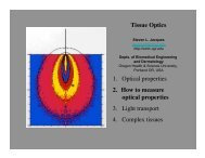
1. Introduction<br />
In recent years, optical imaging modalities received considerable attention of researchers and are moving toward clinical<br />
applications. Optical properties of soft biological tissue such as <strong>scattering</strong> and absorption are related to its morphology<br />
and biochemical composition. Optical imaging modalities, such as photoacoustic (PA) and acousto optics (AO) provides<br />
very useful structural and functional information about tissue physiology [1-4]. However, <strong>the</strong>se imaging modalities suffer<br />
from quantification problem. The quantification problem is a result of unknown local light fluence in <strong>scattering</strong> <strong>medium</strong>.<br />
Since, images of soft biological tissue obtained with optical imaging technique are weighted with unknown local light<br />
fluence. It is difficult to get quantitative information about µa without knowing <strong>the</strong> local fluence. Several attempts have<br />
been made to compensate for fluence variation in PA imaging [5-7], as well in acousto-optics [4, 8]. Main challenge<br />
remains to predict <strong>the</strong> local fluence for a <strong>medium</strong> with unknown and inhomogeneous optical properties. Previously we<br />
presented a purely experimental strategy to do fluence independent photoacoustic imaging [9]. The strategy proposed<br />
combination of two photoacoustic experiments with illumination at two points and one transmission mode AO<br />
experiment.<br />
We propose a <strong>the</strong>oretical concept for experimentally measuring <strong>the</strong> local light fluence in absolute terms in <strong>scattering</strong><br />
media. The proposed methodology is based on an experimental technique that allows local labeling of light with known<br />
efficiency and detection of labeled light in pure reflection mode. We validate our <strong>the</strong>oretical concept <strong>using</strong> Monte Carlo<br />
simulations, results are presented in numerical experiment section. Experimentally we exploit <strong>the</strong> feasibility of <strong>using</strong><br />
reflection mode AO as techniques to locally tag <strong>the</strong> light. Since tagging efficiency of AO is unknown, <strong>the</strong>refor, <strong>the</strong><br />
proposed expression for local fluence is simplified, with few justifiable assumptions. Our experimental results on<br />
measuring <strong>the</strong> fluence in <strong>scattering</strong> <strong>medium</strong> <strong>using</strong> reflection mode AO are presented in experiment section.<br />
2. Theory<br />
In this section, we describe <strong>the</strong> ma<strong>the</strong>matical model of our proposed methodology. This model is based on two<br />
principles, one photon path reversibly in <strong>scattering</strong> <strong>medium</strong> [9, 10] and local labeling of photons <strong>inside</strong> <strong>the</strong> <strong>scattering</strong><br />
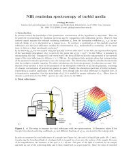
<strong>medium</strong>. Consider a <strong>scattering</strong> <strong>medium</strong> depicted in Fig. 1, where point 1 is on <strong>the</strong> surface and point 2 is <strong>inside</strong> <strong>the</strong><br />
<strong>medium</strong>. The fluence rate ϕ1,2 at point 2 due to injection of light at point 1 with power P1 can be written as,<br />
( ). Where Pr(1,2) is <strong>the</strong> probability per unit area and per unit solid angle that a photon injected at point 1 will<br />
cross <strong>the</strong> aperture placed at point 2, traversing any possible trajectory.<br />
Pl,1<br />
(a) (b)<br />
Ω1<br />
P 1<br />
A 1<br />
A2<br />
Fig. 1. a) Schematic of <strong>medium</strong> and photon trajectories from injection point 1 to detection point 1 through internal point “2” (labeling<br />
volume). b) MC simulation results, estimated light fluence <strong>using</strong> model (squares) and actual simulated fluence (circles).
The photons addressing <strong>the</strong> internal point 2 can be “labeled/modulated” <strong>using</strong> strongly focused ultrasound and detected at<br />
<strong>the</strong> surface of <strong>the</strong> <strong>medium</strong> (AO). Assuming incoming fluence rate ϕ1,2 in internal point 2 (labeling volume) and that all<br />
<strong>the</strong> photons entering <strong>the</strong> labeling volume get labeled (labeling efficiency is unity) and leave without absorption. The<br />
power of detected tagged photons at point 1, can be written as, ( ) By applying photon path reversibility<br />
principle Pr(1,2)= Pr(2,1), and rearranging aforementioned equations for fluence at point 2 and power of tagged photons<br />
measured at 1, we get <strong>the</strong> expression for local fluence rate at point 2,<br />
√ (1)<br />
If we take into account all <strong>the</strong> parameters regarding labeling volume and detection system, <strong>the</strong> expression for <strong>the</strong> absolute<br />
local fluence rate can be written as.<br />
√<br />
√ ( )<br />
This expression for local fluence contains excitation parameter P1, instrumental parameters (Ω1, A1 and A2) and<br />
externally measureable quantity Pl,1. Hence, Eqn. 2 suggests that local light fluence can be measured in optically<br />
inhomogeneous <strong>medium</strong> experimentally. Reflection mode AO is a technique which allows local labeling of light <strong>using</strong><br />
ultrasonic modulation and detection of ultrasonically modulated light in reflection mode [11].<br />
3. Numerical experiment<br />
To proof our methodology we used Monte Carlo simulation program for light transport in turbid media [12]. We<br />
modified <strong>the</strong> program to be able to “label” photons addressing a certain region of interest in <strong>medium</strong>. This region of<br />
interest, we call labeling volume, was considered of spherical shape in our simulations. We simulated a <strong>scattering</strong><br />
<strong>medium</strong> of dimensions 40x40x4 cm 3 , containing a spherical absorber/labeling volume of diameter 1 mm. The optical<br />
properties of background <strong>medium</strong> µs ’ =7.5/cm and µa=0.01/cm were different from <strong>the</strong> optical properties of labeling<br />
volume µs ’ =7.5/cm and µa=1/cm.<br />
In simulations we injected 10^7 photons into <strong>the</strong> <strong>medium</strong> through a circular aperture A1 of diameter 3 mm. Photons<br />
addressing <strong>the</strong> labeling volume during <strong>the</strong>ir random walk through <strong>the</strong> <strong>scattering</strong> <strong>medium</strong> were “labeled”. Labeled<br />
photons leaving <strong>the</strong> <strong>medium</strong> through aperture A1 within <strong>the</strong> opening angle of 25 degrees were detected to simulate<br />
reflection mode AO. We used Eqn. 2 to estimate <strong>the</strong> fluence at <strong>the</strong> position of labeling volume in <strong>scattering</strong> <strong>medium</strong>. The<br />
quantities P1 and Pl,1 power of incident photons and detected labeled photons respectively in Eqn. 2, were replaced by <strong>the</strong><br />
weight of injected photons and detected labeled photons. Results from Monte Carlo simulations presented in Fig. 1b,<br />
show that estimation of <strong>the</strong> local fluence <strong>inside</strong> <strong>the</strong> <strong>scattering</strong> <strong>medium</strong> <strong>using</strong> our proposed methodology.<br />
4. Experimental validation<br />
Acousto optics is a technique that allows such local labeling of light [13], and detection of ultrasonically labeled in<br />
reflection configuration [11]. However, unlike presented <strong>the</strong>oretical model (Eqn. 1) <strong>the</strong> labeling volume of AO is of<br />
complicated shape and its labeling efficiency is unknown. As a result measuring fluence in absolute terms <strong>using</strong><br />
reflection mode AO is not possible at this stage. Therefore, we used Eqn. 1 instead and show that we can measure fluence<br />
variation in <strong>scattering</strong> <strong>medium</strong>. We used speckle contrast detection method to measure ultrasonically modulated<br />
backscattered light []. The speckle contrast decreases in <strong>the</strong> presence of ultrasound. Change in speck contrast (ΔC)<br />
between US ON and OFF is can be measured and it is approximately proportional to <strong>the</strong> intensity of locally<br />
ultrasonically modulated light [14]. This means Eqn. 1 can be written as, √ The experiment was performed on<br />
a soft tissue mimicking cubical phantom of dimensions 3x3x3 mm 3 . The phantom was made of 3% Agar gel and a<br />
dilution of 4% Intralipid (20%), resulting in approximate reduced <strong>scattering</strong> coefficient 7.5 cm -1 . US transducer was<br />
attached to <strong>the</strong> xy translation stage. Speckle contrast change (ΔC) in <strong>the</strong> backscattered light was measured by scanning<br />
<strong>the</strong> ultrasound focus along <strong>the</strong> optical axis (x-axis). To validate our results we measured <strong>the</strong> fluence invasively <strong>using</strong> an<br />
optical fiber.<br />
Fig. 4 shows <strong>the</strong> normalized fluence, measured acousto-optically (red) <strong>using</strong> Eqn. 1 and with <strong>the</strong> fiber (black). Horizontal<br />
axis represents <strong>the</strong> depth from <strong>the</strong> surface along optical axis (x-axis). We measured <strong>the</strong> fluence acousto optically twice,<br />
before (Fig. 2a) and after (Fig. 2b) measuring <strong>the</strong> fluence with <strong>the</strong> fiber, to see if introducing <strong>the</strong> fiber into <strong>the</strong> <strong>medium</strong><br />
effects <strong>the</strong> local fluence, results show no significant change.
Fig. 4. Comparison of acousto optically measured (circles) light fluence with invasively measured (squares) light fluence <strong>using</strong> optical<br />
fiber, in both cases <strong>the</strong> measured signal has been normalized to maximum value of one. a) measurement before inserting <strong>the</strong> fiber into<br />
<strong>medium</strong>, b) measurement after inserting <strong>the</strong> fiber into <strong>medium</strong>.<br />
Results presented in Fig. 4, show that acousto optically measured local light fluence, <strong>using</strong> our proposed methodology, is<br />
in good agreement with invasively measured fluence <strong>using</strong> optical fiber. Presented results are average of fifteen AO<br />
measurements and error bars are obtained by calculating <strong>the</strong> error propagation based on standard deviation in acoustic<br />
measurements. The error in estimated fluence increases with depth as it is expected since SNR in reflection mode AO<br />
becomes lower at higher depth.<br />
We presented a <strong>the</strong>ory that proposes a method to measure <strong>the</strong> local light fluence in <strong>scattering</strong> <strong>medium</strong> experimentally,<br />
without <strong>the</strong> need of any prior knowledge about <strong>the</strong> optical properties of <strong>the</strong> <strong>medium</strong>. Our <strong>the</strong>oretical and numerical<br />
simulation model assumes a simplistic labeling volume and unit tagging efficiency. However, it is not <strong>the</strong> case at<br />
experimental level, when <strong>using</strong> AO as labeling technique. Therefore, we simplified our model (Eqn. 2) to use reflection<br />
mode AO as technique to measure local fluence in <strong>scattering</strong> <strong>medium</strong>, which gives <strong>the</strong> fluence map in relative terms with<br />
an unknown pre-factor. Here in this proceeding, we have shown in real experimental settings that <strong>using</strong> reflection mode<br />
AO local light fluence can be measured. Our present experiments are done on homogeneous <strong>scattering</strong> <strong>medium</strong> and our<br />
goal in future is to investigate <strong>the</strong> applicability of this method in heterogeneous (<strong>scattering</strong> and absorption) <strong>medium</strong>.<br />
5. Reference<br />
[1] R. I. Siphanto, K. K. Thumma, R. G. M. Kolkman, T. G. van Leeuwen, F. F. M. de Mul, J. W. van Neck, L. N.<br />
A. van Adrichem, and W. Steenbergen, Optics Express 13 (2005) 89.<br />
[2] S. Manohar, S. E. Vaartjes, J. C. G. van Hespen, J. M. Klaase, F. M. van den Engh, W. Steenbergen, and T. G.<br />
van Leeuwen, Optics Express 15 (2007) 12277.<br />
[3] X. D. Wang, Y. J. Pang, G. Ku, G. Stoica, and L. H. V. Wang, Optics Letters 28 (2003) 1739.<br />
[4] A. Lev, E. Rubanov, and B. Sfez, Photons Plus Ultrasound: Imaging and Sensing 2005 5697 (2005) 190.<br />
[5] H. F. Zhang, K. Maslov, and L. V. Wang, Photons Plus Ultrasound: Imaging and Sensing 2008: The Ninth<br />
Conference on Biomedical Thermoacoustics, Optoacoustics, and Acoustic-Optics 6856 (2008) T8561.<br />
[6] B. T. Cox, S. R. Arridge, K. P. Kostli, and P. C. Beard, Photons Plus Ultrasound: Imaging and Sensing 2005<br />
5697 (2005) 49.<br />
[7] B. T. Cox, J. G. Laufer, and P. C. Beard, Biomed Opt Express 1 (2010) 201.<br />
[8] A. Bratchenia, R. Molenaar, and R. P. H. Kooyman, Laser Physics 21 (2011) 601.<br />
[9] K. Daoudi, A. Hussain, E. Hondebrink, and W. Steenbergen, Optics Express 20 (2012) 14117.<br />
[10] X. A. Xu, H. L. Liu, and L. V. Wang, Nature Photonics 5 (2011) 154.<br />
[11] A. Lev and B. G. Sfez, Optics Letters 27 (2002) 473.<br />
[12] S. L. Jacques, Photochemistry and Photobiology 67 (1998) 23.<br />
[13] F. A. Marks, H. W. Tomlinson, and G. W. Brooksby, Proceedings of Photon Migration and Imaging in Random<br />
Media and Tissues 1888 (1993) 500.<br />
[14] J. Li, G. Ku, and L. H. V. Wang, Applied Optics 41 (2002) 6030.