PDF (tesi dottorato XXI ciclo Scienze Odontostomatolgiche ... - FedOA
PDF (tesi dottorato XXI ciclo Scienze Odontostomatolgiche ... - FedOA
PDF (tesi dottorato XXI ciclo Scienze Odontostomatolgiche ... - FedOA

You also want an ePaper? Increase the reach of your titles
YUMPU automatically turns print PDFs into web optimized ePapers that Google loves.
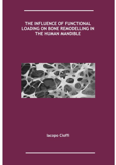
THE INFLUENCE OF FUNCTIONAL<br />
LOADING ON BONE REMODELLING IN<br />
THE HUMAN MANDIBLE<br />
Iacopo Cioffi<br />
1
THE INFLUENCE OF FUNCTIONAL LOADING ON BONE<br />
REMODELLING IN THE HUMAN MANDIBLE<br />
IACOPO CIOFFI<br />
Dottorato di ricerca in <strong>Scienze</strong><br />
Odontostomatologiche <strong>XXI</strong> <strong>ciclo</strong><br />
UNIVERSITA’ DEGLI STUDI DI NAPOLI “FEDERICO II”<br />
2
Promotor:<br />
Prof. Roberto Martina<br />
Copromotors:<br />
Prof. Mauro Farella<br />
Prof. Ambra Michelotti<br />
Prof. Geerling Langenbach<br />
Ir. Leo van Ruijven<br />
Prof. Theo van Eijden<br />
Department of Oral Sciences, Section of Orthodontics and<br />
Temporomandibular Disorders, University of Naples “Federico II”,<br />
Italy<br />
Department of Functional Anatomy, Academic Centre for Dentistry<br />
Amsterdam (ACTA), The Netherlands<br />
This thesis was the result of a PhD program attended at the Department of Oral<br />
Sciences, Section of Orthodontics and Temporomandibular Disorders, University of<br />
Naples “Federico II”. The study described in this thesis was performed at the<br />
Department of Oral Sciences, Section of Orthodontics and Temporomandibular<br />
Disorders, University of Naples “Federico II”, and at the Department of Functional<br />
Anatomy, Academic Centre for Dentistry Amsterdam (ACTA), The Netherlands.<br />
The studies included in this thesis are part of articles published or submitted for<br />
publication, and are covered by copyrights.<br />
3
CONTENTS<br />
Overview on microcomputed tomography<br />
Regional variations in mineralization and strain<br />
distributions in the cortex of the human mandibular<br />
condyle<br />
The influence of muscular activity on bone remodelling in<br />
the human mandible<br />
References 35<br />
Curriculum vitae et studiorum 39<br />
5<br />
9<br />
23<br />
4
CHAPTER 1<br />
Overview on microcomputed tomography (Micro-CT)<br />
Microtomography is a tecnique that uses x-rays to create cross-sections of 3D-<br />
objects that are used then to recreate a virtual model without destroying the<br />
original one (Fig.1). The term micro is used to indicate that the pixel sizes of the<br />
cross-sections, which are generally in the micrometer range or less. Microcomputed<br />
tomography (microCT) is a high- resolution type of computerized axial tomography.<br />
Typically the spatial resolution of conventional medical CT-scanners is in the range<br />
of 1-2.5 mm, which corresponds to 1-10 cubic mm voxel (volume element) . With<br />
micro-CT, in the most recent releases, resolutions up to few nanometers have been<br />
reached. These systems make use of computers or working station, as servers, and<br />
are able provide high resolution models, which can be easily assessed in three<br />
dimensions. The X-ray tube (open or sealed) produces a conic beam of electron<br />
that penetrates the object to be analyzed, and a digital signal is interpreted by the<br />
2D detector as a Digital Radiograph image. The object is positioned on a precision<br />
rotational stage and an image is acquired during the rotation at a constant step.<br />
The scan usually covers a rotation of 360 degrees, but for specific applications a<br />
limited angle scan<br />
Fig.1 Scheme of computed tomography<br />
5
can be performed. Microcomputed tomography has been successfully used for<br />
different purposes, such as the study of porous or cavity-containing objects<br />
(metallic foams, electronics, stones, wood and composite polymers) or for biologic<br />
investigations. On the whole, these systems have been widely used for the study of<br />
hard tissues because of the high linear attenuation coefficient of the calcified bone<br />
and dental matrices. In the recent years, this technique has shown to be promising<br />
in the study of trabecular and cortical bone morphology following bone loss in<br />
osteoporotic patients or in animal models of osteoporosis. In bone biology,<br />
microCTs are widely used for the measurement of the characteristics of the<br />
trabecular, cortical, and canalicular network. Indeed, bone histomorphometry and<br />
microarchitecture can be easily assessed in order to estimate the mechanical<br />
characteristics of bone tissue in different locations. Three dimensional (3D)<br />
modelling and analysis reconstruction of specimens can be obtained with several<br />
softwares. The ANT software (Skyscan - Aartselaar, Belgium), was successfully used<br />
for the studies presented in this thesis. The program allows reconstruction of<br />
objects from 2D slices, after thresholding. The reconstructed 3D models are<br />
obtained by a surface-rendering algorithm. Four different 3D models can be<br />
reconstructed and made visible on the computer screen simultaneously, thus<br />
offering the possibility to combine several images. In addition, the program offers<br />
facilities for 360° model rotations in all directions, displacements, lightening<br />
effects and colouring of the desired structures. A very interesting facility for the<br />
study of porous structures was the possibility to make the virtual models semi-<br />
transparent. Another interesting possibility was to obtain 2D reslices of the objects<br />
across a plane, positioned in a specified direction. Morphometric measurements<br />
can be done on 2D images and 3D models with software.<br />
Micro-CT Specifications<br />
In the studies presented in this thesis, the following micro-CT system was used.<br />
Micro-CT 40 SCANCO<br />
Type Destktop Cone-Beam MicroCT Scanner (Fig.2)<br />
No specimen preparation required<br />
Non-destructive<br />
X-Ray Microfocus X-Ray-source<br />
5 µm Spot Size<br />
6
50-70 kVp, 8 W (160 µA)<br />
Detector 2048x252 elements, 24 µm pitch<br />
Resolution 6 µm nominal, 9 µm (10% MTF @ 12 mm Ø)<br />
Image Matrix variable from 512 x 512 to 2048x20<br />
Specimen Size up to 38 mm Ø<br />
max. Scan Length 80 mm<br />
Computer hp 64bit Itanium IA64<br />
multiprocessor systems<br />
Memory: 48 GB<br />
Disk: 1 TB disk tower<br />
Optional PostScript printers<br />
Software Complete 64-bit imaging solution<br />
- Data acquisition<br />
- Online/Offline Reconstruction<br />
- Sophisticated 2D/3D evaluation<br />
- 3D-visualization/animation<br />
- Archiving<br />
Density Calibratioon<br />
Database<br />
Browser-access (web-based access)<br />
Electrical 100-240 V/50-60 Hz<br />
300 W<br />
Physical Scanner weight: 130 kg<br />
Scanner dimensions (WxDxH): 85 x 41 x 72 cm<br />
7
Fig.2 Scanco MicroCT scanner (MicroCT 40)<br />
8
CHAPTER 2<br />
Regional variations in mineralization and strain<br />
distributions in the cortex of the human mandibular<br />
condyle<br />
Abstract<br />
The strain (i.e. deformation) history influences the degree of mineralization of<br />
cortical bone (DMB) as well as its osteonal microstructure. This study aimed to<br />
examine the relationships of stress and strain distributions with the variations in<br />
DMB and the osteonal orientations in the cortical bone of the human mandibular<br />
condyle. It was hypothesized that strains are inversely proportional to local DMB<br />
and that the principal strains are oriented parallel to the osteons. To test this, ten<br />
human mandibular condyles were scanned in a microCT system. Finite element<br />
models were created in order to simulate static clenching. Within each condyle, 18<br />
volumes of interest were selected to analyze regional differences in DMB, stress<br />
and strains. Subchondral bone showed a lower equivalent strain (2652±612 µε) as<br />
compared to the anterior (p=0.030) and posterior cortex (p=0.007) and was less<br />
mineralized. Contrary to our hypothesis, the results show that strains correlated<br />
positively with regional variations in DMB (r=0.750, pb0.001). In the anterior and<br />
the posterior cortex, the first principal strain was parallel to the cortical surface<br />
and oriented supero-inferiorly with a fan-like shape. In subchondral bone, the first<br />
and the second principal strain were parallel to the surface and oriented antero-<br />
posteriorly and medio- laterally, respectively. It was concluded that the strain<br />
distributions, by themselves, cannot explain the regional differences found in DMB.<br />
In agreement with our second hypothesis, the orientation of the osteonal network<br />
of the mandibular condyle was closely related to the strain orientations. The<br />
results of this study suggest that the subchondral and the cortical bone are<br />
structured to ensure an optimal load distribution within the mandibular condyle<br />
and have a different mechanical behaviour. Subchondral bone plays a major role in<br />
the transmission of the strains to the anterior and posterior cortex, while these<br />
9
ensure an optimal transmission of the strains within the condylar neck and,<br />
eventually, to the mandibular ramus.<br />
INTRODUCTION<br />
Bone is a dynamic tissue capable of adapting its structure to local mechanical<br />
stimuli by continuous bone renewal [1–3].It has been noted that strains occurring<br />
within bone affect this turnover and hence bone macrostructure (i.e. shape and<br />
size of bones) and microstructure (i.e. osteons, plates, rods etc.) by initiating cell-<br />
mediated remodelling [4,5]. Consequently, bone morphology and internal<br />
architecture strongly depend on the deformation history [6–8]. Except for the bone<br />
structure, the deformation history also influences the mineralization of bone since<br />
the average age of bone tissue is inversely proportional to the remodelling rate,<br />
and the mineralization is proportional to the bone age. Therefore, bone mineral<br />
content is commonly believed to be inversely proportional to the rate of<br />
remodelling [9]. For cortical bone, the relationship between mechanical loading<br />
and mineralization has been investigated in several bones. Regional variations in<br />
cortical bone mineralization have been related to the amount or to the mode (i.e.<br />
tensile/compressive) of locally occurring strains [10–12]. A significant lower ash<br />
mineral content was found in the highly strained caudal compression cortex of the<br />
sheep radius [12]. Differences in mineral content between tension/compression<br />
cortices were found also in the mule deer calcaneus [10]. In the human femur, the<br />
mineralization of cortical bone was found to vary among sites subjected to<br />
different loading conditions [11]. Recently, in the human mandibular condyle,<br />
differences in DMB have been found between cortical regions (i.e. anterior,<br />
posterior and sub- chondral) [13]. These variations were also attributed to the<br />
remodelling process. Although several studies have investigated the stress and<br />
strain distributions in the mandible [14–18], the relationship between strains and<br />
bone mineralization in the cortex of the human mandibular condyle has not been<br />
described. Hence, it is not certain to what extent the observed regional variations<br />
in DMB are dependent on the locally occurring strains. Previous studies [19–21]<br />
have shown that the orientation of the cortical canals is parallel to locally<br />
occurring strains. In human mandibular condyle, the orientation of the canals,<br />
which is medio-lateral in subchondral bone and supero-inferior in the anterior and<br />
the posterior cortex, suggests an accordingly oriented principal direction of the<br />
10
strains [22]. However, the data available regarding the strain orientations in this<br />
location [14,15,17] are not sufficient to detect the relation between cortical canal<br />
orientations and strain directions. The aim of the current study was to analyze the<br />
potential relationship between regional variations in DMB with stress and strain<br />
distributions in the cortical bone of the human mandibular condyle and to<br />
determine the orientation of the strains occurring during functional loading. It was<br />
hypothesized that strains correlate inversely to DMB and that the principal strains<br />
are parallel to the cortical canals. For this purpose, ten mandibular condyles were<br />
analyzed with a microCT scanner. The reconstructed bone tissue was used to<br />
generate finite element (FE) models which include the DMB and its inhomogenous<br />
distribution. These models were used to analyze regional differences in DMB, von<br />
Mises stress, equivalent strain, strain orientations and strain energy density in<br />
various cortical bone locations and to calculate the condylar displacements.<br />
Materials and methods<br />
Condyles<br />
Ten left mandibular condyles were obtained from ten embalmed human male<br />
cadavers (mean age SD: 69.8±14.4 years, range: 43 to 92 years). On average, the<br />
subjects had 10.2±4.5 teeth in the upper jaw and 11.5±2.6 in the lower jaw. No<br />
signs of relevant malocclusion or condylar erosions were found. The condyles were<br />
separated from the mandible at the transition from the neck to the ramus with a<br />
hand saw; condylar bone marrow was left in situ. Specimens were fixated in 60%<br />
formalin. The use of the condyles conforms to a written protocol that was reviewed<br />
and approved by the Department of Anatomy and Embryology of the Academic<br />
Medical Center of the University of Amsterdam.<br />
MicroCT<br />
Three-dimensional reconstructions of the cortical bone of the condyles were<br />
obtained using a high-resolution microCTsystem (µCT 40, ScancoMedical AG,<br />
Bassersdorf, Switzerland). Each condyle was positioned to be scanned in frontal<br />
slices. The scan resolution was 15 µm, the beam energy 45 kV, which corresponds<br />
to an effective energy of approximately 24 keV [23]. To minimize noise, an<br />
11
integration time of 1250 ms was used. To minimize beam-hardening artefacts, the<br />
system was equipped with a specially developed algorithm (Scanco Medical AG,<br />
Bassersdorf, Switzerland) and an aluminium filter (0.5 mm) to remove the softest<br />
rays. To assess the amount of noise and the effect of beam hardening<br />
quantitatively, homogeneous K2HPO4 solutions with different concentrations were<br />
scanned [23] using the same settings as described above. Noise level and the effect<br />
of beam hardening were maximally 6% and 7%, respectively, which was well below<br />
natural variations in mineralization.<br />
The degree of mineralization (DMB) of each volume element (voxel) was computed<br />
from the attenuation coefficient using a linear relation, which was calibrated with<br />
a phantom containing hydroxyapatite densities of 0, 100, 200, 400 and 800<br />
mg/cm3.<br />
Finite element analysis<br />
With a voxel conversion technique [24], finite element meshes were created with<br />
30-µm brick elements. This size was sufficiently less than the requirement of one-<br />
fourth of the mean trabecular thickness [25]. On average, the meshes consisted of<br />
38 million elements. The procedure used to define the joint load was described<br />
previously [26]. A custom-made program was used to define a joint load resembling<br />
static clenching. A result of this procedure is shown in Fig. 1. All joint loads were<br />
scaled to obtain a total force of 300 N for every condyle [27]. Finally, the nodes in<br />
the saw-plane were fixed. The tissue modulus (Et) was approximated from the DMB<br />
value of the corresponding microCT voxel according to Log E t =−8.58+4.05*log [Ca]<br />
[28]. The concentration of [Ca] was recalculated to the concentration of<br />
hydroxyapatite [HA] or DMB by multiplying [Ca] by a factor 2.5 (approximately 40%<br />
of hydroxyapatite consists of calcium) and subsequently by multiplying this by 2<br />
g/cm3 [29], which is the bone tissue density. The finite element models were<br />
solved using 32 parallel processors on an SGI Origin 3800 and specific software [24].<br />
Volumes of interest<br />
Three regions were defined within each condyle, i.e. anterior cortex, posterior<br />
cortex and subchondral bone. To define these regions, each condyle was first<br />
divided into four supero-inferior parts. In the upper part, the bone between the<br />
12
apical points of lateral and medial poles was selected as the subchondral region.<br />
The 15% most medial and 15% most lateral parts of the condyle were discarded. In<br />
the third part from the top, the cortex was divided in an anterior and a posterior<br />
region (Fig. 1). To analyze medio-lateral differences, each of the three defined<br />
regions was divided into six equal sub-regions fromthe lateral to themedial border<br />
(Fig. 1). In each sub-region a volume of interest (VOI) containing only cortical bone<br />
was selected. In total, 18 VOIs were selected in each condyle. In order to check<br />
whether the distance from the surface had an effect on the results, in the sub-<br />
regions B and D (Fig. 1) deep and superficial layers were distinguished. Two VOIs<br />
were finally selected at the medial and lateral pole to calculate the displacements<br />
occurring in antero-posterior, supero-inferior and medio-lateral direction.<br />
Fig. 1. Finite element model of the human mandibular condyle. The selection of the sub-regions for both<br />
subchondral and cortical bone was limited within the black lines. The letters indicate the six cortical and<br />
subchondral sub-regions (A to F; A, most lateral sub-region, F, most medial). The saw plane is indicated by<br />
the light blue arrow. The color bar indicates the relative magnitude of load.<br />
13
Bone surface orientation<br />
In order to relate strain orientation to the direction of the local surface of the<br />
cortex, the latter was determined manually. For this purpose, in each sub-region a<br />
plane was fitted through three selected points of the bone surface. The normal<br />
vector of these planes was considered the local normal vector of the cortex. Angles<br />
between the local normal vectors and the strain vectors were determined for all<br />
anterior, posterior and subchondral sub-regions.<br />
Statistical analysis<br />
Means and standard errors of mean of DMB, principal strains, von Mises stress,<br />
equivalent strain and strain energy density (SED) were calculated for each sub-<br />
region. These parameters were also determined for the three regions by combining<br />
the six sub-regional results. Grand means and standard deviations were then<br />
calculated over all ten condyles. A general linear model (repeated measures) was<br />
used to identify regional and sub-regional differences and to test whether the<br />
distance from the surface had an effect on the results. Regression analyses were<br />
performed to identify correlations between local variations in DMB of cortical bone<br />
and local variations in stresses and strains. Tests were performed using SPSS<br />
statistical software package (SPSS Inc., Chichago, IL, USA, version 12.0.1).<br />
Results<br />
General results<br />
The total bone volume was (mean±standard error of mean) 748.4±34.2 mm3. The<br />
average Young's modulus was 8.3± 0.8 GPa, as calculated from Currey's model [28].<br />
The cortical and the trabecular volume were 77% and 23%, respectively.<br />
During simulated static clenching, the condyle was mainly bent in posterior<br />
direction. In addition, a larger compliance occurred medially, resulting in torsion<br />
around the condylar neck. At the medial apex, the antero-posterior displacement<br />
was 0.23± 0.04 mm (laterally: 0.12±0.02 mm) and the supero-inferior displacement<br />
was 0.09±0.03 mm (laterally: 0.01±0.02 mm).<br />
14
Fig. 2. Finite element models of ten human mandibular condyles showing the von Mises stress distributions.<br />
The color bar indicates the amount of stress (MPa).<br />
The medio-lateral displacement was relatively small (0.07± 0.02 mm medially<br />
versus 0.05±0.03 mm laterally). The distributions of von Mises stress for the mid-<br />
sagittal cross-sections for all ten condyles are shown in Fig. 2.<br />
Regional differences<br />
The DMB was lower in the subchondral (1023±25 mg/cm3) than in the posterior<br />
region (1087±30 mg/cm3 , p=0.044). No significant differences were found between<br />
the anterior (1102± 38 mg/cm3 ) and the posterior cortex and between the anterior<br />
and the subchondral regions (Fig. 3). In the posterior cortex, the von Mises stress<br />
(26.5±2.5 MPa) was higher than in the anterior (19.7±2.6 MPa, p=0.035) and<br />
subchondral (14.9±1.0 MPa, p=0.002) regions (Fig. 3). No significant differences<br />
were found between the anterior and the subchondral region. The strain energy<br />
density in the subchondral region was lower (0.020±0.003 J/m3 , p=0.008) than in<br />
the posterior cortex (0.035±0.007 J/m3 ). No significant differences were found<br />
between the anterior (0.024±0.002 J/m3) and the posterior regions (Fig. 3). The<br />
first principal strain was oriented supero-inferiorly and was tensile (positive) in the<br />
anterior cortex (3083±463 µε) and compressive (negative) in the posterior cortex<br />
(−3698±539 µε). In the subchondral region, it was oriented antero-posteriorly and<br />
15
compressive (−1550±381 µε). The magnitudes of the second principal strain were<br />
−2250±376 µε, 2000±313 µε and 1070± 251 µε for the anterior, posterior and<br />
subchondral region respectively. Finally, the third principal strain was −372±80 µε<br />
(anterior region), 680±162 µε (posterior region) and 208± 110 µε (subchondral<br />
region). The equivalent strain was lower (p=0.030) in the subchondral region<br />
(2652±612 µε) than in the anterior (5187±790 µε) and the posterior cortex<br />
(5561±805 µε, p=0.007). No significant differences were found between the<br />
anterior and the posterior regions (Fig. 3).<br />
Fig. 3. Regional differences in the degree of mineralization (DMB), stress and strain and strain energy<br />
density (SED) of the anterior (dark gray), posterior (light gray) and subchondral (white) regions. Significant<br />
differences between regions are indicated by the P values above the bars. The error lines above the bars<br />
indicate the standard error of mean.<br />
16
Sub-regional differences<br />
The stress and the strains, as well as the DMB, were not influenced by the depth of<br />
the volumes of interest selected (i.e. the DMB, the stress and the strains did not<br />
vary significantly within the cortical thickness). The sub-region B in the anterior<br />
cortex corresponding to the posterior part of the mandibular notch (Fig. 1) had the<br />
highest DMB, but the sub-regional differences were not significant (Fig. 4). Also the<br />
von Mises stress, the equivalent strain and the strain energy density did not show<br />
significant sub-regional differences for all the three regions. In all 18 sub-regions,<br />
the orientation of the first principal strain was parallel to the surface of the<br />
cortical bone. In the anterior cortex (Fig. 5), this orientation was fan-shaped (i.e.<br />
the strain vectors were oriented significantly more laterally in the lateral sub-<br />
regions A and B and medially in the medial sub- regions D, E and F). In the medial<br />
sub-regions, the orientation showed the least interindividual variation (sub-regions<br />
D, E, F). Similarly to the anterior region, in the posterior cortex the first principal<br />
strain was also fan-shaped with very low interindividual variation. The second and<br />
the third components had varying orientations in both the regions. In the<br />
subchondral region both the first and the second principal strain were parallel to<br />
the bone surface. The first principal strain was oriented antero-posteriorly in each<br />
of the sub-regions. The second component was oriented medio-laterally (Fig. 5).<br />
Seen from the top, both components were slightly rotated clockwise. The<br />
negligible third component was oriented perpendicular to the articular surface for<br />
all sub-regions.<br />
Regression analysis<br />
Regional variation in DMB was correlated to local variation in the equivalent strain<br />
(r=0.750, pb0.001, Fig. 6), von Mises stress (r=0.593, p=0.009), strain energy<br />
density (r=0.625, p=0.006) and principal strains (1st component r=0.690, p=0.002;<br />
2nd component r=0.765, pb0.001). No significant correlations were found between<br />
histomorphometric parameters of trabecular bone (e.g. trabecular separation,<br />
trabecular thickness, bone volume fraction) and the stresses or strains occurring in<br />
cortical bone regions for regional as well as sub-regional values. No age-effect on<br />
DMB was found.<br />
17
Fig. 4. Sub-regional differences in the degree of mineralization (DMB), stress and strain and strain energy<br />
density (SED) of the anterior (dark gray), posterior (light gray) and subchondral (white) regions. The error<br />
lines above the bars indicate the standard error of mean.<br />
Fig. 5. Orientation of the first principal strain in the condyle. The projections of the first principal strains<br />
on the frontal plane are depicted for both the anterior and the posterior sub-regions. For the subchondral<br />
region, the projections on the horizontal plane of both the 1st (compression) and the 2nd principal strain<br />
(tension) are shown. L: lateral pole, M: medial pole. Significant differences between sub-regions are<br />
indicated by the P values. A three-dimensional motion representation of the 1st, 2 nd and 3rd principal<br />
strains is available at http://www.ortodonzia.unina.it/3d.htm.<br />
18
Discussion<br />
To our knowledge, this is the first study that analyses the regional mechanical<br />
behaviour of the cortical bone in the human mandibular condyle by means of FE<br />
models which include the mineral distribution of the specimens as scanned with a<br />
microCT. As described earlier [15,26], the displacements at the medial and lateral<br />
poles indicated that the condyle was slightly bent posteriorly and inferiorly during<br />
simulated static clenching. The largest compliance occurredmedially, suggesting<br />
torsion around a vertical axis through the neck. This is likely due to the condylar<br />
morphology as its lateral pole is mostly supported by the mandibular ramus and the<br />
condylar neck. The DMB values fall within the ranges found recently in human<br />
mandibular condyles [13] although the DMB in the anterior region was found to be<br />
lower. This difference results probably from a slightly different location definition.<br />
Indeed, in that study the anterior region included a part of the pterygoid fovea,<br />
which probably has different bone material properties in comparison with<br />
surrounding bone. In accordance with previous studies [30], a significantly lower<br />
DMB was found in the subchondral bone than in the posterior cortical region.<br />
The distribution of stresses and strains in the mandibular condylar cortex had<br />
topographic characteristics. Once loaded, higher stress occurred in the posterior<br />
region. Similar to earlier studies, where strains were measured in vitro [15,17],<br />
tension was largely observed anteriorly and compression posteriorly. As compared<br />
to previous studies in which strain gauges were used [15,31], the relatively high<br />
strains found in our study might be due to the different measurement technique<br />
[15,31,32], but also to the relatively low Young's modulus of the FE models [33].<br />
A positive correlation was found between DMB and both stress and strain. This is<br />
contrary to the hypothesis that a lower DMB corresponds to higher strain. A positive<br />
correlation between DMB and von Mises stress might be explained by the fact that<br />
voxels with high DMB have a high Young's modulus and consequently support the<br />
larger stresses in the tissue. These voxels, however, should also have a lower<br />
strain. Hence, the positive correlation between both stress and strain and DMB<br />
cannot be explained by the differences in Young's modulus only. Since the<br />
subchondral bone is less mineralized and has significant lower strains (Figs. 3, 6), it<br />
is likely to be responsible for the positive correlation found between the DMB and<br />
the equivalent strain within all the condyle. Therefore, the strains cannot explain<br />
the regional variations found in bone mineral content [13,14]. This suggests that<br />
there are other factors, not included in our analysis, that may keep subchondral<br />
19
one less mineralized and stiff although lower stresses and strains occur. In<br />
accordance to previous analyses in which strains were found to be parallel to the<br />
vertical axis of the condyle [14,15], the first principal strainwas parallel to the<br />
cortical surface in both the anterior and posterior regions, and oriented supero-<br />
inferiorly with a fan-like shape (Fig. 5). This matches exactly the orientation found<br />
for the canal network [22]. The least variation occurred in the antero-medial (D, E,<br />
F) and in the posterior sub-<br />
regions (Fig. 5). This might be explained by the condylar surface, which is<br />
relatively flat at these sites. Conversely, the higher variation in the antero-lateral<br />
sub-regions (A, B, C) is presumably caused by the mandibular notch. The fan-<br />
shaped orientation of the principal strains in the anterior and the posterior cortex,<br />
together with the absence of variation from the exterior to the inner layers of the<br />
tissue, denotes that cortical bone may play an important role in the transmission of<br />
the joint forces to the mandibular ramus. In subchondral bone, the first principal<br />
strain (compression) was antero-posteriorly oriented and slightly angled in medial<br />
direction. This is in contrast to the medio-lateral orientation of canals, which<br />
resemble exactly the orientation of the second (tension) principal strain (Fig. 5). It<br />
is important to note that the shear stress that occurred at the<br />
cartilaginous/subchondral bone interface may have caused a tension in the<br />
underlying bone in the antero-posterior direction [34]. Therefore, the absence of<br />
the cartilage in the model likely resulted in an overestimation of the compression<br />
in the antero-posterior direction in this region. Furthermore, the tilted orientation<br />
of the strains occurring in the subchondral region might be due to the higher<br />
displacement occurring in medial sub-regions. Surprisingly, with a load oriented<br />
perpendicular to the subchondral region, both the first and the second principal<br />
strains in this location were parallel to the surface. Presumably, as in a gothic<br />
arch, most of the joint load is transferred through the convex shape of the<br />
subchondral bone to the anterior and the posterior cortex. The support from the<br />
underlying trabecular bone seems to be less relevant since the third principal<br />
strain, which pointed out of the surface, was negligible. These findings support the<br />
hypothesis that subchondral bone is primarily involved in the transmission of joint<br />
force from the articular cartilage to the cortical bone in the<br />
condyle [35,36] and suggest that subchondral bone is more important in the<br />
distribution of the load within the mandibular condylar cortex than the underlying<br />
cancellous bone. The orientation of the principal strains clearly indicates that both<br />
20
cortical and subchondral bone are primarily involved in the distribution of the<br />
strains within the condyle. It is important to note that, for this study, attempts<br />
were made<br />
to exclude all factors, other than the applied load, which might increase the<br />
amount of variation in the analyzed parameters. Firstly, only specimens from male<br />
subjects were used to exclude any effects that might have resulted from<br />
postmenopausal hormonal changes [9]. Only subjects with full or almost full<br />
dentition were selected. In order to reduce the partial volume effect, voxels of the<br />
bone surface were neglected. The selection of the anterior and posterior VOIs was<br />
limited to the lower three- quarters of the condyles to be certain of including<br />
cortical bone. The lower quarter was discarded as the boundary condition imposed<br />
on the sawing plane is not realistic. The selection of subchondral VOIs was limited<br />
to the upper third of the condyles although local boundaries might cause a partial<br />
effect in this region. However, these boundaries resemble natural conditions. The<br />
15% most medial and 15% most lateral parts were also excluded because at these<br />
sites the selection of the VOIs was difficult because of the very thin cortical shell.<br />
It is important to remember that the compression in antero-posterior direction in<br />
subchondral region was likely overestimated in this study because of the absence of<br />
the cartilage in the model. Finally, the absence of interindividual correlations, as<br />
well as the low interindividual variation of the DMB and the trabecular<br />
morphometric parameters (bone volume fraction: 18.9±1.1%; trabecular thickness:<br />
0.21±0.01 mm; trabecular separation: 0.81±0.02 mm; trabecular number:<br />
0.89±0.03 mm−1), suggests that interindividual variations were minimal.<br />
In conclusion, this study has provided a high-detailed description of the strains<br />
occurring within different locations of the cortical bone of the mandibular condyle<br />
during functional loading, revealing that stresses, strains and DMB have topographic<br />
characteristics. Contrary to the hypothesis, the strains correlated positively with<br />
the regional variation in DMB. This correlation was largely due to the DMB and the<br />
equivalent strain of subchondral bone. However, the strains cannot explain the<br />
differences in DMB found between cortical locations. The distribution and the<br />
orientation of the strains, together with the orientation of the cortical canals,<br />
suggest that the subchondral and the cortical bone have a different mechanical<br />
behaviour and are structured to ensure an optimal load distribution within the<br />
human mandibular condyle. In particular, subchondral bone, which is more elastic,<br />
through the convex shape of the condyle, seems to be largely involved in the<br />
21
transmission of the strains to the anterior and posterior cortex, while these ensure<br />
an optimal transmission of the strains within the condylar neck and, eventually, to<br />
the mandibular ramus.<br />
22
CHAPTER 3<br />
Abstract<br />
The influence of muscular activity on bone<br />
remodelling in the human mandible<br />
Bone remodelling, as well as, its degree of mineralization, is affected by muscular<br />
activity. In this study bone remodelling at the attachment site of lateral pterygoid<br />
muscles was assessed, by measuring the degree of mineralization of bone (DMB), in<br />
order to test to which extent muscular activity might influence bone turnover in a<br />
certain location. Ten left mandibular condyles were obtained from ten embalmed<br />
human male cadavers (mean age ± SD: 69.8±14.4 years, range: 43 to 92 years). A<br />
high-resolution microCT system was used to obtain three-dimensional<br />
reconstructions of the condyles. For each condyle the attachment site of the<br />
lateral pterygoid muscle was identified, and the degree of mineralization measured<br />
in that location and compared to a control region were no muscle was attached. At<br />
the attachment site the DMB was lower (1036.5±70.3 mg HA/cm 3 ) than in the<br />
posterior control region (1079.3±62.3 mg HA/cm 3 , p=0.003). The mineralization in<br />
the lateral subregions of the attachment(1052.2±74 mg HA/cm 3 ) was significantly<br />
higher (p=0.016) than in the medial subregions (1004±66.8 mg HA/cm 3 ).<br />
The result of this study show that bone remodelling is higher at the attachment site<br />
of the lateral pterygoid muscle. Hence, muscular activity sensibly affect bone<br />
turnover.<br />
INTRODUCTION<br />
Bone is a dynamic tissue capable of adapting its structure to local mechanical<br />
stimuli and repairing micro damage [3]. The multi-cellular mechanism responsible<br />
for the adaptation, known as bone remodelling, allows for an optimal protection<br />
against failure.<br />
23
Bone remodelling determines the mineral properties of bones. The Degree of<br />
Mineralization of Bone (DMB) is inversely proportional to the remodelling rate [9].<br />
In fact, as a consequence of higher remodelling rates, the life span of osteogenic<br />
cells is lower, as well as the deposit of mineral content. Therefore, variations in<br />
DMB are considered valid indicators of the amount of remodelling.<br />
Changes in strain frequencies, magnitudes, and types are related to regional<br />
differences in remodelling rates [9]. For instance, it has been shown that the side<br />
of a bone, which is loaded compressively has a higher mineral density than the side<br />
which is subjected to tensile loading [10]. Also, regional differences in<br />
mineralization of long bones have been related to topographical differences in<br />
mechanical loading [11]<br />
The characteristics of bone are affected by muscular activity. Indeed, regional<br />
vartiation in bone architecture and mineralization have been related to different<br />
sport and daily activities. [37,38].<br />
Recently the DMB of the human mandibular condyle has been reported to be<br />
hetereogeneous, and the differences in DMB between the anterior and posterior<br />
cortices have been suggested to be related to differences in bone remodelling<br />
between condylar surfaces [39]. This, in turn, was attributed to the activity of the<br />
lateral pterygoid muscle, which is attached at the anterior surface of the condyle.<br />
In this study we aimed to measure the DMB at the attachment site of the lateral<br />
pterygoid muscle in order to test to which extent muscular attachment might<br />
influence bone remodelling, Since the activity of lateral pterygoid muscle has been<br />
suggested to be involved in mandibular growth, the relation between muscular<br />
activity and bone remodelling at this site might be of importance in view of clinical<br />
orthopaedic correction of mandibular deficiency.<br />
The DMB of cortical bone at the attachment site of ten lateral pterygoid muscles of<br />
ten mandibular condyles was measured by means of a micro-CT device. The sites of<br />
muscular attachment were identified and contoured and three-dimensional<br />
distribution of mineralization within each condyle was measured.<br />
It was hypothesized that DMB at the attachment site of the lateral pterygoid<br />
muscle was lower than in the control region. Since the enthesis of the human<br />
lateral pterygoid muscle is etereogeneous in its histological structures (references),<br />
differences in DMB within the attachment sites were expected.<br />
24
Materials and Methods<br />
Condyles<br />
Ten left mandibular condyles were obtained from ten embalmed human male<br />
cadavers (mean age ± SD: 69.8±14.4 years, range: 43 to 92 years). These specimens<br />
have been used previously [39]. On average, the subjects had 10.2±4.5 teeth in the<br />
upper jaw and 11.5±2.6 in the lower jaw. No signs of relevant malocclusion or<br />
condylar erosions were found. The condyles were separated from the mandible at<br />
the transition from the neck to the ramus with a hand saw; condylar bone marrow<br />
was left in situ. The specimens were fixated in 60% formalin.<br />
Bony attachments of upper and lower heads of the lateral pterygoid muscle were<br />
identified and dissected (fig.1) . The bone was preserved and muscle tissue, as well<br />
as the articular capsule, was left in situ. Using cyanoacrylate (Histoacryl blue,<br />
Braun Melsungen AG Melsungen,Germany), slices of radiopaque markers (hand<br />
rolled guttapercha points for dental use, size #30, Demedis, Dusseldorf, Germany)<br />
were glued to the external face of the muscle close to the bone surface at the<br />
medial, lateral and inferior boundaries of the attachment zone.<br />
The use of the condyles conforms to a written protocol that was reviewed and<br />
approved by the Department of Anatomy and Embryology of the Academic Medical<br />
Center of the University of Amsterdam.<br />
25
Fig.1 Bony attachment of the lateral pterygoid muscle<br />
MicroCT<br />
A high-resolution microCT system was used to obtain three-dimensional<br />
reconstructions of the condyles (µCT 40, Scanco Medical AG, Bassersdorf,<br />
Switzerland). Each condyle was positioned to be scanned in frontal slices. The scan<br />
resolution was 30 µm, the beam energy 55 kV. To minimize noise, an integration<br />
time of 1250 ms was used. To minimize beam-hardening artefacts, the system was<br />
equipped with a specially developed algorithm (Scanco Medical AG, Bassersdorf,<br />
Switzerland) and an aluminium filter (0.5 mm) was used to remove the softest rays.<br />
To assess the amount of noise and the effect of beam hardening quantitatively,<br />
homogeneous K 2HPO 4 solutions with different concentrations were scanned [23]<br />
using the same settings as described above.<br />
Noise level and the effect of beam hardening were maximally 6% and 7%,<br />
respectively, which was well below natural variations in mineralization.<br />
26
The degree of mineralization (DMB) of each volume element (voxel) was computed<br />
from the attenuation coefficient using a linear relation, which was calibrated with<br />
a phantom containing hydroxyapatite densities of 0, 50, 200, 800 and 1200<br />
mg/cm3.<br />
Volumes of interest<br />
In each condyle two regions were defined, namely the attachment region of the<br />
lateral pterygoid muscle, and a control region at the posterior site.<br />
To define the attachment site, each condyle was first divided into an anterior and<br />
posterior part. A plane passing trough the most posterior radiopaque marker was<br />
used to define the two regions (see Fig 2).<br />
In the anterior part, the muscle attachment zone was delimited inferiorly, medially<br />
and laterally by the radiopaque markers. The upper limit of the anterior surface<br />
was considered as the superior boundary of the attachment site. To analyze sub-<br />
regional differences, the attachment area was further divided into eight sub-<br />
regions as shown in fig. 2b.<br />
For the control region a part of the posterior cortex was used, where no muscles or<br />
ligaments are attached to the cortex. To define this region, each condyle was<br />
divided into four supero-inferior zones. In the third one from the top, the control<br />
region was selected.<br />
In each sub-region a volume of interest (VOI) containing only cortical bone was<br />
selected. To avoid surface artefacts, the two most superficial layers of voxels in<br />
each VOI were discarded.<br />
27
Fig. 2 Selection of the Volumes of Interest at the attachement site of the lateral pterygoid muscle and<br />
control region.<br />
Means and standard deviations of DMB were calculated for each VOI. The same<br />
parameters were also determined for the two regions by combining the sub-<br />
regional results. Grand means and standard deviations were then calculated over<br />
all ten condyles. One tailed paired Student T-test was used to compare the DMB of<br />
the attachment sites with the control regions. A general linear model (repeated<br />
measures) was used to identify mediolateral and superoinferior differences within<br />
the attachment site. Statistical analysis was performed using SPSS Software<br />
(version 12.0.1Inc., Chichago, IL, USA,).<br />
Results<br />
The regional differences in DMB are shown in fig.3. At the attachment site the DMB<br />
was lower (1036.5±70.3 mg HA/cm 3 ) than in the posterior control region<br />
(1079.3±62.3 mg HA/cm 3 , p=0.003).<br />
A representation of the three-dimensional distribution of mineralization is given in<br />
Fig. 3<br />
On average, the DMB increased in medio-lateral and in supero-inferior directions.<br />
28
The mineralization in the lateral subregions C-F (1052.2±74 mg HA/cm 3 ) was<br />
significantly higher (p=0.016) than in the medial subregions A-D (1004±66.8 mg<br />
HA/cm 3 ). The sub-regions G-H showed a higher mineralization (1062±27.7 mg<br />
HA/cm 3 ; p=0.049) as compared to the sub-regions D-E-F (1027.5±28.3 mg HA/cm 3 ).<br />
No significant differences were found between the sub-regions A-B-C (1028.6±18.5<br />
mg HA/cm 3 ) and the lower subregions (Fig. 4). Inter-regional differences explain<br />
7.4% of the total variation. Inter-individual differences explain 63.3% of the total<br />
variation.<br />
Fig.3 DMB at the attachment site and in control region<br />
29
Fig.4 Mediolateral and super-inferior differences in DMB within the attachment site<br />
Discussion<br />
To our knowledge this is the first study that analyzed the DMB at the attachment<br />
site of the lateral pterygoid muscle in human mandibles. The results of the current<br />
study suggest that remodelling rate, as assessed by DMB, at the attachment site of<br />
the lateral pterygoid is higher than in the posterior control region, where no<br />
muscle or tendon is attached. This finding is in accordance to previous studies<br />
which showed a relationship between muscular activity and bone remodelling.<br />
However, the novelty of our study is that it is the first one that related directly and<br />
anatomically bone remodelling and muscular attachment site.<br />
Different explanations might be given for the results found. First, the pulling of the<br />
lateral pterygoid muscle at this site might determine increased bone turnover,<br />
hence lower mineralization. Another possible explanation for this result might be<br />
related to the anterior-inferiorly directed load that during function is exerted in<br />
mandibular condyle. This, in turn, might cause anterior bulging of the condyle,<br />
30
which may determine differences in bone turnover between the two sites<br />
investigated.<br />
Differences in cortical bone mineralization were found between the sub-regions of<br />
the attachment site. In particular, a slightly increase in DMB in medio-lateral<br />
direction has been found. The histological characteristics of the enthesis of the<br />
lateral pterygoid muscle might explain the differences found in bone<br />
mineralization. Indeed, within the attachment zone of the lateral pterygoid muscle<br />
at the pterygoid fovea of the neck of the mandible a transition from a chondral to<br />
a periosteal structure in the tendon enthesis has been identified in four steps in a<br />
cranial-caudal direction. In particular, long tendon fibers of the cranialmost muscle<br />
fibers insert into a layerof fibrocartilaginous tissue immediately below the<br />
attachment of the mandibular joint capsule. Moreover, few tendon fibers insert<br />
immediately to the bone, while fibers below this area insert immediately to the<br />
bone and in part they interweave with the collagen fibrils of the periosteum. The<br />
caudalmost tendon fibers are completely interwoven with collagen fibrils<br />
orientated in parallel to the bone surface and elastic fibers of the periosteum [40].<br />
Differences were also found within the structure of the proximal patellar enthesis<br />
[41]. These differences have been related to unequal force transmission from bone<br />
to tendon.<br />
It is important to note that, for this study, attempts were made to exclude all<br />
factors which might increase the amount of variation in the analyzed parameters.<br />
Firstly, only specimens from male subjects were used to exclude any effects that<br />
might have resulted from postmenopausal hormonal changes [9]. Only subjects with<br />
full or almost full dentition were selected. In order to reduce the partial volume<br />
effect, voxels of the bone surface were neglected. The selection of the anterior<br />
and posterior VOIs was limited only to cortical bone.<br />
In conclusion, our study has shown that the cortical bone at the attachement site<br />
of the lateral pterygoid muscle is less mineralized than a control region where no<br />
muscle or tendon is attached. Finally topographic changes in bone mineralization<br />
between the sub-regions of the attachment site were found.<br />
The results of the present study might be of particular interest in view of possible<br />
application of functional therapies in orthopaedic correction of mandibular<br />
deficiency. Indeed, our results show that muscles might considerably effect bone<br />
remodelling at a certain site.<br />
31
Fig. 5 Rapresentation of the three-dimensional distribution of DMB in ten condyles.<br />
33
References<br />
[1] Turner CH. Three rules for bone adaptation to mechanical stimuli. Bone<br />
1998;23:399–407.<br />
[2] Huiskes R. If bone is the answer, then what is the question? J Anat 2000;197:<br />
145–56.<br />
[3] Klein-Nulend J, Bacabac RG, Mullender MG. Mechanobiology of bone tissue.<br />
Pathol Biol 2005;53:576–80.<br />
[4] Burger EH, Klein-Nulend J. Responses of bone cells to biomechanical forces in<br />
vitro. Adv Dent Res 1999;13:93–8.<br />
[5] Robling AG, Castillo AB, Turner CH. Biomechanical and molecular regulation of<br />
bone remodelling. Annu Rev Biomed Eng 2006;8:455–98.<br />
[6] Rubin CT, Lanyon LE. Static vs dynamic loads as an influence on bone<br />
remodelling. J Biomech 1984;17:897–905.<br />
[7] Carter DR. Mechanical loading history and skeletal biology. J Biomech<br />
1987;20:1095–109.<br />
[8] Skedros JH, Hunt KJ, Bloebaum RD. Relationships of loading history and<br />
structural and material characteristics of bone: development of the mule deer<br />
calcaneus. J Morphol 2004;259:281–307.<br />
[9] Meunier PJ, Boivin G. Bone mineral density reflects bone mass but also the<br />
degree of mineralization of bone: therapeutic implications. Bone 1997;21: 373–7.<br />
34
[10] Skedros JG, Bloebaum RD, Mason MW, Bramble DM. Analysis of a<br />
tension/compression skeletal system: possible strain-specific differences in the<br />
hierarchical organization of bone. Anat Rec 1994;239:396–404.<br />
[11] Loveridge N, Power J, Reeve J, Boyde A. Bone mineralization density and<br />
femoral neck fragility. Bone 2004;35:929–41.<br />
[12] Lanyon LE, Magee PT, Baggot DG. The relationship of functional stress and<br />
strain to the process of bone remodelling. An experimental study on the sheep<br />
radius. J Biomech 1979;12:593–600.<br />
[13] Renders GAP, Mulder L, van Ruijven LJ, van Eijden TMGJ. Degree and<br />
distribution of mineralization in the human mandibular condyle. Calcif Tissue Int<br />
2006;79:190–6.<br />
[14] Korioth TWP,RomillyDP,HannamAG. 3-dimensional finite-element stress-<br />
analysis of the dentate human mandible. Am J Phys Anthropol 1992;88: 69–96.<br />
[15] Trockmorton GS, Dechow PC. In vitro strain measurement in the condylar<br />
process of the human mandible. Arch Oral Biol 1994;39:853–67.<br />
[16] Daegling DJ, Hylander WL. Biomechanics of torsion in human mandible. J Am<br />
Phys Anthropol 1998;105:73–87.<br />
[17] Meyer C, Kahn JL, Boutemi P, Wilk A. Photoelastic analysis of bone<br />
deformation in the region of the mandibular condyle during mastication.<br />
J Craniomaxilofac Surg 2002;30:160–9. [18] Vollmer D, Meyer U, Joos U, Vegh A,<br />
Piffko J. Experimental and finite element study of a humanmandible. J<br />
Craniomaxillofac Surg 2000;28:91–6.<br />
[19] Lanyon LE, Bourn S. The influence of mechanical function on the development<br />
and remodeling of the tibia. An experimental study in sheep. J Bone Joint Surg Am<br />
1979;61:263–73.<br />
35
[20] Petrtyl H, Hert J, Fiala P. Spatial organization of haversian bone in man. J<br />
Biomech 1996;29:161–9.<br />
[21] Smit TH, Burger EH, Huyghe JM. A case for strain-induced fluid flow as a<br />
regulator of BMU-coupling and osteonal alignment. J Bone Miner Res 2002;17:2021–<br />
9.<br />
[22] Renders GAP, Mulder L, van Ruijven LJ, van Eijden TMGJ. Porosity of human<br />
mandibular condylar bone. J Anat 2007;210:239–48.<br />
[23] Mulder L, Koolstra JH, van Eijden TMGJ. Accuracy of microCT in the<br />
quantitative determination of the degree and distribution of mineralization in<br />
developing bone. Acta Radiol 2004;45:769–77.<br />
[24] van Rietbergen B, Weinans H, Huiskes R, Odgaard A. A new method to<br />
determine trabecular bone elastic properties and loading using micro- mechanical<br />
finite-element models. J Biomech 1995;28:69–81.<br />
[25] Niebur GL, Yuen JC, Hsia AC, Keaveny TM. Convergence behavior of high-<br />
resolution finite element models of trabecular bone. J Biomech Eng 1999;121:629–<br />
35.<br />
[26] van Ruijven LJ, Mulder L, van Eijden TMGJ. Variations in mineralization affect<br />
the stress and strain distributions in cortical and trabecular bone. J Biomech<br />
2007;40:1211–8.<br />
[27] Koolstra JH, van Eijden TMGJ, Weijs WA, Naeije M. A 3-dimensional<br />
mathematicalmodel of the humanmasticatory systempredictingmaximum possible<br />
bite forces. J Biomech 1988;21:563–76.<br />
[28] Currey JD. What determines the bending strength of compact bone? J Exp Biol<br />
1999;202:2495–503.<br />
[29] Giesen EBW, Ding M, Dalstra M, van Eijden TMGJ. Architectural measures of<br />
the cancellous bone of the mandibular condyle identified by principal components<br />
analysis. Calcif Tissue Int 2003;73:225–31.<br />
36
[30] Choi K, Kuhn JL, Ciarelli MJ, Goldstein SA. The elastic moduli of human<br />
subchondral, trabecular, and cortical bone tissue and the size-dependency of<br />
cortical bone modulus. J Biomech 1990;23:1103–13.<br />
[31] Burr DB, Milgrom C, Fyhrie D, Forwood M, Nyska M, Finestone A, Hoshaw S,<br />
Saiag E, Simkin A. In vivo measurement of human tibial strains during vigorous<br />
activity. Bone 1996;18:405–10.<br />
[32] Nicolella DP, Bonewald LF, Moravit DE, Lankford J. Measurement of<br />
microstructural strain in cortical bone. Eur J Morphol 2005;42:23–9.<br />
[33] van Ruijven LJ, Giesen EBW, Farella M, van Eijden TMGJ. Prediction of<br />
mechanical properties of the cancellous bone of the mandibular condyle. J Dent<br />
Res 2003;82:819–23.<br />
[34] Ateshian GA, LaiWM, ZhuWB,Mow VC. An asymptotic solution for the contact of<br />
two biphasic cartilage layers. J Biomech 1994;27:1347–60.<br />
[35] Giesen EBW, van Eijden TMGJ. The three-dimensional cancellous bone<br />
architecture of the human mandibular condyle. J Dent Res 2000;79:957–63.<br />
[36] van Eijden TMGJ, van der Helm PN, van Ruijven LJ,Mulder L. Structural<br />
and mechanical properties of mandibular condylar bone. J Dent Res 2006;85:33–7.<br />
[37] Colletti LA, Edwards J, Gordon L, Shary J, Bell NH. The effects of muscle-<br />
building exercise on bone mineral density of the radius, spine, and hip in young<br />
men. Calcif Tissue Int. 1989, 45:12-14.<br />
[38] Karlsson MK, Johnell O, Obrant KJ. Bone mineral density in professional ballet<br />
dancers. Bone Miner. 1993, 21: 163-169.<br />
[39] Cioffi I, van Ruijven LJ, Renders GA, Farella M, Michelotti A, van Eijden TM.<br />
Regional variation and strain distributions in the cortex of the human mandibular<br />
condyle. Bone 2007, 41: 1051-1058.<br />
37
[40] Hems T, Tillman B. The enthesis of the human masticatory muscles. Anat<br />
Embryol 2000; 202: 201-208<br />
[41] Regional variations in human patellar trabecular architecture and the structure<br />
of the proximal patellar tendon enthesis. Toumi H,Higashiyama I, Suzuki D, Kumai<br />
T, Bydder G, McGonagle D, Emery P, Fairclough J, Benjamin M. J Anat 2006: 208-<br />
47-57<br />
38
Iacopo Cioffi, DDS Iacopo Cioffi, DDS<br />
Phd Programme in Oral Sciences<br />
University of Naples “Federico II” Italy<br />
e-mail iacopo.cioffi@unina.it<br />
phone +39 0817462192<br />
mobile +39 3487711084<br />
Iacopo Cioffi was born in Vico Equense, Italy, in 1982. In 2005<br />
he graduated cum laude in Dentistry (DDS).<br />
From 2003 to 2005, he attended as a volunteer at the Section<br />
of Orthodontics and Temporomandibular disorders of<br />
University of Naples "Federico II" (Department of Dental, Oral<br />
and Maxillo-Facial Sciences). Since October 2005 he is a PhD student in Oral<br />
Sciences.<br />
His clinical interests are limited to the orthodontic practice. He has also attended<br />
numerous national and international congresses.<br />
His research interests are mainly focused on the effect of orthodontic treatment on<br />
bone microstructure and web protocols for clinical trials management.<br />
He attended for ten months the Department of Functional Anatomy of the<br />
Academic Centre for Dentistry – Amsterdam, The Netherlands (Chairman: Prof. Dr.<br />
TMGJ van Eijden) where he investigated the mechanical properties and the<br />
remodelling process in the cortical bone of the human mandibular condyle. Since<br />
August 2006, he is an active member of the European Orthodontic Society.<br />
39
ORAL PRESENTATIONS AND POSTERS<br />
• Farella M, Chiodini P, Cioffi I, Tagliaferri R, Martina R. A web-based<br />
multicenter randomized controlled clinical trial in orthodontics<br />
ORAL PRESENTATION<br />
Second International Meeting – “methodological Issues in Oral Health<br />
Research: Assessing and Improving Data Quality” – April 19,21, 2006 –<br />
Ghent, Belgium – Universiteit Gent, Katholieke Universiteit Leuven – UCL<br />
• Farella M, Michelotti A, Cioffi I. The use of a new web-based interface to<br />
carry out multicentre RCTs using RDC/TMD criteria<br />
ORAL PRESENTATION<br />
International RDC/TMD Consortium 6° Annual Meeting – Brisbane,<br />
Australia June 28, 2006<br />
• Fragnito R, Farella M, Cioffi I, Michelotti A, Martina R. Test diagnostici<br />
in odontoiatria e loro ruolo nelle decisioni cliniche<br />
POSTER MULTIMEDIALE, XIII Congresso Nazionale del “Collegio dei<br />
Docenti” di Odontoiatria. Roma, 5-8 Aprile 2006. Attestato di merito.<br />
• Tagliaferri, R, Cioffi, I, Martina, R. The relationship between mandibular<br />
side shift and unilateral posterior cross-bite<br />
POSTER European Orthodontic Society Congress. Wien 2006<br />
• Cioffi I. Sviluppo di un’interfaccia web per la realizzazione di una<br />
sperimentazione clinica multicentrica<br />
ORAL PRESENTATION<br />
XII Simposio delle Scuole Ortodontiche. Firenze, 30 Marzo 2007<br />
Awarded as best graduation thesis in orthodontics in 2006<br />
• Cioffi I, van Ruijven LJ, Tagliaferri R, Avecone S, Michelotti A.<br />
Microtomografia computerizzata per l’analisi microscopica<br />
tridimensionale della mandibola umana. POSTER, XIV Congresso Nazionale<br />
del “Collegio dei Docenti” di Odontoiatria. Roma, 18-21 Aprile 2007.<br />
Attestato di merito.<br />
• Borrelli R, Stellato A, Cioffi I. Prevalence of parafunctions in subjects<br />
affected by temporomandibular disorders. POSTER PRESENTATION. XX<br />
Congresso internazionale della Società Italiana di Ortodonzia. Napoli 24-27<br />
Ottobre 2007<br />
• Paduano S, Cioffi I, Paciolla G. Evaluation of the 3D position of the<br />
impacted canine during daily clinical inspection. POSTER PRESENTATION.<br />
XX Congresso internazionale della Società Italiana di Ortodonzia. Napoli<br />
24-27 Ottobre 2007<br />
• Albano A, Leone P, Cioffi I,Tagliaferri R, Manzo P. Self-Ligating<br />
Brackets: Biomechanics and Clinical Improvements. International meeting<br />
SIDO-SFODF. Venice, Italy. May 8-11 2008<br />
40
• Albano A, Pellegrino G, Valletta R, Cioffi I, Manzo P. Vantaggi derivanti<br />
dall’utilizzo della maschera di Delaire associata a trazione modificata<br />
secondo la Scuola di Specializzazione di Napoli. International meeting<br />
SIDO-SFODF. Venice, Italy. May 8-11 2008. POSTER PRESENTATION<br />
• Michelotti A, Capuozzo R, Cioffi I, Festa P, Farella M. The relationship<br />
between chronic pain in patient suffering from TMDs and weathe<br />
conditions. European Academy of Craniomandibular disorders EACD,<br />
International Congress, Zurich, Switzerland, September 28-21, 2008.<br />
POSTER PRESENTATION<br />
• Cioffi I. Basi Biologiche delle terapie funzionali. I Summit Orthodontic<br />
school, Gaeta, Italy- 18-20 September 2008. ORAL PRESENTATION.<br />
• Cioffi I, Festa P, Capuozzo R. Variazioni giornaliere e settimanali del<br />
dolore in pazienti affetti da dolore miofasciale cronico. Diurnal and<br />
weekly variations of pain in patients affected by chronic myofascial pain.<br />
XIX International Congress SIDO, Italian Orthodontic Society. Florence,<br />
Italy, 20-22 November 2008. POSTER PRESENTATION<br />
• Festa P, Cioffi I, Landino D. Effetti di un’interferenza occlusale<br />
sperimentale sui contatti non funzionali nei soggetti con abitudini<br />
parafunzionali. Effects of experimental occlusal interference in<br />
parafunztional subjects. XIX International Congress SIDO, Italian<br />
Orthodontic Society. Florence, Italy, 20-22 November 2008. POSTER<br />
PRESENTATION.<br />
• Cioffi I, Michelotti M, Farella M. Influenza dei fattori meteorologici sul<br />
dolore cronico oro-facciale. Influence of meteorological factors on chronic<br />
orofacial pain. XIX International Congress SIDO, Italian Orthodontic<br />
Society. Florence, Italy, 20-22 November 2008. ORAL PRESENTATION.<br />
• Tagliaferri R, Mariniello A, Cioffi I. Influenza dell’esperienza clinica sulla<br />
diagnosi cefalometrica ortodontica. Influence of clinical experience on<br />
cephalometric orthodontic diagnosis. XIX International Congress SIDO,<br />
Italian Orthodontic Society. Florence, Italy, 20-22 November 2008. ORAL<br />
PRESENTATION.<br />
PUBLICATIONS in journals indexed in SCI<br />
• Cioffi I, van Ruijven LJ, Renders GA, Farella M, Michelotti A, van<br />
Eijden TM. Regional variation and strain distributions in the cortex of<br />
the human mandibular condyle. Bone 2007, 41: 1051-1058.<br />
• Paduano S, Cioffi I, Iodice G, Rapuano A, Silva R. Time Efficiency of<br />
Self-ligating vs coventional brackets in orthodontics: effect of<br />
appliances and ligating systems. Progress in Orthodontics 2008;<br />
9(2):30-36.<br />
41
• Cioffi I, Martina R, Michelotti A, Chiodini P, Tagliaferri R, Farella M.<br />
Web-based Randomized controlled trials in orthodontics. 2008.<br />
Evidence Based Dentistry, accepted for publication<br />
• Martina R, Cioffi I, Tagliaferri R, Michelotti A, Farella M.<br />
Relationship between molar dentoalveolar and craniofacial heights in<br />
children. 2008. Submitted to “Progress in Orthodontics”<br />
• Michelotti A, Cioffi I, Festa P, Scala G, Martina R, Farella M. Oral<br />
parafunctions as risk factors for TMDs. Submitted to Journal of Oral<br />
Rehabilitation<br />
OTHER SCIENTIFIC PUBLICATIONS<br />
• Cioffi I, Tagliaferri R, Farella M, Martina R. Sviluppo di un’<br />
interfaccia Web per studi clinici controllati multicentrici. Mondo<br />
Ortodontico 2007; 6: 1-6.<br />
• Cioffi I, Festa P, Scala G, Paduano S, Michelotti A. Prevalenza di<br />
parafunzioni orali in soggetti affetti da disordini temporomandibolari.<br />
Ortognatodonzia Italiana 2008; 15: 3-7.<br />
INDICATORS OF ESTEEM<br />
• Referee for the Journal of Biomechanics (IF 2.8)<br />
• Referee for the Journal of Oral Sciences (IF2.07)<br />
• Active member of the European Orthodontic society (EOS)<br />
• Member of the Italian Orthodontic Society (SIDO)<br />
COLLABORATIONS<br />
• Department of Oral Function and Oral Rehabilitation, Section of<br />
Functional Anatomy. Academic Centre For Dentistry(ACTA),<br />
Amsterdam, The Netherlands (Ir. LJ van Ruijven, Pr. Dr. WA Weijs,<br />
Drs GA Renders, Dr. G. Langenbach)<br />
• Department of Biostatistics, Faculty of Medicine. Seconda Università<br />
di Napoli, Naples, Italy (Dr. Paolo Chiodini)<br />
• Department of Electronic Engineering and Informatics, Università<br />
della Calabria, Catanzaro, Italy (Dr.Ir.Arrigo Palumbo)<br />
• School of Dentistry, Università della Calabria, Catanzaro, Italy (Prof.<br />
S. Paduano, Dr.ssa R. Silva)<br />
• Clinic for Masticatory Disorders and Complete Dentures, Centre for<br />
Oral Medicine, Dental and Maxillofacial Surgery, University of Zurich,<br />
Zurich, Switzerland (Prof. Mauro Farella)<br />
42