Stages of Embryonic Development of the Zebrafish
Stages of Embryonic Development of the Zebrafish
Stages of Embryonic Development of the Zebrafish

Create successful ePaper yourself
Turn your PDF publications into a flip-book with our unique Google optimized e-Paper software.
282 KIMMEL ET AL.<br />
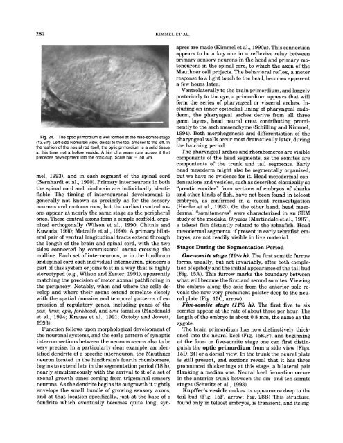
Fig. 24. The optic primordium is well formed at <strong>the</strong> nine-somite stage<br />
(13.5 h). Left-side Nomarski view, dorsal to <strong>the</strong> top, anterior to <strong>the</strong> left. In<br />
<strong>the</strong> fashion <strong>of</strong> <strong>the</strong> neural rod itself, <strong>the</strong> optic primordium is a solid tissue<br />
at this time, not a hollow vesicle. A hint <strong>of</strong> a seam runs across it that<br />
precedes development into <strong>the</strong> optic cup. Scale bar = 50 pm.<br />
mel, 1993), and in each segment <strong>of</strong> <strong>the</strong> spinal cord<br />
(Bernhardt et al., 1990). Primary interneurons in both<br />
<strong>the</strong> spinal cord and hindbrain are individually identi-<br />
fiable. The timing <strong>of</strong> interneuronal development is<br />
generally not known as precisely as for <strong>the</strong> sensory<br />
neurons and motoneurons, but <strong>the</strong> earliest central ax-<br />
ons appear at nearly <strong>the</strong> same stage as <strong>the</strong> peripheral<br />
ones. These central axons form a simple scaffold, orga-<br />
nized orthogonally (Wilson et al., 1990; Chitnis and<br />
Kuwada, 1990; Metcalfe et al., 1990): A primary bilat-<br />
eral pair <strong>of</strong> ventral longitudinal tracts extend through<br />
<strong>the</strong> length <strong>of</strong> <strong>the</strong> brain and spinal cord, with <strong>the</strong> two<br />
sides connected by commissural axons crossing <strong>the</strong><br />
midline. Each set <strong>of</strong> interneurons, or in <strong>the</strong> hindbrain<br />
and spinal cord each individual interneuron, pioneers a<br />
part <strong>of</strong> this system or joins to it in a way that is highly<br />
stereotyped (e.g., Wilson and Easter, 1991), apparently<br />
matching <strong>the</strong> precision <strong>of</strong> motor axonal pathfinding in<br />
<strong>the</strong> periphery. Notably, when and where <strong>the</strong> cells de-<br />
velop and where <strong>the</strong>ir axons extend correlate closely<br />
with <strong>the</strong> spatial domains and temporal patterns <strong>of</strong> ex-<br />
pression <strong>of</strong> regulatory genes, including genes <strong>of</strong> <strong>the</strong><br />
pax, krox, eph, forkhead, and wnt families (Macdonald<br />
et al., 1994; Krauss et al., 1991; Oxtoby and Jowett,<br />
1993).<br />
Function follows upon morphological development <strong>of</strong><br />
<strong>the</strong> neuronal systems, and <strong>the</strong> early pattern <strong>of</strong> synaptic<br />
interconnections between <strong>the</strong> neurons seems also to be<br />
very precise. In a particularly clear example, an iden-<br />
tified dendrite <strong>of</strong> a specific interneuron, <strong>the</strong> Mauthner<br />
neuron located in <strong>the</strong> hindbrain’s fourth rhombomere,<br />
begins to extend late in <strong>the</strong> segmentation period (18 h),<br />
nearly simultaneously with <strong>the</strong> arrival to it <strong>of</strong> a set <strong>of</strong><br />
axonal growth cones coming from trigeminal sensory<br />
neurons. As <strong>the</strong> dendrite begins its outgrowth it tightly<br />
envelops <strong>the</strong> small bundle <strong>of</strong> growing sensory axons,<br />
and at that location specifically, just at <strong>the</strong> base <strong>of</strong> a<br />
dendrite which eventually becomes quite long, syn-<br />
apses are made (Kimmel et al., 1990a). This connection<br />
appears to be a key one in a reflexive relay between<br />
primary sensory neurons in <strong>the</strong> head and primary mo-<br />
toneurons in <strong>the</strong> spinal cord, to which <strong>the</strong> axon <strong>of</strong> <strong>the</strong><br />
Mauthner cell projects. The behavioral reflex, a motor<br />
response to a light touch to <strong>the</strong> head, becomes apparent<br />
a few hours later.<br />
Ventrolaterally to <strong>the</strong> brain primordium, and largely<br />
posteriorly to <strong>the</strong> eye, a primordium appears that will<br />
form <strong>the</strong> series <strong>of</strong> pharyngeal or visceral arches. In-<br />
cluding an inner epi<strong>the</strong>lial lining <strong>of</strong> pharyngeal endo-<br />
derm, <strong>the</strong> pharyngeal arches derive from all three<br />
germ layers, head neural crest contributing promi-<br />
nently to <strong>the</strong> arch mesenchyme (Schilling and Kimmel,<br />
1994). Both morphogenesis and differentiation <strong>of</strong> <strong>the</strong><br />
pharyngeal walls occur most dramatically later, during<br />
<strong>the</strong> hatching period.<br />
The pharyngeal arches and rhombomeres are visible<br />
components <strong>of</strong> <strong>the</strong> head segments, as <strong>the</strong> somites are<br />
compontents <strong>of</strong> <strong>the</strong> trunk and tail segments. Early<br />
head mesoderm might also be segmentally organized,<br />
but we have no evidence for it. Head mesodermal con-<br />
densations and vesicles, such as described classically as<br />
“preotic somites” from sections <strong>of</strong> embryos <strong>of</strong> sharks<br />
and o<strong>the</strong>r kinds <strong>of</strong> fish, have not been found in teleost<br />
embryos, as confirmed in a recent reinvestigation<br />
(Horder et al., 1993). On <strong>the</strong> o<strong>the</strong>r hand, head meso-<br />
dermal so mi to meres^' were characterized in an SEM<br />
study <strong>of</strong> <strong>the</strong> medaka, Oryzias (Martindale et al., 1987),<br />
a teleost fish distantly related to <strong>the</strong> zebrafish. Head<br />
mesodermal segments, if present in early zebrafish em-<br />
bryos, are not readily visible in live material.<br />
<strong>Stages</strong> During <strong>the</strong> Segmentation Period<br />
One-somite stage (10% h). The first somitic furrow<br />
forms, usually, but not invariably, after both comple-<br />
tion <strong>of</strong> epiboly and <strong>the</strong> initial appearance <strong>of</strong> <strong>the</strong> tail bud<br />
(Fig. 15A). This furrow marks <strong>the</strong> boundary between<br />
what will become <strong>the</strong> first and second somites. Viewing<br />
<strong>the</strong> embryo along <strong>the</strong> axis from <strong>the</strong> anterior pole re-<br />
veals <strong>the</strong> now very prominent polster deep to <strong>the</strong> neu-<br />
ral plate (Fig. 15C, arrow).<br />
Five-somite stage (112h h). The first five to six<br />
somites appear at <strong>the</strong> rate <strong>of</strong> about three per hour. The<br />
length <strong>of</strong> <strong>the</strong> embryo is about 0.8 mm, <strong>the</strong> same as <strong>the</strong><br />
zygote.<br />
The brain primordium has now distinctively thick-<br />
ened into <strong>the</strong> neural keel (Fig. 15E,F), and beginning<br />
at <strong>the</strong> four- or five-somite stage one can first distin-<br />
guish <strong>the</strong> optic primordium from a side view (Figs.<br />
15D, 24) or a dorsal view. In <strong>the</strong> trunk <strong>the</strong> neural plate<br />
is still present, and sections reveal that it has three<br />
pronounced thickenings at this stage, a bilateral pair<br />
flanking a median one. Neural keel formation occurs<br />
in <strong>the</strong> anterior trunk between <strong>the</strong> six- and ten-somite<br />
stages (Schmitz et al., 1993).<br />
Kupffer’s vesicle makes its appearance deep to <strong>the</strong><br />
tail bud (Fig. 15F, arrow; Fig. 28B) This structure,<br />
found only in teleost embryos, is transient, and its sig-