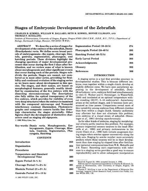
Stages of Embryonic Development of the Zebrafish
Stages of Embryonic Development of the Zebrafish
Stages of Embryonic Development of the Zebrafish

You also want an ePaper? Increase the reach of your titles
YUMPU automatically turns print PDFs into web optimized ePapers that Google loves.
DEVELOPMENTAL DYNAMICS 2032553’10 (1995)<br />
<strong>Stages</strong> <strong>of</strong> <strong>Embryonic</strong> <strong>Development</strong> <strong>of</strong> <strong>the</strong> <strong>Zebrafish</strong><br />
CHARLES B. KIMMEL, WILLIAM W. BALLARD, SETH R. KIMMEL, BONNIE ULLMANN, AND<br />
THOMAS F. SCHILLING<br />
Institute <strong>of</strong> Neuroscience, University <strong>of</strong> Oregon, Eugene, Oregon 97403-1254 (C.B.K., S.R.K., B.U., T.F.S.); Department <strong>of</strong><br />
Biology, Dartmouth College, Hanover, NH 03755 (W.W.B.)<br />
ABSTRACT We describe a series <strong>of</strong> stages for<br />
development <strong>of</strong> <strong>the</strong> embryo <strong>of</strong> <strong>the</strong> zebrafish, Danio<br />
(Brachydanio) rerio. We define seven broad peri-<br />
ods <strong>of</strong> embryogenesis-<strong>the</strong> zygote, cleavage, blas-<br />
tula, gastrula, segmentation, pharyngula, and<br />
hatching periods. These divisions highlight <strong>the</strong><br />
changing spectrum <strong>of</strong> major developmental pro-<br />
cesses that occur during <strong>the</strong> first 3 days after fer-<br />
tilization, and we review some <strong>of</strong> what is known<br />
about morphogenesis and o<strong>the</strong>r significant events<br />
that occur during each <strong>of</strong> <strong>the</strong> periods. <strong>Stages</strong> sub-<br />
divide <strong>the</strong> periods. <strong>Stages</strong> are named, not num-<br />
bered as in most o<strong>the</strong>r series, providing for flexi-<br />
bility and continued evolution <strong>of</strong> <strong>the</strong> staging series<br />
as we learn more about development in this spe-<br />
cies. The stages, and <strong>the</strong>ir names, are based on<br />
morphological features, generally readily identi-<br />
fied by examination <strong>of</strong> <strong>the</strong> live embryo with <strong>the</strong><br />
dissecting stereomicroscope. The descriptions<br />
also fully utilize <strong>the</strong> optical transparancy <strong>of</strong> <strong>the</strong><br />
live embryo, which provides for visibility <strong>of</strong> even<br />
very deep structures when <strong>the</strong> embryo is examined<br />
with <strong>the</strong> compound microscope and Nomarski<br />
interference contrast illumination. Photomicro-<br />
graphs and composite camera lucida line draw-<br />
ings characterize <strong>the</strong> stages pictorially. O<strong>the</strong>r<br />
char-<br />
figures chart <strong>the</strong> development <strong>of</strong> distinctive<br />
acters used as staging aid signposts.<br />
0 1995 Wiley-Liss, Inc.<br />
Key words: Danio rerio, Morphogenesis, Embryogenesis,<br />
Zygote, Cleavage, Blastula,<br />
Gastrula, Segmentation, Pharyngula,<br />
Hatching<br />
CONTENTS<br />
Introduction<br />
Organization<br />
Procedures<br />
Temperature and Standard<br />
<strong>Development</strong>al Time<br />
Zygote Period (0-% h)<br />
Cleavage Period (3/4-21/4 h)<br />
Blastula Period (21/451/4 h)<br />
Gastrula Period (5%-10 h)<br />
0 1995 WILEY-LISS, INC.<br />
253<br />
254<br />
254<br />
260<br />
260<br />
261<br />
263<br />
268<br />
Segmentation Period (10-24 h) 274<br />
Pharyngula Period (24-48 h) 285<br />
Hatching Period (48-72 h) 298<br />
Early Larval Period 303<br />
Acknowledgments 303<br />
Glossary 303<br />
References 309<br />
INTRODUCTION<br />
A staging series is a tool that provides accuracy in<br />
developmental studies. This is because different embryos,<br />
even toge<strong>the</strong>r within a single clutch, develop at<br />
slightly different rates. We have seen asynchrony appearing<br />
in <strong>the</strong> development <strong>of</strong> zebrafish, Danio<br />
(Brachydanio) rerio, embryos fertilized simultaneously<br />
in vitro (C. Walker and G. Streisinger, in Westerfield,<br />
1994) and incubated at an optimal temperature without<br />
crowding (28.S0C, 5-10 embryos/ml). Asynchrony<br />
arises at <strong>the</strong> earliest stages, and it becomes more pronounced<br />
as time passes. Comparisons reveal more <strong>of</strong><br />
this variability among embryos from different clutches<br />
than from within a single clutch. Genetic uniformity<br />
may alleviate but does not eliminate this problem;<br />
even embryos <strong>of</strong> a clonal strain <strong>of</strong> zebrafish (Streisinger<br />
et al., 1981) develop asynchronously.<br />
As for o<strong>the</strong>r kinds <strong>of</strong> embryos, staging by morphological<br />
criteria partly resolves this problem. For example,<br />
primary trigeminal sensory neurons in <strong>the</strong> head (Metcalfe<br />
et al., 1990) and primary motoneurons in <strong>the</strong><br />
trunk (Eisen et al., 1986) both initiate axogenesis during<br />
stages when somites appear successively along <strong>the</strong><br />
body axis. Staging by somite number more accurately<br />
predicts where <strong>the</strong>se neurons will be in <strong>the</strong>ir development<br />
than does staging by elapsed time after fertilization<br />
(personal communications from W.K. Metcalfe and<br />
J.S. Eisen). Recording one’s experiments with reference<br />
to a staging series provides a good way to ensure<br />
reproducibility and to allow subsequent incorporation<br />
Received August 22, 1994; accepted January 4, 1995.<br />
Address reprint requestsicorrespondence to Charles B. Kimmel, In-<br />
stitute <strong>of</strong> Neuroscience, 1254 University <strong>of</strong> Oregon, Eugene, OR<br />
97403-1254.<br />
Thomas F. Schilling is now at <strong>the</strong> Department <strong>of</strong> Molecular Em-<br />
bryology, Imperial Cancer Research Fund, Lincoln’s Inn Fields, Lon-<br />
don WCZA 3PX, UK.
254 KIMMEL ET AL.<br />
<strong>of</strong> new observations and details. A series based on<br />
morphology can also facilitate communication; <strong>the</strong> de-<br />
scriptor “18-somite embryo” has more meaning than<br />
“18-hour-old embryo,” particularly in cross-species<br />
comparisons.<br />
An earlier staging series for zebrafish, although less<br />
complete than <strong>the</strong> present one, fairly accurately por-<br />
trays <strong>the</strong> first third (or 1st day) <strong>of</strong> embryonic develop-<br />
ment, and includes useful sets <strong>of</strong> photographs (Hisaoka<br />
and Battle, 1958; Hisaoka and Firlit, 1960). Warga and<br />
Kimmel (1990) briefly described stages <strong>of</strong> <strong>the</strong> blastula<br />
and gastrula. Preliminary versions <strong>of</strong> this series were<br />
circulated and published in The <strong>Zebrafish</strong> Book (Wester-<br />
field, 1994). The present version substantially revises,<br />
corrects, and expands <strong>the</strong> earlier ones. An up-to-date<br />
version is available in electronic form on <strong>the</strong> Internet<br />
World Wide Web server: “http:/zfish.uoregon.edu.<br />
ORGANIZATION<br />
We have named <strong>the</strong> stages ra<strong>the</strong>r than numbering<br />
<strong>the</strong>m as in most o<strong>the</strong>r series, because named stages are<br />
more flexible and easier to remember or recognize than<br />
numbered ones. A stage defines more than an instant<br />
in time, it is merely a device for approximately locating<br />
a part <strong>of</strong> <strong>the</strong> continuum <strong>of</strong> development. With named<br />
stages one can easily add detail as it is learned, and<br />
intercalate new stages into <strong>the</strong> series as necessary for<br />
a particular study without resorting to cumbersome<br />
devices like decimal numbers, minuses, and pluses. For<br />
example, here we describe a five-somite stage, and next<br />
a 14-somite stage, but a particular study might use<br />
eight stages in between (six-somite, seven-somite, and<br />
so on), and one will immediately understand <strong>the</strong>ir<br />
meaning. We emphasize that we do not mean to ex-<br />
clude <strong>the</strong>se extra stages, wherever <strong>the</strong>y are in <strong>the</strong> se-<br />
ries, by ignoring <strong>the</strong>m.<br />
As an aid to communicating a broader perspective<br />
toward development, we group <strong>the</strong> stages into larger<br />
time-blocks called periods (Table 11, and summarize in<br />
<strong>the</strong> text <strong>the</strong> principal events during <strong>the</strong>se periods. Spe-<br />
cialized terms are defined in <strong>the</strong> glossary, and boldface<br />
type in <strong>the</strong> text highlights terms important for staging.<br />
Table 2 collects brief descriptions <strong>of</strong> all <strong>the</strong> stages, and<br />
Figure 1 shows corresponding sketches. One can use<br />
<strong>the</strong>se two sources to approximately locate a stage <strong>of</strong><br />
interest, and <strong>the</strong>n find additional detail as necessary in<br />
<strong>the</strong> text and o<strong>the</strong>r figures.<br />
Staging<br />
PROCEDURES<br />
One can determine <strong>the</strong> approximate developmental<br />
stage <strong>of</strong> a living embryo by examining it with a dis-<br />
secting stereo-microscope, generally with transmitted<br />
(not epi- or incident) illumination and high magnifica-<br />
tion (about 50 x ). During <strong>the</strong> segmentation period <strong>the</strong><br />
tail elongates, and if <strong>the</strong> embryo is left within its<br />
chorion, <strong>the</strong> tail eventually curves over <strong>the</strong> trunk and<br />
head so as to obscure <strong>the</strong> view. When this has hap-<br />
pened, one must remove <strong>the</strong> embryo from its chorion to<br />
TABLE 1. Periods <strong>of</strong> Early <strong>Development</strong><br />
Period h DescriDtion<br />
Zygote 0 The newly fertilized egg through <strong>the</strong><br />
completion <strong>of</strong> <strong>the</strong> first zygotic cell<br />
cycle<br />
Cleavage<br />
Blastula<br />
3/4 Cell cycles 2 through 7 occur rapidly<br />
and synchronously<br />
2 1/4 Rapid, metasynchronous cell cycles<br />
(8, 9) give way to leng<strong>the</strong>ned,<br />
asynchronous ones at <strong>the</strong><br />
midblastula transition; epiboly<br />
<strong>the</strong>n begins<br />
Gastrula 5 v4 Morphogenetic movements <strong>of</strong><br />
involution, convergence, and<br />
extension form <strong>the</strong> epiblast,<br />
hypoblast, and embryonic axis;<br />
through <strong>the</strong> end <strong>of</strong> epiboly<br />
Segmentation 10 Somites, pharyngeal arch primordia,<br />
and neuromeres develop; primary<br />
organogenesis; earliest movements;<br />
<strong>the</strong> tail appears<br />
Pharyngula 24 Phylotypic-stage embryo; body axis<br />
straightens from its early curvature<br />
about <strong>the</strong> yolk sac; circulation,<br />
pigmentation, and tins begin<br />
development<br />
Hatching 48 Completion <strong>of</strong> rapid morphogenesis <strong>of</strong><br />
primary organ systems; cartilage<br />
development in head and pectoral<br />
tin; hatching occurs asynchronously<br />
Early larva 72 Swim bladder inflates; food-seeking<br />
and active avoidance behaviors<br />
stage it, ei<strong>the</strong>r manually with #5 watchmaker’s for-<br />
ceps, or by treatment with a proteolytic enzyme (Wes-<br />
terfield, 1994). Late in <strong>the</strong> pharyngula period <strong>the</strong> em-<br />
bryo, if removed from <strong>the</strong> chorion, swims away in<br />
response to touch; this can be prevented by anes<strong>the</strong>sia<br />
in 0.003% tricaine (3-amino benzoic acid ethyl ester;<br />
Sigma Chemical Co.) at pH 7 (Westerfield, 1994). Re-<br />
peated anes<strong>the</strong>sia and rinsing appears to slightly but<br />
significantly retard subsequent development (see leg-<br />
end to Fig. 2).<br />
Nomarski Optics<br />
A compound microscope equipped with Nomarski dif-<br />
ferential interference contrast optics and objectives in-<br />
<strong>the</strong> range <strong>of</strong> 25 x to 40 x reveals details o<strong>the</strong>rwise not<br />
visible in <strong>the</strong> live preparation and permits <strong>the</strong> most<br />
critical staging. For instance, Nomarski optics permits<br />
<strong>the</strong> most accurate counts <strong>of</strong> cells in <strong>the</strong> blastula and <strong>of</strong><br />
somites during segmentation. Moreover, “prim” stages<br />
refer to <strong>the</strong> position <strong>of</strong> a structure, <strong>the</strong> primordium <strong>of</strong><br />
<strong>the</strong> posterior lateral line, that one must use Nomarski<br />
optics to see for <strong>the</strong> sharpest determination <strong>of</strong> stages<br />
during much <strong>of</strong> <strong>the</strong> pharyngula period. The embryo is<br />
anes<strong>the</strong>tized, removed from <strong>the</strong> chorion, and mounted<br />
between bridged coverslips (Westerfield, 1994), for ob-<br />
servation and later recovery.<br />
It is always better to stage embryos while <strong>the</strong>y are<br />
alive ra<strong>the</strong>r than after killing and fixation. For exam-<br />
ple, staging during <strong>the</strong> straightening and hatching pe-<br />
riods calls for a comparison <strong>of</strong> sizes <strong>of</strong> <strong>the</strong> head and <strong>the</strong>
TABLE 2. <strong>Stages</strong> <strong>of</strong> <strong>Embryonic</strong> <strong>Development</strong>a<br />
Stage h HB Description<br />
Zygote period<br />
1-cell<br />
Cleavage period<br />
2-cell<br />
4-cell<br />
8-cell<br />
16-cell<br />
32-cell<br />
64-cell<br />
Blastula period<br />
128-cell<br />
256-cell<br />
512-cell<br />
lk-cell<br />
High<br />
Oblong<br />
Sphere<br />
Dome<br />
30%-epiboly<br />
Gastrula period<br />
50%-epiboly<br />
Germ-ring<br />
Shield<br />
75%-epiboly<br />
90%-epiboly<br />
Bud<br />
Segmentation period<br />
1-somite<br />
5-somite<br />
14-somite<br />
20-somite<br />
26-somite<br />
Pharyngula period<br />
Prim-5<br />
Prim- 15<br />
Prim-25<br />
High-pec<br />
Hatching period<br />
Long-pec<br />
Pec-fin<br />
Protruding-mouth<br />
0<br />
3/4<br />
1<br />
1 '/4<br />
1%<br />
13/4<br />
2<br />
2 '/4<br />
2%<br />
2%<br />
3<br />
31/3<br />
3 2/3<br />
4<br />
41/3<br />
42/3<br />
5 '/4<br />
52/3<br />
6<br />
8<br />
9<br />
10<br />
10%<br />
112/3<br />
16<br />
19<br />
22<br />
24<br />
30<br />
36<br />
42<br />
48<br />
60<br />
72<br />
12<br />
3<br />
4<br />
5<br />
6<br />
7<br />
8<br />
9<br />
10<br />
11<br />
12<br />
13<br />
14<br />
15<br />
16<br />
17<br />
18<br />
19<br />
20<br />
Cytoplasm streams toward animal pole to form <strong>the</strong> blastodisc<br />
Partial cleavage<br />
2 x 2 array <strong>of</strong> blastomeres<br />
2 x 4 array <strong>of</strong> blastomeres<br />
4 x 4 array <strong>of</strong> blastomeres<br />
2 regular tiers (horizontal rows) <strong>of</strong> blastomeres, sometimes in 4 x 8 array<br />
3 regular tiers <strong>of</strong> blastomeres<br />
5 blastomere tiers; cleavage planes irregular<br />
7 blastomere tiers<br />
9 tiers <strong>of</strong> blastomeres; NO: YSL forms<br />
11 tiers <strong>of</strong> blastomeres; NO: single row <strong>of</strong> YSL nuclei; slight blastodisc cell cycle asynchrony<br />
> 11 tiers <strong>of</strong> blastomeres; beginning <strong>of</strong> blastodisc flattening; NO: YSL nuclei in two rows; substantial<br />
division asynchrony<br />
Flattening produces an elliptical shape; NO: multiple rows <strong>of</strong> YSL nuclei<br />
Spherical shape; flat border between blastodisc and yolk<br />
Shape remains spherical; yolk cell bulging (doming) toward animal pole as epiboly begins<br />
Blastoderm an inverted cup <strong>of</strong> uniform thickness; margin reaches 30% <strong>of</strong> distance between <strong>the</strong> animal<br />
and vegetal poles<br />
Blastoderm remains uniform in thickness<br />
Germ ring visible from animal pole; 50%-epiboly<br />
<strong>Embryonic</strong> shield visible from animal pole, 50%-epiboly<br />
Dorsal side distinctly thicker; epiblast, hypoblast, evacuation zone visible<br />
Brain rudiment thickened; notochord rudiment distinct from segmental plate<br />
Tail bud prominent; notochord rudiment distinct from neural keel; early polster; midsagittal groove in<br />
anterior neural keel; 100%-epiboly<br />
First somite furrow<br />
Polster prominent; optic vesicle, Kupffer's vesicle<br />
EL = 0.9 mm; otic placode; brain neuromeres, v-shaped trunk somites; YE barely forming; NO:<br />
pronephric duct<br />
EL = 1.4 mm; YE/YB > 0.5 and < 1; muscular twitches; lens, otic vesicle, rhombic flexure;<br />
hindbrain neuromeres prominent; tail well extended<br />
EL = 1.6 mm; HTA = 125"; side-to-side flexures; otoliths; Prim-3<br />
EL = 1.9 mm; HTA = 120"; OVL = 5; YE/YB = 1; early pigmentation in retina and skin; median<br />
fin fold; red blood cells on yolk, heartbeat<br />
EL = 2.5 mm; HTA = 95"; OVL = 3; YEiYB > 1; YB/HD = 2; early touch reflex and reduced<br />
spontaneous movements; retina pigmented; dorsal stripe to somite 12; weak circulation; caudal<br />
artery halfway to end <strong>of</strong> tail; caudal vein braided; shallow pectoral fin buds; straight tail; NO:<br />
cellular degeneration at end <strong>of</strong> tail; circulation in aortic arch 1<br />
EL = 2.7 mm; HTA = 75"; OVL = 1; PF(HIW) = 3h; early motility; tail pigmentation and ventral<br />
stripe filling out; strong circulation; single aortic arch pair; caudal artery is 3/4 <strong>of</strong> <strong>the</strong> way to <strong>the</strong><br />
end <strong>of</strong> tail; pericardium not swollen; NO: PF apical ectodermal ridge<br />
EL = 2.9 mm; HTA = 55"; OVL < 1 and > %; YEiYB = 1.5; YB/HD < 1.3; PF(H/W) = 1;<br />
dechorionated embryos rest on side after swimming; YE remaining cylindrical; PF apical ridge<br />
prominent; early lateral stripe; complete dorsal stripe; xanthophores in head only; iridophores in<br />
retina only; pericardium prominent; NO: heart chambers; segmental blood vessels; mandibular and<br />
hyoid arches; foregut developments; olfactory cilia; thickened otic vesicle walls<br />
EL = 3.1 mm; HTA = 45"; OVL = PF(H/W) = 2; resting dorsal up; YE beginning to taper; PF<br />
pointed; dorsal and ventral stripes meet at tail; ca. 6 melanophores in lateral stripe; iridophores<br />
plentiful on retina; distinct yellow cast to head; NO: circulation in 2-4 aortic arches and in<br />
segmental vessels; olfactory cilia beating; semicircular canals; neuromasts<br />
EL = 3.3 mm; HTA = 35"; movements too rapid to resolve; YB tapering into YE; up to 10<br />
melanophores in lateral stripe; PF flattened into fin shape with prominent circulation; iridophore<br />
retinal ring fills out; iridophores in dorsal stripe; NO: PF cartilage and actinotrichia; gut tract; 2<br />
chambers in otic vesicle; early jaw cartilages; circulation in 5-6 aortic arches; mouth remaining<br />
small and open at ventral location midway between eyes<br />
EL = 3.5 mm; HTA = 25"; wide open mouth protruding anterior to eye; iridophores in yolk stripe;<br />
eye half covered by iridophores; dorsal body as yellow as head; NO: ail1 slits and filament buds: '<br />
cartilage in branchial arch 1 and 5; opercuhm covers <strong>the</strong> branchial arch 1 or 2; clerithrum<br />
"EL, embryo length; PF, pectoral fin; h, hours <strong>of</strong> development at 28.5"C; HB, approximate stage no. in <strong>the</strong> Hisaoka and Battle<br />
(1958) zebrafish staging series (reasonably accurate through HB stage 20); HD, head diameter in dorsal view; NO, Nomarski<br />
optics; H/W, heightiwidth; Prim, Prim stages refer to <strong>the</strong> no. <strong>of</strong> <strong>the</strong> myotome to which <strong>the</strong> leading end <strong>of</strong> <strong>the</strong> posterior lateral<br />
line prirnordium has advanced; YB, yolk ball; YE, yolk extension; YSL, yolk syncytial layer; HTA, head-trunk angle; OVL, otic<br />
vesicle length.
1 -cell<br />
0.2 h<br />
16-cell<br />
1.5 h<br />
256-cell<br />
2.5 h<br />
2-cell<br />
0.75 h<br />
32-cell<br />
1.75 h<br />
512-cell<br />
2.75 h<br />
4-cell<br />
l h<br />
64-cell<br />
2h<br />
1 k-cell<br />
3h<br />
Fig. 1 (Legend to Fig. 1 appears on page 259)<br />
8-cell<br />
1.25 h<br />
128-cell<br />
2.25 h<br />
high<br />
3.3 h
oblong<br />
3.7 h<br />
50%-epiboly<br />
5.3 h<br />
90%-epiboly<br />
9h<br />
sphere<br />
4h<br />
germ ring<br />
5.7 h<br />
bud<br />
10 h<br />
Fig. 1 (continued).<br />
dome<br />
4.3 h<br />
shield<br />
6h<br />
3-somite<br />
11 h<br />
I<br />
30%-epi boly<br />
4.7 h<br />
75%-epiboly<br />
8h<br />
6-somite<br />
12 h
258 KIMMEL ET AL.<br />
10-somite<br />
14 h<br />
26-somite<br />
22 h<br />
14-somite<br />
16 h<br />
prim-6<br />
25 h<br />
Fig. 1 (continued)<br />
18-somite<br />
ia h<br />
prim-1 6<br />
31 h<br />
21-somite<br />
19.5 h<br />
prim-22<br />
35 h
high pec<br />
42 h<br />
STAGES OF EMBRYONIC DEVELOPMENT OF THE ZEBRAFISH 259<br />
long pec<br />
48 h<br />
Fig. 1. Camera lucida sketches <strong>of</strong> <strong>the</strong> embryo at selected stages. The<br />
animal pole is to <strong>the</strong> top for <strong>the</strong> early stages, and anterior is to <strong>the</strong> top<br />
later, except for <strong>the</strong> two animal polar (AP) views shown below <strong>the</strong>ir side<br />
view counterparts for germ-ring and shield gastrulas. Face views are<br />
shown during cleavage and blastula stages. After shield stage, <strong>the</strong> views<br />
are <strong>of</strong> <strong>the</strong> embryo's left side, but before <strong>the</strong> shield arises one cannot<br />
reliably ascertain which side is which. Pigmentation is omitted. Arrow-<br />
heads indicate <strong>the</strong> early appearance <strong>of</strong> some key diagnostic features at<br />
yolk mass, and differential shrinkage during fixation<br />
distorts <strong>the</strong> normal relationship. Never<strong>the</strong>less, if pres-<br />
ervation is good enough, one can fairly reliably stage<br />
fixed and whole-mounted embryos (e.g., immunola-<br />
beled ones) using o<strong>the</strong>r criteria. One cannot easily<br />
stage an embryo after it is sectioned.<br />
pec fin<br />
60 h<br />
protruding<br />
mouth<br />
72 h<br />
<strong>the</strong> following stages: 1 k-cell: YSL nuclei. Dome: <strong>the</strong> doming yolk syncy-<br />
tium. Germ ring: germ ring. Shield: embryonic shield. 75Oh-epiboly: Bra-<br />
chet's cleft. 90%-epiboly: blastoderm margin closing over <strong>the</strong> yolk plug.<br />
Bud: polster. 3-somite: third somite. 6-somite: eye primordium (upper<br />
arrow), Kupffer's vesicle (lower). 10-somite: otic placode. 21-somite: lens<br />
primordium. Prim-6: primordium <strong>of</strong> <strong>the</strong> posterior lateral line (on <strong>the</strong> dorsal<br />
side), hatching gland (on <strong>the</strong> yolk ball). Prim-16: heart. High-pec: pectoral<br />
fin bud. Scale bar = 250 pn.<br />
Photographs<br />
The accompanying photographs are <strong>of</strong> living em-<br />
bryos, anes<strong>the</strong>tized for <strong>the</strong> later stages. The original<br />
photographs were made as color slides (Kodak Ektach-<br />
rome 160T DX), and <strong>the</strong> black and whites plates are<br />
reproduced from internegatives. Sets <strong>of</strong> copies <strong>of</strong> <strong>the</strong>
260 KIMMEL ET AL.<br />
original color slides are available at cost from <strong>the</strong> authors.<br />
The lower-magnification views, showing <strong>the</strong> entire<br />
embryo, were made using a Zeiss STEM1 stereo<br />
dissecting microscope: The embryo is mounted on a depression<br />
slide, without an overlying coverslip, in standard<br />
embryo medium (Westerfield, 1994) containing<br />
1.5-3% methyl cellulose to allow positioning as desired.<br />
The higher-magnification views, showing parts<br />
<strong>of</strong> embryos, were taken with Nomarski optics (generally<br />
a Zeiss 4 0 water-immersion ~<br />
objective) using a<br />
Zeiss UEM compound microscope. The embryo was<br />
mounted between coverslips, sometimes in 1% agar, for<br />
positioning.<br />
TEMPERATURE AND STANDARD<br />
DEVELOPMENTAL TIME<br />
We convert staging information to "standard devel-<br />
opmental time," as designated by <strong>the</strong> letter "h," and<br />
defined as normalized hours after fertilization at<br />
28.5"C, an optimal temperature <strong>of</strong> incubation (G. Strei-<br />
singer, unpublished experiments). Incubation at an-<br />
o<strong>the</strong>r temperature will change developmental rate, as<br />
may be useful in particular studies, e.g., to bring em-<br />
bryos to two different stages at <strong>the</strong> same time for het-<br />
erochronic transplantation. Comparisons <strong>of</strong> embryos<br />
raised at different temperatures should be made with<br />
caution, because <strong>the</strong>re is no assurance that all features<br />
<strong>of</strong> <strong>the</strong> embryo coordinately change <strong>the</strong>ir rates <strong>of</strong> devel-<br />
opment when <strong>the</strong> temperature is changed. Never<strong>the</strong>-<br />
less, embryos appear to develop normally if <strong>the</strong>y are<br />
m<br />
fi,<br />
L<br />
m<br />
b<br />
75%<br />
S<br />
d<br />
1 kc<br />
64c<br />
8C<br />
Pm<br />
Pf<br />
ID<br />
hp<br />
~ 2 5<br />
~ 1 5<br />
P5<br />
20s<br />
14s<br />
1s<br />
I A<br />
0 2 4 6 8 1 0 1 2<br />
t B<br />
12<br />
1 i 90<br />
0 10 20 30 40 50 60 70 80 90<br />
Hours <strong>of</strong> development<br />
kept within an eight degree range, between 25°C (per-<br />
haps a few degrees lower; see Schirone and Gross,<br />
1968) and 33°C. Incubating <strong>the</strong>m for long periods above<br />
or below <strong>the</strong>se extremes may produce abnormalities.<br />
Figure 2 shows <strong>the</strong> rates <strong>of</strong> development at <strong>the</strong> ex-<br />
tremes, and <strong>the</strong> legend to this figure provides a simple<br />
formula permitting one to estimate when an embryo<br />
developing at any temperature within this range will<br />
reach a given stage.<br />
ZYGOTE PERIOD (0-3/4 h)<br />
The newly fertilized egg is in <strong>the</strong> zygote period until<br />
<strong>the</strong> first cleavage occurs (Fig. 3), about 40 minutes af-<br />
ter fertilization. The zygote is about 0.7 mm in diame-<br />
ter at <strong>the</strong> time <strong>of</strong> fertilization. We include only a single<br />
stage; however many changes are occurring, and one<br />
Fig. 2. Rates <strong>of</strong> development for embryos incubated at 25°C (solid<br />
circles) and 33°C (open circles). A: <strong>Development</strong> showing selected<br />
stages through <strong>the</strong> bud stage (occurring at 10 h at <strong>the</strong> standard temper-<br />
ature <strong>of</strong> 28.5%). B: <strong>Development</strong> through <strong>the</strong> end <strong>of</strong> embryogenesis, <strong>the</strong><br />
protruding-mouth stage (occurring at 72 h at <strong>the</strong> standard temperature).<br />
The stages (8c through b for A, and s through pm for B) are positioned<br />
along <strong>the</strong> ordinate according to when <strong>the</strong>y occur at 28.5"C, <strong>the</strong> standard<br />
incubation temperature. Because <strong>of</strong> this presentation, a plot <strong>of</strong> develop-<br />
ment at 28.5"C would be expected to yield a straight line passing through<br />
<strong>the</strong> origin, and with a slope <strong>of</strong> 1 .O. We determined this to be <strong>the</strong> case, in<br />
fact, by watching sets <strong>of</strong> control embryos developing at 28.5" during<br />
collection <strong>of</strong> <strong>the</strong> data for this experiment (not shown). Notice in <strong>the</strong> figures<br />
that developmental rates at both 25°C and 33°C are also approximately<br />
linear, and that <strong>the</strong> slopes <strong>of</strong> <strong>the</strong> lines at each temperature are <strong>the</strong> same<br />
in A and B. Hence developmental rate varies as a linear function <strong>of</strong><br />
incubation temperature, and a simple calculation allows one to determine<br />
approximately when embryos developing at any temperature between<br />
<strong>the</strong> extremes will reach a desired stage <strong>of</strong> interest: H, = h/(0.055T -<br />
0.57), where H, = hours <strong>of</strong> development at temperature T, and h =<br />
hours <strong>of</strong> development to reach <strong>the</strong> stage at 28.5", as set out in Table 1.<br />
For example, computation <strong>of</strong> development to <strong>the</strong> 20-somite stage (oc-<br />
curring at 19 h at <strong>the</strong> standard temperature) yields 23.6 hours at 25"C,<br />
and 15 hours at 33°C. These times do not differ significantly from those<br />
observed (23.5 hours and 14.5 hours). Stage abbreviations (and hours<br />
<strong>of</strong> development to reach <strong>the</strong> stage at 28.5" in paren<strong>the</strong>ses): 8c, 8-cell<br />
(1.25 h); 64c, 64 cell (2 h); 1 kc, 1 k-cell (3 h); d, dome (4.3 h); s, shield (6<br />
h); 75%, 75%-epiboly (8 h); b, bud (10 h); Is, I-somite (10.3 h); 14s,<br />
14-somite (16 h); 205 20-somite (19 h); p5, prim-5 (24 h); p15, prim-I5<br />
(30 h); p25, prim-25 (36 h); hp, high-pec (42 h); Ip, long-pec (48 h); pf,<br />
pec-fin (60 h); pm, protruding-mouth (72 h). Method: Observations were<br />
made on embryos developing from several natural spawnings, separated<br />
in each case into groups at each <strong>of</strong> <strong>the</strong> temperatures (25.0%. 28.5"C,<br />
and 33.0"C) f 0.2"C. Each group generally included 20-30 embryos,<br />
never less than 6. They were incubated in covered 150-mi beakers, in<br />
water baths regulated so that <strong>the</strong> temperatures within <strong>the</strong> beakers <strong>the</strong>m-<br />
selves were as desired. We assigned a time point for a particular stage<br />
when we judged that <strong>the</strong> majority <strong>of</strong> embryos in a given group had<br />
reached that stage. We used several defining characteristics for <strong>the</strong><br />
stage, not just <strong>the</strong> most obvious one, and discarded malformed embryos.<br />
Fur<strong>the</strong>r, we took care to assure that embryos taken at intervals from <strong>the</strong><br />
beakers for staging were not developing differently from control embryos<br />
kept separately without being disturbed. This is because staging, partic-<br />
ularly at <strong>the</strong> later time points, necessarily involves some handling <strong>of</strong> <strong>the</strong><br />
embryos that could retard development (e.g., pipetting, dechorionation,<br />
tricaine anes<strong>the</strong>sia, mounting in observation chambers), particularly if<br />
done repeatedly. In fact during <strong>the</strong>se studies we observed that repeated<br />
anes<strong>the</strong>sia and rinsing (3 times over <strong>the</strong> period between 20 h and 48 h)<br />
significantly retard development: Treated embryos looked 2-6 h younger<br />
than control embryos at <strong>the</strong> long-pec stage.
Fig. 3. The zygote period. A: The zygote within its uplifted chorion, a<br />
few minutes after fertilization. B: The dechorionated zygote with <strong>the</strong> an-<br />
imal pole to <strong>the</strong> top, about 10 min after fertilization. Yolk-free cytoplasm<br />
has begun to segregate to <strong>the</strong> animal pole. Scale bar = 250 pm.<br />
could easily subdivide <strong>the</strong> period (see Hisaoka and Bat-<br />
tle, 1958).<br />
STAGES OF EMBRYONIC DEVELOPMENT OF THE ZEBRAFISH 261<br />
<strong>Stages</strong> During <strong>the</strong> Zygote Period<br />
One-cell stage (0 h). The chorion swells and lifts<br />
away from <strong>the</strong> newly fertilized egg (Fig. 3A). Fertili-<br />
zation also activates cytoplasmic movements, easily ev-<br />
ident within about 10 minutes. Nonyolky cytoplasm<br />
begins to stream toward <strong>the</strong> animal pole, segregating<br />
<strong>the</strong> blastodisc from <strong>the</strong> clearer yolk granule-rich vege-<br />
tal cytoplasm (Fig. 3B). This segregation continues<br />
during early cleavage stages.<br />
CLEAVAGE PERIOD (3/4-21/4 h)<br />
After <strong>the</strong> first cleavage <strong>the</strong> cells, or blastomeres, di-<br />
vide at about 15-minute intervals (Figs. 4, 5). The cy-<br />
toplasmic divisions are meroblastic; <strong>the</strong>y only incom-<br />
pletely undercut <strong>the</strong> blastodisc, and <strong>the</strong> blastomeres, or<br />
a specific subset <strong>of</strong> <strong>the</strong>m according to <strong>the</strong> stage (Kim-<br />
me1 and Law, 1985a), remain interconnected by cyto-<br />
plasmic bridges. The six cleavages that comprise this<br />
period frequently occur at regular orientations (Figs. 6,<br />
7) so that one can see how many blastomeres are<br />
present are by <strong>the</strong>ir arrangement; counting <strong>the</strong>m is<br />
unneccessary.<br />
One can be quite accurate about staging during this<br />
period, as well as <strong>the</strong> early part <strong>of</strong> <strong>the</strong> next one, by<br />
using Nomarski optics to subdivide each cell cycle. All<br />
<strong>the</strong> cells <strong>of</strong> <strong>the</strong> blastodisc procede through <strong>the</strong>ir cell<br />
cycles synchronously, or nearly so. Nuclei are present<br />
and visible during about <strong>the</strong> first half <strong>of</strong> each cycle, i.e.,<br />
during interphase, and <strong>the</strong> nuclear shapes change sys-<br />
tematically (see below, Fig. 10). The nuclei are globu-<br />
lar during very early interphase, become spherical by<br />
late interphase, and <strong>the</strong>n, as <strong>the</strong> cells enter mitosis,<br />
<strong>the</strong>ir nuclei take on an ellipsoidal shape shortly before<br />
disappearing during prophase. The long axis <strong>of</strong> <strong>the</strong> el-<br />
lipsoid predicts <strong>the</strong> orientation <strong>of</strong> <strong>the</strong> following cleav-<br />
age. Mitotic chromosomes, present during <strong>the</strong> o<strong>the</strong>r<br />
half <strong>of</strong> <strong>the</strong> cleavage cell cycle, are more difficult to vi-<br />
sualize. Near <strong>the</strong> end <strong>of</strong> mitosis, and heralding <strong>the</strong> cy-<br />
toplasmic division, <strong>the</strong> blastomeres become more<br />
rounded in shape.<br />
<strong>Stages</strong> During <strong>the</strong> Cleavage Period<br />
Two-cell stage (3h h). The first cleavage furrow,<br />
ending <strong>the</strong> first zygotic cell cycle, is vertically oriented,<br />
as is usual until <strong>the</strong> 32-cell stage. The furrow arises<br />
near <strong>the</strong> animal pole and progresses rapidly toward <strong>the</strong><br />
vegetal pole, passing through only <strong>the</strong> blastodisc and<br />
not <strong>the</strong> yolky region <strong>of</strong> <strong>the</strong> egg (Fig. 4A). Near <strong>the</strong><br />
bottom <strong>of</strong> <strong>the</strong> blastodisc <strong>the</strong> furrow changes to a hori-<br />
zontal orientation to undercut <strong>the</strong> blastodisc in <strong>the</strong><br />
fashion described originally by Wilson (1889) for <strong>the</strong><br />
sea bass, but still leaves <strong>the</strong> cells only partly cleaved<br />
from <strong>the</strong> underlying yolky region. The two blastomeres<br />
are <strong>of</strong> equal size and appear o<strong>the</strong>rwise undistinguished<br />
from one ano<strong>the</strong>r.<br />
The following several cleavages are strictly oriented<br />
relative to <strong>the</strong> first one. However, <strong>the</strong> eventual axes <strong>of</strong><br />
body symmetry (i.e., <strong>the</strong> dorsal-ventral and anterior-<br />
posterior axes) apparently cannot be predicted with<br />
any certainty from <strong>the</strong> orientation <strong>of</strong> <strong>the</strong> cleavage<br />
(Kimmel and Warga, 1987; Abdelilah et al., 1994;<br />
Helde et al., 19941, despite some reports to <strong>the</strong> contrary<br />
(Strehlow and Gilbert, 1993; Strehlow et al., 1994).<br />
Four-cell stage (1 h). The two blastomeres cleave<br />
incompletely (Fig. 4B) and in a single plane that passes<br />
through <strong>the</strong> animal pole at right angles to <strong>the</strong> plane <strong>of</strong><br />
<strong>the</strong> first cleavage. Hence, cycle 3 begins with four blas-<br />
tomeres in a 2 x 2 array. A view from <strong>the</strong> animal pole<br />
(“animal polar view,” cartooned in Fig. 6) reveals that<br />
<strong>the</strong> blastodisc is ellipsoidal in shape. The second cleav-<br />
age plane is oriented along <strong>the</strong> longer axis.<br />
Eight-cell stage (I1% h). Cleavages ending cycle 3,<br />
still incomplete, occur in two separate planes, parallel<br />
to <strong>the</strong> first one, and on ei<strong>the</strong>r side <strong>of</strong> it. They cut <strong>the</strong><br />
blastodisc into a 2 x 4 array <strong>of</strong> blastomeres. As <strong>the</strong>
262<br />
Fig. 4. Embryos during <strong>the</strong> cleavage period. Face views, except for B,<br />
which shows <strong>the</strong> embryo twisted about <strong>the</strong> animal-vegetal axis, roughly<br />
45" from <strong>the</strong> face view. A: Two-cell stage (0.75 h). 8: Four-cell stage (1<br />
-<br />
Ik<br />
512<br />
5 16<br />
8 '<br />
4 -<br />
2 .<br />
-<br />
.<br />
256 - I<br />
u) -<br />
I<br />
a, 128 .<br />
--. I<br />
0<br />
I<br />
6 64 - --. I<br />
I<br />
32 .<br />
1- n<br />
-.<br />
--. I<br />
- I<br />
I--. I<br />
10<br />
I --. I<br />
--- I<br />
I<br />
I ,<br />
1- - I --.<br />
-<br />
I- ,<br />
I<br />
KIMMEL ET AL.<br />
h). C: Eight-cell stage (1.25 h). D: Sixteen-cell stage (1.5 h). E: Thirty-two<br />
cell stage (1.75 h). F: Sixty-four cell stage (2 h). Scale bar = 250 pm.<br />
Fig. 5, Idealized blastomere number as a function <strong>of</strong> time after fertil-<br />
Fig. 6. Diagrammatic animal polar views <strong>of</strong> <strong>the</strong> planes <strong>of</strong> <strong>the</strong> first five<br />
ization (at 29.5"C), for <strong>the</strong> time when divisions Occur fairly synchronously, cleavages. The Outer ClrCk iS <strong>the</strong> yolk, and <strong>the</strong> inner ellipse in A is <strong>the</strong><br />
before <strong>the</strong> midblastula transition at <strong>the</strong> tenth cycle.<br />
uncleaved blastodisc. B-F show successive cleavages, with odd numbered<br />
cleavages cutting <strong>the</strong> short axis <strong>of</strong> <strong>the</strong> blastodisc and even-numbered<br />
ones cutting <strong>the</strong> long axis. Reprinted from Kimmel et al. (1991) with<br />
permission <strong>of</strong> Wiley-Liss, a division <strong>of</strong> John Wiley & Sons, Inc.<br />
dechorionated embryo usually lies in a dish, <strong>the</strong> four-<br />
cell aspect, ra<strong>the</strong>r than <strong>the</strong> two-cell aspect, faces <strong>the</strong><br />
observer. This "face" view (Kimmel and Law, 1985a) is<br />
4<br />
4<br />
E<br />
along <strong>the</strong> odd-numbered cleavage planes (furrows 1<br />
and 3 are visible; Fig. 7). The dechorionated embryo<br />
tends to lie in <strong>the</strong> same orientation through late blas-<br />
F
LZ2<br />
( ,250 w,<br />
Fig. 7. Camera lucida drawings <strong>of</strong> face views during cleavage, show-<br />
ing lineal relationships <strong>of</strong> <strong>the</strong> blastomeres from <strong>the</strong> four-cell stage through<br />
<strong>the</strong> 64-cell stage. Blastomere names are according to cell lineage, and<br />
also indicate cell positions. Each name represents two cells in <strong>the</strong> em-<br />
bryo, one visible in <strong>the</strong> figure and one that would be visible by rotating <strong>the</strong><br />
embryo through 180” about <strong>the</strong> animal-vegetal axis (i.e., only two cells<br />
are shown here at <strong>the</strong> four-cell stage). Cell L is to <strong>the</strong> left <strong>of</strong> <strong>the</strong> first<br />
cleavage plane, and cell R is to <strong>the</strong> right at <strong>the</strong> four-cell stage. (Hence <strong>the</strong><br />
designations L and R are not with reference to <strong>the</strong> future left and right<br />
sides <strong>of</strong> <strong>the</strong> embryo; see text.) Cell L divides to generate L1, by conven-<br />
tion <strong>the</strong> sister closer to <strong>the</strong> animal pole, and L2. L1 divides to generate<br />
L11 (closer to <strong>the</strong> animal pole) and L12, and so on. The deep cells are not<br />
in <strong>the</strong> drawing for <strong>the</strong> 64-cell stage. Reproduced with permission from<br />
Academic Press from Kimmel and Law (1985a).<br />
tula stages, and this view is <strong>the</strong> one shown in <strong>the</strong> first<br />
part <strong>of</strong> Figure 1 (through <strong>the</strong> high stage), Figure 4<br />
(except for Fig. 4B), and Figure 7.<br />
16-cell stage (1% h). The fourth set <strong>of</strong> cleavages<br />
also occur along two planes, parallel to and on ei<strong>the</strong>r<br />
side <strong>of</strong> <strong>the</strong> second one, and produces a 4 x 4 array <strong>of</strong><br />
STAGES OF EMBRYONIC DEVELOPMENT OF THE ZEBRAFISH 263<br />
J<br />
cells. Use care to distinguish this stage from <strong>the</strong> eight-<br />
cell stage, because <strong>the</strong>y look similar in face view (Fig.<br />
4C,D).<br />
For <strong>the</strong> first time some <strong>of</strong> <strong>the</strong> cells now become com-<br />
pletely cleaved from <strong>the</strong> o<strong>the</strong>rs. These “complete” cells<br />
are <strong>the</strong> four most central blastomeres, <strong>the</strong> quartet that<br />
is entirely surrounded by o<strong>the</strong>r cells in Figure 6E.<br />
Their complete cleavage occurs near <strong>the</strong> end <strong>of</strong> <strong>the</strong> 16-<br />
cell stage because <strong>of</strong> <strong>the</strong> way <strong>the</strong> cleavage furrows un-<br />
dercut <strong>the</strong> blastodisc from <strong>the</strong> center, going outward<br />
toward <strong>the</strong> blastodisc margin. Indeed, <strong>the</strong> undercutting<br />
furrows still do not reach <strong>the</strong> margin, and <strong>the</strong> 12 cells<br />
surrounding <strong>the</strong>se four central ones, <strong>the</strong> so-called mar-<br />
ginal blastomeres, remain connected to <strong>the</strong> yolk cell by<br />
cytoplasmic bridges (Kimmel and Law, 1985a). From<br />
this stage onward until <strong>the</strong> midblastula period <strong>the</strong><br />
cleavages completely partition most or all <strong>of</strong> <strong>the</strong> non-<br />
marginal blastomeres, but still incompletely partition<br />
<strong>the</strong> marginal ones.<br />
32-cell stage (1% h). The cleavages ending cycle 5<br />
<strong>of</strong>ten occur along four parallel planes, ra<strong>the</strong>r than two,<br />
lying between those <strong>of</strong> <strong>the</strong> first and third cycles. How-<br />
ever, oblique orientations <strong>of</strong> <strong>the</strong> furrows are now com-<br />
mon. Frequently <strong>the</strong> 32 blastomeres <strong>of</strong> this stage are<br />
present in a 4 x 8 array, but o<strong>the</strong>r regular patterns, as<br />
well as irregular ones involving one or more <strong>of</strong> <strong>the</strong><br />
blastomeres, also occur (Kimmel and Law, 198513). In a<br />
side view one usually sees two tiers, or horizontal rows,<br />
<strong>of</strong> blastomeres between <strong>the</strong> margin and <strong>the</strong> animal pole<br />
(Figs. 4E, 7). This is because <strong>the</strong> plane <strong>of</strong> <strong>the</strong> blastodisc<br />
is curved; marginal cells are more vegetal, and <strong>the</strong>y lie<br />
partly in front <strong>of</strong> <strong>the</strong> nonmarginal ones positioned<br />
closer to <strong>the</strong> animal pole.<br />
64-cell stage (2 h). Cleavages ending <strong>the</strong> sixth cycle<br />
pass horizontally, so that in an animal polar view <strong>the</strong><br />
blastomere array may look similar to <strong>the</strong> 32-cell stage,<br />
although <strong>the</strong> cells entering cycle 7 are smaller. From<br />
<strong>the</strong> side <strong>the</strong> cell mound looks distinctly higher (Figs. 1,<br />
4F, 7). For <strong>the</strong> first time some <strong>of</strong> <strong>the</strong> blastomeres com-<br />
pletely cover o<strong>the</strong>r ones. The buried cells, or deep cells,<br />
each arise as one <strong>of</strong> <strong>the</strong> two daughters <strong>of</strong> <strong>the</strong> four cen-<br />
tral blastomeres that were present at <strong>the</strong> 32-cell stage.<br />
The o<strong>the</strong>r daughter remains superficial, in <strong>the</strong> topmost<br />
tier <strong>of</strong> what is now <strong>the</strong> enveloping layer (EVL) <strong>of</strong> <strong>the</strong><br />
blastodisc. During <strong>the</strong> same cleavage <strong>the</strong> horizontal<br />
divisions <strong>of</strong> marginal blastomeres present at cycle 6<br />
produce two EVL sister cells, and in a face view <strong>of</strong><br />
<strong>the</strong> 64-cell stage one sees three tiers <strong>of</strong> EVL cells (Fig.<br />
7).<br />
BLASTULA PERIOD (2%-51/4 h)<br />
We use <strong>the</strong> term blastula to refer to <strong>the</strong> period when<br />
<strong>the</strong> blastodisc begins to look ball-like, at <strong>the</strong> 128-cell<br />
stage, or eighth zygotic cell cycle, and until <strong>the</strong> time <strong>of</strong><br />
onset <strong>of</strong> gastrulation, ca. cycle 14. Important processes<br />
occur during this blastula period; <strong>the</strong> embryo enters<br />
midblastula transition (MBT), <strong>the</strong> yolk syncytial layer<br />
(YSL) forms, and epiboly begins. Epiboly continues<br />
during <strong>the</strong> gastrulation period.
264 KIMMEL ET AL.<br />
Fig. 8. Face views <strong>of</strong> embryos during <strong>the</strong> blastula period. A: 256-cell<br />
stage (2.5 h). B: High stage (3.3 h). C: Transition between <strong>the</strong> high and<br />
oblong stages (3.5 h). D: Transition between <strong>the</strong> oblong and sphere<br />
“Stereoblastula” would be a more appropriate term<br />
than blastula to describe <strong>the</strong> period, for it means no<br />
blastocoele is present, which is <strong>the</strong> case (Fig. 8). Only<br />
small irregular extracellular spaces exist between <strong>the</strong><br />
deep cells <strong>of</strong> <strong>the</strong> blastodisc. The orientation <strong>of</strong> <strong>the</strong><br />
cleavage planes is indeterminate, and <strong>the</strong>y are much<br />
less regularly arranged than <strong>the</strong>y were during <strong>the</strong><br />
cleavage period. The daughter cells from <strong>the</strong>se later<br />
cleavages slip out <strong>of</strong> what have been ra<strong>the</strong>r neat rows,<br />
in ei<strong>the</strong>r side or top views, so that blastomere size is<br />
more useful than blastomere position in determining<br />
<strong>the</strong> stage during this period. The blastodisc looks<br />
slightly ellipsoidal in animal polar view (particularly<br />
during <strong>the</strong> early part <strong>of</strong> <strong>the</strong> period).<br />
Early on, <strong>the</strong> cells <strong>of</strong> <strong>the</strong> blastula continue to divide<br />
synchronously, at <strong>the</strong> same rhythm as before (Figs. 5,<br />
9). More accurately, <strong>the</strong> cleavages during <strong>the</strong> early<br />
blastula period are “meta~yn~hr~n~~~” because <strong>the</strong> mitoses<br />
do not all occur at quite <strong>the</strong> same time. Instead, a<br />
wave crosses <strong>the</strong> blastodisc at <strong>the</strong> end <strong>of</strong> each cell cycle.<br />
Cells near <strong>the</strong> animal pole enter <strong>the</strong> wave first, and <strong>the</strong><br />
marginal cells enter <strong>the</strong> wave last. Generally <strong>the</strong> wave<br />
passes through <strong>the</strong> blastodisc obliquely, such that,<br />
stages (3.8 h). E: Dome stage (4.3 h). F: 30%-epiboly stage (4.7 h). Scale<br />
bar = 250 pm.<br />
from a face view, one can observe <strong>the</strong> wave pass across<br />
<strong>the</strong> field <strong>of</strong> cells along <strong>the</strong> long axis <strong>of</strong> <strong>the</strong> blastoderm,<br />
as it also passes from <strong>the</strong> animal pole to <strong>the</strong> margin.<br />
Cell cycle leng<strong>the</strong>ning marks <strong>the</strong> onset <strong>of</strong> <strong>the</strong> mid-<br />
blastula transition (MBT; Kane and Kimmel, 1993;<br />
Fig. 9A). Not all <strong>of</strong> <strong>the</strong> cycles begin to leng<strong>the</strong>n syn-<br />
chronously or to <strong>the</strong> same extent (Fig. 9B), and at any<br />
time point after MBT some cells are in interphase and<br />
have nuclei easily visualized with Nomarski optics<br />
(Fig. 10A,D), while o<strong>the</strong>r cells are in mitosis (and have<br />
no nuclei; Fig. 10C,E): Asynchrony among <strong>the</strong> blas-<br />
tomeres is thus immediately apparent from <strong>the</strong> mor-<br />
phology. The MBT begins during <strong>the</strong> tenth cell cycle<br />
(512-cell stage), two cycles earlier than in Xenopus<br />
(Newport and Kirshner, 1982a,b), but o<strong>the</strong>rwise <strong>the</strong><br />
essentials <strong>of</strong> <strong>the</strong> MBT, its features, and how its onset is<br />
controlled appear <strong>the</strong> same in <strong>the</strong> two species (Kane<br />
and Kimmel, 1993). As interphases leng<strong>the</strong>n, cells be-<br />
come motile, and RNA syn<strong>the</strong>sis increases over back-<br />
ground levels.<br />
Time-lapse analysis reveals that after <strong>the</strong> onset <strong>of</strong><br />
<strong>the</strong> MBT <strong>the</strong> deep cells are motile during <strong>the</strong>ir longer<br />
interphases (Kane and Kimmel, 1993). The deep cell
STAGES OF EMBRYONIC DEVELOPMENT OF THE ZEBRAFISH 265<br />
-<br />
8 7 8 9 10 I1 12 .- 13 .-<br />
Cycle Number<br />
“:1 I<br />
5 20<br />
6<br />
0<br />
~ [<br />
8 9 10 11 12<br />
Cycle Number<br />
Fig. 9. Cell cycle leng<strong>the</strong>ning and loss <strong>of</strong> synchrony at <strong>the</strong> midblastula<br />
transition, beginning at <strong>the</strong> tenth zygotic cell cycle (512-cell stage). The<br />
three different symbols in A represent data collected by following fields <strong>of</strong><br />
cells in three different embryos. The distributions in B represent cycle<br />
lengths <strong>of</strong> individual cells followed in a field within a single embryo. Re-<br />
produced from Kane and Kimmel (1993) with permission from Company<br />
<strong>of</strong> Biologists Ltd.<br />
movements appear to be unoriented, at least as seen<br />
from <strong>the</strong> surface.<br />
The marginal tier <strong>of</strong> blastomeres in <strong>the</strong> early blas-<br />
tula have a unique fate. They lie against <strong>the</strong> yolk cell<br />
and remain cytoplasmically connected to it throughout<br />
cleavage. Beginning during cycle 10 (Kimmel and Law,<br />
1985b), <strong>the</strong> marginal cells undergo a collapse, releas-<br />
ing <strong>the</strong>ir cytoplasm and nuclei toge<strong>the</strong>r into <strong>the</strong> imme-<br />
diately adjoining cytoplasm <strong>of</strong> <strong>the</strong> yolk cell. Thus arises<br />
<strong>the</strong> yolk syncytial layer (YSL) a feature prominent<br />
with Nomarski optics, and important for staging (Fig.<br />
10). After <strong>the</strong> YSL forms, <strong>the</strong> EVL cells that were in<br />
<strong>the</strong> second blastodisc tier now lie at <strong>the</strong> marginal po-<br />
sition. They look like <strong>the</strong>ir former neighbors did, but<br />
with <strong>the</strong> important difference that <strong>the</strong>se new marginal<br />
blastomeres are nonsyncytial: For <strong>the</strong> first time, none<br />
<strong>of</strong> <strong>the</strong> blastomeres have cytoplasmic bridges to o<strong>the</strong>r<br />
cells, except those present for a short time between<br />
newly divided sibling-cell pairs.<br />
Although <strong>the</strong> YSL forms just at <strong>the</strong> time <strong>of</strong> <strong>the</strong> MBT,<br />
<strong>the</strong> regulatory control <strong>of</strong> <strong>the</strong>se two events appears to be<br />
distinct; MBT alone depends on <strong>the</strong> nuclear-cytoplas-<br />
mic ratio (Kane, 1991). The YSL nuclei continue to<br />
undergo mitotic divisions in <strong>the</strong> midblastula, but <strong>the</strong><br />
nuclear divisions are unaccompanied by cytoplasmic<br />
ones, and <strong>the</strong> yolk cell remains uncleaved and syncy-<br />
tial. As in <strong>the</strong> blastodisc, <strong>the</strong> YSL division cycles<br />
leng<strong>the</strong>n during <strong>the</strong> midblastula period, but not as<br />
much, and <strong>the</strong> YSL nuclei continue to divide metasyn-<br />
chronously, not asynchronously. After about three cy-<br />
cles, and coinciding with <strong>the</strong> beginning <strong>of</strong> epiboly, <strong>the</strong><br />
YSL divisions abruptly cease (Kane et al., 1992; see<br />
also Trinkaus, 1992). The YSL nuclei now begin to en-<br />
large; possibly meaning that <strong>the</strong>y are actively tran-<br />
scribing RNA.<br />
The YSL, an organ unique to teleosts, may be ex-<br />
traembryonic, making no direct contribution to <strong>the</strong><br />
body <strong>of</strong> <strong>the</strong> embryo. At first <strong>the</strong> YSL has <strong>the</strong> form <strong>of</strong> a<br />
narrow ring around <strong>the</strong> blastodisc edge (Fig. 101, but<br />
soon (within two division cycles) it spreads underneath<br />
<strong>the</strong> blastodisc, forming a complete “internal’’ syncy-<br />
tium (<strong>the</strong> I-YSL), that persists throughout embryogen-<br />
esis. In this position, between <strong>the</strong> embryonic cells and<br />
<strong>the</strong>ir yolk stores, <strong>the</strong> I-YSL might be presumed to be<br />
playing a nutritive role. Ano<strong>the</strong>r portion <strong>of</strong> it, <strong>the</strong><br />
E-YSL, is transiently “external” to <strong>the</strong> blastodisc edge<br />
during epiboly. Indeed, work with <strong>the</strong> teleost Fundulus<br />
shows that this region, <strong>the</strong> E-YSL, appears to be a ma-<br />
jor motor for epiboly (Trinkaus, 1984).<br />
Epiboly, beginning in <strong>the</strong> late blastula (Solnica-<br />
Krezel and Driever, 1994), is <strong>the</strong> thinning and spread-<br />
ing <strong>of</strong> both <strong>the</strong> YSL and <strong>the</strong> blastodisc over <strong>the</strong> yolk<br />
cell, as you might model by pulling a knitted ski cap<br />
over your head. Eventually, at <strong>the</strong> end <strong>of</strong> <strong>the</strong> gastrula<br />
period, <strong>the</strong> yolk cell becomes engulfed completely. Dur-<br />
ing <strong>the</strong> early stages <strong>of</strong> this morphogenetic movement<br />
<strong>the</strong> blastodisc thins considerably, changing from a<br />
high-piled cell mound (Fig. 8B) to a cup-shaped cell<br />
multilayer <strong>of</strong> nearly uniform thickness (Fig. 8F). This<br />
is accomplished by <strong>the</strong> streaming outward, toward <strong>the</strong><br />
surface, <strong>of</strong> <strong>the</strong> deepest blastomeres. As <strong>the</strong>y move, <strong>the</strong>y<br />
mix fairly indiscriminately among more superficial<br />
cells along <strong>the</strong>ir way (Wilson et a1.,1993). Active cell<br />
repacking by <strong>the</strong>se so-called radial intercalations<br />
(Keller, 1980) may be a part <strong>of</strong> <strong>the</strong> driving force <strong>of</strong> early<br />
epiboly. The intercalations do not drive deep cells into<br />
<strong>the</strong> EVL, which remains a compartmentalized mono-<br />
layer (Kimmel et al., 1990b). Additionally, deep blas-<br />
tomeres at <strong>the</strong> margin mix toge<strong>the</strong>r to a considerably<br />
lesser extent than do <strong>the</strong> central ones (Helde et al.,<br />
1994), where, as can be seen from Figure 8, <strong>the</strong> pile is<br />
much thicker before epiboly begins. This relative lack<br />
<strong>of</strong> mixing among marginal blastomeres may have sig-<br />
nificance with respect to how pattern is established<br />
during early development. This is because <strong>the</strong> meso-<br />
derm will arise from <strong>the</strong>se nonmixing marginal deep<br />
cells (see <strong>the</strong> fate map below, Fig. 14). Presuming that<br />
events specifying mesoderm begin to occur before epi-<br />
boly, as <strong>the</strong>y seem to do in Xenopus, <strong>the</strong>n lack <strong>of</strong> mix-<br />
ing in <strong>the</strong> marginal deep cell population during epiboly<br />
could be important to maintain regions with different<br />
cellular “specifications” (Kimmel et al., 1991) or iden-<br />
tities.<br />
The yolk cell prominently changes shape at <strong>the</strong> same<br />
time that <strong>the</strong> radial intercalations occur. The I-YSL<br />
surface bulges or “domes” toward <strong>the</strong> animal pole, this<br />
change in shape being <strong>the</strong> clearest sign that epiboly is<br />
beginning (Fig. 8E), and serving to substantially in-<br />
crease <strong>the</strong> area <strong>of</strong> contact between <strong>the</strong> I-YSL and <strong>the</strong><br />
blastodisc’s inner surface. As <strong>the</strong> yolk cell domes, it
266 KIMMEL ET AL.<br />
Fig. 10. Formation <strong>of</strong> <strong>the</strong> yolk syncytial layer (YSL), viewed with<br />
Nomarski optics. A: A marginal blastomere during <strong>the</strong> tenth interphase<br />
(512-cell stage; <strong>the</strong> arrow indicates its nucleus). B: The nucleus disap-<br />
pears as <strong>the</strong> cell enters <strong>the</strong> tenth mitosis, a special one because <strong>the</strong>re is<br />
no cytokinesis. The daughter nuclei <strong>the</strong>n reappear in a common cyto-<br />
plasm during interphase 11. During very early interphase <strong>the</strong> nuclei are<br />
globular (C), and later, just before entering mitosis again <strong>the</strong>y are large<br />
occupies territory simultaneously vacated by <strong>the</strong> deep<br />
blastomeres during <strong>the</strong>ir radial intercalations; yolk<br />
cell doming could exert force that drives <strong>the</strong> deep cells<br />
outward. Whatever its cause, epiboly clearly involves<br />
<strong>the</strong> blastodisc and yolk cell alike, and begins quite rap-<br />
idly. As epiboly continues during <strong>the</strong> next several<br />
hours, <strong>the</strong> E-YSL continues to advance across <strong>the</strong> yolk<br />
ahead <strong>of</strong> <strong>the</strong> cells that are riding upon it. In Fundulus,<br />
at least, desmosomes serve to attach <strong>the</strong> E-YSL to <strong>the</strong><br />
EVL margin (Betchaku and Trinkaus, 1978), and <strong>the</strong><br />
and football-shaped (D). The YSL nuclei continue to disappear during<br />
mitoses (E; mitosis 11; note that interphase nuclei are present just in <strong>the</strong><br />
blastoderm cells and not <strong>the</strong> YSL, meaning that <strong>the</strong> blastoderm and YSL<br />
are no longer in synchrony), and reappear during interphases (F; inter-<br />
phase 12) for several mitotic cycles, always within a common syncytium.<br />
Reproduced with permission from Academic Press from Kimmel and Law<br />
(1985b). Scale bar = 50 pm.<br />
spreading YSL appears to pull <strong>the</strong> EVL along behind<br />
it.<br />
Epiboly appears to depend on functional microtu-<br />
bules (Strahle and Jesuthasan, 1993) and might be un-<br />
der control <strong>of</strong> early-acting zygotic genes (Kane, 1991).<br />
The earliest-expressed genes identified so far code for<br />
regionally localized putative transcription factors, and<br />
begin expression in <strong>the</strong> late blastula (e.g., <strong>the</strong> gene no<br />
tail, Schulte-Merker et al., 1992; goosecoid, Stachel et<br />
al., 1993; snaill, Thisse et al., 1993).
Changes also occur in <strong>the</strong> EVL during <strong>the</strong> blastula<br />
period. At <strong>the</strong> end <strong>of</strong> <strong>the</strong> cleavage period <strong>the</strong>re are more<br />
EVL cells than deep cells, but with successive divisions<br />
<strong>the</strong> EVL cells become vastly outnumbered. The EVL<br />
flattens, its cells thinning and stretching markedly to<br />
eventually form a tightly sealed (Bennett and Trink-<br />
aus, 1970) epi<strong>the</strong>lial monolayer that becomes increas-<br />
ingly difficult to see. By <strong>the</strong> late blastula period, EVL<br />
cell cycles are longer and less synchronous than deep<br />
cell cycles (Kane et al., 1992). At <strong>the</strong> same time <strong>the</strong><br />
EVL cells become lineage-restricted; <strong>the</strong>ir cell divi-<br />
sions generate daughters that are always within <strong>the</strong><br />
EVL (Kimmel et al., 1990b). However, <strong>the</strong>ir develop-<br />
mental potential does not seem to be restricted during<br />
<strong>the</strong> late blastula period; if <strong>the</strong>y are transplanted singly<br />
among <strong>the</strong> deep cells, <strong>the</strong>ir descendants acquire new<br />
fates (Ho, 1992a).<br />
<strong>Stages</strong> During <strong>the</strong> Blastula Period<br />
128-cell stage (2% h). The eighth cycle begins with<br />
128 blastomeres arranged as a high mound <strong>of</strong> cells, a<br />
solid half ball perched on <strong>the</strong> yolk cell. In contrast to<br />
earlier cleavages, <strong>the</strong> cleavage furrows that bring <strong>the</strong><br />
64-cell stage to an end generally occur so irregularly<br />
that with few exceptions one cannot after this time<br />
deduce a blastomere’s cellular ancestry from its posi-<br />
tion. One can, however, distinguish whe<strong>the</strong>r a view<br />
from <strong>the</strong> side corresponds to a view along <strong>the</strong> earlier<br />
odd-numbered cleavage planes (<strong>the</strong> face view, de-<br />
scribed above) or <strong>the</strong> even-numbered ones because <strong>of</strong><br />
<strong>the</strong> oblong shape <strong>of</strong> <strong>the</strong> blastodisc, which remains as<br />
described for <strong>the</strong> four-cell stage. As seen from a face<br />
view, <strong>the</strong> EVL cells line up in about five irregular tiers<br />
between <strong>the</strong> animal pole and <strong>the</strong> margin.<br />
256-cell stage (2% h). A face view (Fig. 8A) at <strong>the</strong><br />
end <strong>of</strong> <strong>the</strong> eighth set <strong>of</strong> cleavages reveals <strong>the</strong> EVL cells<br />
in about seven irregular tiers. The EVL cells thin out<br />
considerably during <strong>the</strong> interphase <strong>of</strong> this ninth cycle.<br />
The deep cells substantially outnumber those <strong>of</strong> <strong>the</strong><br />
EVL. The ninth cleavage divisions are <strong>the</strong> last highly<br />
synchronous, or metasynchronous, ones to occur.<br />
512-cell stage (2% h). Here begins midblastula<br />
transition; cell cycles leng<strong>the</strong>n gradually during <strong>the</strong><br />
next several divisions (Fig. 9A). In face view, about<br />
nine somewhat irregular tiers <strong>of</strong> EVL blastomeres oc-<br />
cur between <strong>the</strong> margin and animal pole. During <strong>the</strong><br />
last part <strong>of</strong> this stage and particularly as <strong>the</strong>y enter <strong>the</strong><br />
tenth mitosis, <strong>the</strong> marginal blastomeres (<strong>the</strong> first-tier<br />
EVL cells) begin to lose <strong>the</strong>ir lower borders where <strong>the</strong>y<br />
join <strong>the</strong> yolk cell (Fig. 10A,B). This morphogenetic<br />
change signals <strong>the</strong> beginning <strong>of</strong> formation <strong>of</strong> <strong>the</strong> YSL;<br />
<strong>the</strong> marginal cells specifically do not undergo cytoki-<br />
nesis following <strong>the</strong> tenth mitosis. This mitosis occurs<br />
with enough synchrony that one can still find a minute<br />
or so <strong>of</strong> time when all cells, including <strong>the</strong> yolk cell, are<br />
in mitosis (i.e., <strong>the</strong> cells have no interphase nuclei; Fig.<br />
lOC). The 512-cell stage is <strong>the</strong> last cycle when this is<br />
possible.<br />
lk-cell stage (3 h). One can usually, but not always,<br />
STAGES OF EMBRYONIC DEVELOPMENT OF THE ZEBRAFISH 267<br />
see a YSL (Fig. lOD), irregular in form and containing<br />
a total <strong>of</strong> about 20 nuclei within a single ring around<br />
<strong>the</strong> blastodisc margin. There are fewer than 1,024 blas-<br />
tomeres after division 10 because <strong>the</strong> first-tier (mar-<br />
ginal) EVL cells from <strong>the</strong> previous stage joined to-<br />
ge<strong>the</strong>r in <strong>the</strong> YSL. Moreover, because <strong>of</strong> <strong>the</strong> manner <strong>of</strong><br />
YSL formation, <strong>the</strong> cells making up <strong>the</strong> first EVL tier<br />
at this stage are descendants <strong>of</strong> those that were in <strong>the</strong><br />
second tier a stage earlier.<br />
In some embryos, <strong>the</strong> YSL forms over <strong>the</strong> course <strong>of</strong><br />
two cycles, sometimes beginning a stage earlier and<br />
o<strong>the</strong>r times a stage later than <strong>the</strong> lk stage.<br />
About 11 tiers <strong>of</strong> cells occupy <strong>the</strong> EVL. The 11th set<br />
<strong>of</strong> mitoses occurs as a discernable wave passing<br />
through <strong>the</strong> blastodisc, and is <strong>the</strong> last one to do so.<br />
During this wave, for <strong>the</strong> first time, many <strong>of</strong> <strong>the</strong> cells<br />
can be seen to be out <strong>of</strong> phase with <strong>the</strong>ir neighbors;<br />
some have and some do not have interphase nuclei.<br />
High stage (3% h). This stage marks <strong>the</strong> end <strong>of</strong> <strong>the</strong><br />
period during which <strong>the</strong> blastodisc perches “high” upon<br />
<strong>the</strong> yolk cell (Fig. 8B,C). There is still a pinched ring, a<br />
constriction, where <strong>the</strong> marginal cells meet <strong>the</strong> YSL.<br />
One distinguishes <strong>the</strong> high stage from earlier ones by<br />
<strong>the</strong> appearance and numbers <strong>of</strong> both blastodisc cells<br />
and YSL nuclei. In side view <strong>the</strong>re are substantially<br />
more than 11 tiers <strong>of</strong> EVL cells visible between <strong>the</strong><br />
margin and <strong>the</strong> animal pole. In any region <strong>of</strong> <strong>the</strong> blas-<br />
todisc throughout this whole stage <strong>the</strong>re are some cells<br />
in interphase and o<strong>the</strong>rs in mitosis. Most blastodisc<br />
cells complete zygotic cell cycle 12, whereas a few com-<br />
plete cycle 13 during this stage.<br />
The YSL still has <strong>the</strong> form <strong>of</strong> a thin ring, but its<br />
outer edge, i.e., <strong>the</strong> edge away from <strong>the</strong> blastodisc, is<br />
now much smoo<strong>the</strong>r in contour. The YSL nuclei reap-<br />
pear from <strong>the</strong>ir second division without cytokinesis<br />
(<strong>the</strong>ir 11th zygotic division) generally all still external<br />
to <strong>the</strong> blastodisc margin, and more tightly packed to-<br />
ge<strong>the</strong>r (Fig. 10F).<br />
Oblong stage (3% h). The animal-vegetal axis <strong>of</strong><br />
<strong>the</strong> blastula shortens, with <strong>the</strong> blastodisc compressing<br />
down upon <strong>the</strong> yolk cell, as one could imagine to result<br />
from a uniform increase in tension at <strong>the</strong> surface. The<br />
constriction at <strong>the</strong> blastodisc margin that has been<br />
present since <strong>the</strong> elevation <strong>of</strong> nonyolky cytoplasm dur-<br />
ing <strong>the</strong> one-cell stage diminishes (Fig. 8C) and <strong>the</strong>n<br />
disappears. Eventually <strong>the</strong> blastula acquires a smooth-<br />
ly outlined ellipsoidal shape, as viewed from <strong>the</strong> side,<br />
and <strong>the</strong> stage is named for this oblong shape. The blas-<br />
tomeres are dividing highly asynchronously, many <strong>of</strong><br />
<strong>the</strong>m being in cycle 12 or 13. The EVL is by now ex-<br />
tremely thin, and careful observation with Nomarski is<br />
needed to detect its presence.<br />
The YSL is spreading deep underneath <strong>the</strong> blasto-<br />
disc, and spreading away from it as well. During <strong>the</strong>ir<br />
interphases <strong>the</strong> YSL nuclei form several rows, some<br />
nuclei invariably in <strong>the</strong> I-YSL.<br />
Sphere stage (4 h). Continued shortening along <strong>the</strong><br />
animal-vegetal axis generates a late blastula <strong>of</strong> smooth<br />
and approximately spherical shape. The overall shape
268 KIMMEL ET AL.<br />
<strong>the</strong>n changes little <strong>the</strong> next several hours, well into <strong>the</strong><br />
period <strong>of</strong> gastrulation, but cell rearrangements that<br />
begin now seem to occur more rapidly than at any o<strong>the</strong>r<br />
time in development. One distinguishes this stage from<br />
<strong>the</strong> dome stage that comes next by <strong>the</strong> appearance, at<br />
a very deep plane <strong>of</strong> focus, <strong>of</strong> <strong>the</strong> face between <strong>the</strong> lower<br />
part <strong>of</strong> <strong>the</strong> blastodisc and <strong>the</strong> upper part <strong>of</strong> <strong>the</strong> yolk<br />
cell, <strong>the</strong> I-YSL. At sphere stage specifically this inter-<br />
face is flat, or nearly flat (Fig. 8D).<br />
Many <strong>of</strong> <strong>the</strong> cells <strong>of</strong> <strong>the</strong> blastodisc are in cycle 13, and<br />
in <strong>the</strong> EVL this cycle is <strong>of</strong>ten extremely long, such that<br />
mitoses occur very rarely in <strong>the</strong> EVL. The E-YSL is<br />
<strong>of</strong>ten noticeably thinner than earlier, and most YSL<br />
nuclei are internal. The last <strong>of</strong> <strong>the</strong> YSL nuclear divi-<br />
sions occur. Frequently, dechorionated embryos begin<br />
to be oriented animal pole-upward, ra<strong>the</strong>r than lying<br />
on <strong>the</strong>ir sides as earlier.<br />
Dome stage (4% h). Deep to <strong>the</strong> blastodisc <strong>the</strong><br />
I-YSL surface begins to dome toward <strong>the</strong> animal pole<br />
(Fig. 8E). This prominent and rapidly occurring change<br />
in <strong>the</strong> interface between <strong>the</strong> yolk cell and <strong>the</strong> blastodisc<br />
represents <strong>the</strong> first sure sign that epiboly is beginning.<br />
EVL cells are in a long cycle 13, and many deep cells<br />
enter cycle 14. The E-YSL begins to narrow (Solnica-<br />
Krezel and Driever, 1994). The YSL nuclei are postmi-<br />
totic; i.e., in contrast to when <strong>the</strong>y were in mitotic cy-<br />
cles just preceding dome stage, <strong>the</strong> nuclei are now<br />
always visible. They begin to enlarge, and one can see<br />
<strong>the</strong>m quite easily.<br />
3O%-epiboly stage (4% h). Epiboly, including<br />
doming <strong>of</strong> <strong>the</strong> yolk cell, produces a blastoderm, as we<br />
may now call it, <strong>of</strong> nearly uniform thickness (Fig.<br />
8F). It now consists <strong>of</strong> a highly flattened EVL<br />
monolayer (hard to see except by focusing exactly in<br />
<strong>the</strong> plane <strong>of</strong> <strong>the</strong> blastoderm surface with Nomarski<br />
optics), and a deep cell multilayer (DEL) about four<br />
cells thick.<br />
The extent to which <strong>the</strong> blastoderm has spread over<br />
across <strong>the</strong> yolk cell provides an extremely useful stag-<br />
ing index from this stage until epiboly ends. We define<br />
percent-epiboly to mean <strong>the</strong> fraction <strong>of</strong> <strong>the</strong> yolk cell<br />
that <strong>the</strong> blastoderm covers; percent-coverage would be<br />
a more precise term for what we mean to say, but per-<br />
cent-epiboly immediately focuses on <strong>the</strong> process and is<br />
in common usage. Hence, at 30%-epiboly <strong>the</strong> blasto-<br />
derm margin is at 30% <strong>of</strong> <strong>the</strong> entire distance between<br />
<strong>the</strong> animal and vegetal poles, as one estimates along<br />
<strong>the</strong> animal-vegetal axis.<br />
The blastoderm thickness is not exactly uniform in<br />
many embryos at this stage. Observing <strong>the</strong> marginal<br />
region from a side view, as <strong>the</strong> blastula is rotated about<br />
<strong>the</strong> animal-vegetal axis will reveal one region along<br />
<strong>the</strong> margin that is noticeably thinner and flatter than<br />
elsewhere. This particular region will become <strong>the</strong> dor-<br />
sal side <strong>of</strong> <strong>the</strong> embryo (Schmitz and Campos-Ortega,<br />
1994). We observe <strong>the</strong> asymmetry most easily using<br />
low-magification Nomarski optics. To distinguish <strong>the</strong><br />
dorsal side with absolute confidence, however, one<br />
needs to await shield stage (6 h).<br />
GASTRULA PERIOD (51/4-10 h)<br />
Epiboly continues, and in addition, <strong>the</strong> morphoge-<br />
netic cell movements <strong>of</strong> involution, convergence, and<br />
extension occur, producing <strong>the</strong> primary germ layers<br />
and <strong>the</strong> embryonic axis.<br />
The beginning <strong>of</strong> involution defines <strong>the</strong> onset <strong>of</strong> gas-<br />
trulation, and, so far as we have been able to tell, this<br />
occurs at 50%-epiboly (Fig. 11A). As a consequence,<br />
within minutes <strong>of</strong> reaching 50%-epiboly a thickened<br />
marginal region termed <strong>the</strong> germ ring appears, nearly<br />
simultaneously all around <strong>the</strong> <strong>the</strong> blastoderm rim (Fig.<br />
11B, arrow in C). Convergence movements <strong>the</strong>n, nearly<br />
as rapidly, produce a local accumulation <strong>of</strong> cells at one<br />
position along <strong>the</strong> germ ring, <strong>the</strong> so-called embryonic<br />
shield (Fig. 11D, arrow in E). During <strong>the</strong>se events,<br />
epiboly temporarily arrests, but after <strong>the</strong> shield forms,<br />
epiboly continues; <strong>the</strong> margin <strong>of</strong> <strong>the</strong> blastoderm ad-<br />
vances around <strong>the</strong> yolk cell to cover it completely (Fig.<br />
11F-L). The advance occurs at a nearly constant rate,<br />
over an additional 15% <strong>of</strong> <strong>the</strong> yolk cell each hour, and<br />
providing a useful staging index during most <strong>of</strong> gas-<br />
trulation (Fig. 12).<br />
Just as <strong>the</strong>re was no blastocoele during <strong>the</strong> blastula<br />
period, <strong>the</strong>re is no archenteron in <strong>the</strong> gastrula. Nei<strong>the</strong>r<br />
is <strong>the</strong>re a blastopore; DEL cells involute at <strong>the</strong> blasto-<br />
derm margin, which thus plays <strong>the</strong> role <strong>of</strong> a blastopore.<br />
Involution produces <strong>the</strong> germ ring by folding <strong>the</strong> blas-<br />
toderm back upon itself. Hence, within <strong>the</strong> germ ring<br />
<strong>the</strong>re are two germ layers: The upper, <strong>the</strong> epiblast,<br />
continues to feed cells into <strong>the</strong> lower, <strong>the</strong> hypoblast,<br />
throughout gastrulation. Note that <strong>the</strong>se terms, epi-<br />
blast and hypoblast, are also used to describe layers <strong>of</strong><br />
<strong>the</strong> avian embryonic blastoderm, but <strong>the</strong> layers so-<br />
named seem to be altoge<strong>the</strong>r different in <strong>the</strong>se two<br />
kinds <strong>of</strong> vertebrate embryos.<br />
As a morphogenetic movement, involution at <strong>the</strong><br />
blastoderm margin seems particularly well docu-<br />
Fig. 11. <strong>Development</strong> during <strong>the</strong> gastrula period. Left side views, ex-<br />
cept where noted, with anterior up and dorsal to <strong>the</strong> left. A: 50%-epiboly<br />
stage (5.25 h). B: Germ ring stage (5.7 h). C: Animal pole view <strong>of</strong> <strong>the</strong><br />
germ ring stage; <strong>the</strong> arrow indicates <strong>the</strong> germ ring; <strong>the</strong> embryonic shield<br />
will probably develop from <strong>the</strong> flattened region <strong>of</strong> <strong>the</strong> ring at <strong>the</strong> lower<br />
right. D: Shield stage (6 h). The embryonic shield, marking <strong>the</strong> dorsal<br />
side, is visible as a thickening <strong>of</strong> <strong>the</strong> germ ring to <strong>the</strong> left. E: Animal pole<br />
view <strong>of</strong> <strong>the</strong> shield stage; <strong>the</strong> arrow indicates <strong>the</strong> embryonic shield. F:<br />
70%-epiboly stage (7.7 h). The dorsal side <strong>of</strong> <strong>the</strong> blastoderm, to <strong>the</strong> left,<br />
is thicker than <strong>the</strong> ventral side, to <strong>the</strong> right. The anterior axial hypoblast,<br />
or prechordal plate (arrow), extends nearly to <strong>the</strong> animal pole. G: 70%-<br />
epiboly stage, ventral view, but tipped slightly forward anteriorly to reveal<br />
<strong>the</strong> now well-delineated axial hypoblast (arrow) <strong>of</strong> <strong>the</strong> prechordal plate.<br />
H: 75%-epiboly stage (8 h). The arrow indicates <strong>the</strong> thin evacuation zone<br />
on <strong>the</strong> ventral side. I: 80%-epiboly stage (8.4 h), dorsal view. The arrows<br />
indicate <strong>the</strong> boundaries between axial mesoderm in <strong>the</strong> midline, and <strong>the</strong><br />
paraxial mesoderm flanking to ei<strong>the</strong>r side. J: 90%-epiboly stage (9 h).<br />
The tail bud (arrow) becomes visible in some embryos at this stage. K:<br />
90%-epiboly stage, ventral view. The anterior prechordal plate (compare<br />
with G) enlarges as <strong>the</strong> polster. L: Bud stage (10 h). The arrow shows <strong>the</strong><br />
polster, and <strong>the</strong> arrowhead shows <strong>the</strong> tail bud. A distinctive region just<br />
ventral to <strong>the</strong> tail bud (i.e., just to <strong>the</strong> left in this view) shows where <strong>the</strong><br />
yolk disappears as epiboly ends. Scale bar = 250 pm.
STAGES OF EMBRYONIC DEVELOPMENT OF THE ZEBRAFISH 269<br />
Fig. 11.
270<br />
100<br />
/<br />
90 -<br />
/<br />
/<br />
/<br />
80 -<br />
/<br />
/<br />
70 -<br />
/<br />
/<br />
/<br />
60 -<br />
/<br />
/<br />
/<br />
50<br />
40<br />
30<br />
- .- - -.<br />
-<br />
,<br />
I<br />
I<br />
I<br />
. I 1 . ‘ ’ ‘ ‘ ’ . ’ ’<br />
4 5 6 7 8 9 10<br />
Hours <strong>of</strong> development<br />
Fig. 12. Idealized rate <strong>of</strong> advance <strong>of</strong> <strong>the</strong> blastoderm margin over <strong>the</strong><br />
yolk cell during epiboly (at 28.5”’). Percent epiboly refers to <strong>the</strong> fraction<br />
<strong>of</strong> <strong>the</strong> embryo (blastoderm plus yolk cell) that is covered by <strong>the</strong> blasto-<br />
derm. After 6% h <strong>the</strong> rate is fairly constant, at about 15% epiboly per<br />
hour. We observed groups <strong>of</strong> about a dozen embryos developing to-<br />
ge<strong>the</strong>r to obtain <strong>the</strong> data, and here ignore variability among <strong>the</strong> embryos.<br />
KIMMEL ET AL.<br />
mented for <strong>the</strong> rosy barb (Wood and Timmermans,<br />
1988), a small teleast that develaps in many ways very<br />
similarly to <strong>the</strong> zebrafish. However, recently J.P.<br />
Trinkaus has obtained evidence (unpublished) that in<br />
Fundulus, a somewhat larger embryo, involution may<br />
not occur at all; ra<strong>the</strong>r, cells near but usually not just<br />
at <strong>the</strong> margin move from shallow to deeper positions as<br />
individuals, ra<strong>the</strong>r than in a rolling cell layer. This<br />
movement, termed ingression, could also happen in ze-<br />
brafish (J. Shih and S.E. Fraser, unpublished observa-<br />
tions), an issue for fur<strong>the</strong>r study by high-resolution<br />
time-lapse analysis <strong>of</strong> whole cell fields <strong>of</strong> <strong>the</strong> sort done<br />
by Wood and Timmermans.<br />
As a consequence <strong>of</strong> <strong>the</strong> inward movement, a fissure,<br />
Brachet’s cleft (Ballard, 1980), becomes visible with<br />
Nomarski optics (Fig. 13), or in sectioned material, be-<br />
tween <strong>the</strong> epiblast and hypoblast. We point out <strong>the</strong><br />
location <strong>of</strong> <strong>the</strong> cleft in <strong>the</strong> drawing <strong>of</strong> <strong>the</strong> 75%-epiboly<br />
stage in Figure 1, although one cannot always locate it<br />
easily with <strong>the</strong> dissecting microscope (compare Figs. 11<br />
and 13). Cells may not normally mix between <strong>the</strong> epi-<br />
blast and hypoblast across Brachet’s cleft (although<br />
<strong>the</strong>y are able to so mix an experimental situation; see<br />
below). Cells in <strong>the</strong> two layers are streaming in differ-<br />
ent directions. Except for <strong>the</strong> dorsal region (see below),<br />
<strong>the</strong> epiblast cells generally stream toward <strong>the</strong> margin,<br />
and those reaching <strong>the</strong> margin move inward to enter<br />
<strong>the</strong> hypoblast. Then, as hypoblast cells, <strong>the</strong>y stream<br />
away from <strong>the</strong> margin (Warga and Kimmel, 1990). The<br />
cells remaining in <strong>the</strong> epiblast when gastrulation ends<br />
correspond to <strong>the</strong> definitive ectoderm and will give rise<br />
to such tissues as epidermis, <strong>the</strong> central nervous sys-<br />
tem, neural crest, and sensory placodes. The hypoblast<br />
gives rise to derivatives classically ascribed to both <strong>the</strong><br />
mesoderm and endoderm; Thisse et al. (1993) call <strong>the</strong><br />
same layer “mesendoderm.” Both names emphasize<br />
<strong>the</strong> absence <strong>of</strong> three separate germ layers at any stage<br />
during <strong>the</strong> gastrula period; only two are present. The<br />
hypoblast is only about one or two cells thick around<br />
most <strong>of</strong> <strong>the</strong> circumference <strong>of</strong> <strong>the</strong> embryo (Fig. 13), and<br />
it is presently unknown how this layer subdivides into<br />
endoderm and mesoderm.<br />
In <strong>the</strong> manner <strong>of</strong> lineage restrictions to <strong>the</strong> EVL that<br />
occurred at sphere stage at 4 h, about an hour later<br />
many but not all DEL cells become lineage-restricted<br />
to one or ano<strong>the</strong>r tissue or organ-specific fate. The re-<br />
striction occurs about <strong>the</strong> time that gastrulation begins<br />
(Kimmel and Warga, 1987; Kimmel et al., 1990b,<br />
1994), and as for <strong>the</strong> earlier EVL restriction, this re-<br />
striction <strong>of</strong> DEL cells in <strong>the</strong> early gastrula seems un-<br />
accompanied by cell commitment (Ho and Kimmel,<br />
1993): If DEL cells are transplanted, as single cells,<br />
from <strong>the</strong> marginal region that normally generates me-<br />
sodermal or endodermal fates, to <strong>the</strong> animal pole re-<br />
gion that normally forms ectodermal fates, <strong>the</strong>y plas-<br />
tically regulate both <strong>the</strong>ir morphogenetic movements<br />
and <strong>the</strong>ir eventual fates, appearing indistinguishable<br />
from <strong>the</strong>ir new neighbors in both respects. The ability<br />
to developmentally regulate may involve dynamic reg-<br />
ulation <strong>of</strong> <strong>the</strong> activities <strong>of</strong> zygotically expressed pat-<br />
terning genes: Marginal cells specifically express <strong>the</strong><br />
gene no tail (ntl), <strong>the</strong> homologue <strong>of</strong> mouse TIBrachyury,<br />
that codes a nuclear protein (Schulte-Merker et al.,<br />
1992) essential for normal notochord development<br />
(Halpern et al., 1993; Schulte-Merker et al., 1994).<br />
Cells transplanted from <strong>the</strong> margin to <strong>the</strong> animal pole<br />
rapidly turn <strong>of</strong>f ntl transcription, and cells trans-<br />
planted in <strong>the</strong> opposite direction just as rapidly turn it<br />
on (Schulte-Merker et al., 1992). However, by <strong>the</strong> mid-<br />
dle <strong>of</strong> gastrulation <strong>the</strong> ability <strong>of</strong> <strong>the</strong> cells to regulate so<br />
plastically seems to be lost, because single cells trans-<br />
planted from <strong>the</strong> hypoblast to epiblast at this stage will<br />
rapidly leave <strong>the</strong>ir new positions, re-enter <strong>the</strong> hypo-<br />
blast, and develop a hypoblast-derived fate appropriate<br />
for just where <strong>the</strong>y end up. To exhibit this behavior, <strong>the</strong><br />
transplanted cells have to cross Brachet’s cleft, and<br />
time-lapse analysis reveals that <strong>the</strong>y accomplish this<br />
unusual relocation apparently without impediment<br />
(Ho and Kimmel, 1993).<br />
An organ- and tissue-level fate map is available for<br />
<strong>the</strong> onset <strong>of</strong> gastrulation (Fig. 14). The map is made by<br />
injecting single cells with lineage-tracer dye, and later<br />
ascertaining in what region <strong>of</strong> what organ <strong>the</strong> labeled<br />
descendants <strong>of</strong> <strong>the</strong> injected cell lie, and generally what<br />
differentiated type or types <strong>the</strong>y have formed, as deter-<br />
mined from <strong>the</strong> cell shapes, sizes, and positions. Topo-<br />
logically <strong>the</strong> fate map is broadly equivalent to fate<br />
maps <strong>of</strong> o<strong>the</strong>r chordates, notably amphibians (Keller,<br />
1975; 1976; Dale and Slack, 19871, and also birds (Hat-<br />
ada and Stern, 1994) and mammals (Lawson et al.,<br />
1991). Within <strong>the</strong> DEL, primary germ layers are<br />
mapped according to latitude (position relative to <strong>the</strong><br />
animal pole and <strong>the</strong> margin), and tissue rudiments can<br />
be located with reference to both cell latitude and lon-<br />
gitude (<strong>the</strong> position relative to <strong>the</strong> dorsal midline). An-
Fig. 13. The dorsal side <strong>of</strong> <strong>the</strong> blastoderm in a midgastrula embryo, at<br />
<strong>the</strong> 75%-epiboly stage. Nomarski optics. We positioned <strong>the</strong> embryo with<br />
its vegetal pole up, slightly flattened it between coverslips, and focused to<br />
obtain this optical section roughly midway between <strong>the</strong> animal and veg-<br />
etal poles. Hence <strong>the</strong> view here is along <strong>the</strong> embryonic axis with midline<br />
structures at <strong>the</strong> center. The cellular blastoderm, above, covers <strong>the</strong> yolk<br />
cell, below. The EVL, vanishingly thin at this stage, is invisible (but easily<br />
visible in surface view). The arrows indicate Brachet's cleft, <strong>the</strong> distinctive<br />
boundary between <strong>the</strong> epiblast and hypoblast. The epiblast seems to be<br />
o<strong>the</strong>r notable feature, not apparent in <strong>the</strong> figure, is<br />
that position along <strong>the</strong> dorsal-ventral (DV) axis <strong>of</strong> <strong>the</strong><br />
gastrula generally corresponds to later position along<br />
<strong>the</strong> anterior-posterior (AP) axis <strong>of</strong> <strong>the</strong> pharyngula.<br />
This is <strong>the</strong> case for all three germ layers; eventual AP<br />
cell positions in <strong>the</strong> spinal cord (ectoderm), muscle (me-<br />
soderm), and gut wall (endoderm) all correlate with DV<br />
cell position in <strong>the</strong> early gastrula. Morphogenesis to<br />
make this DV-AP transform is complicated and largely<br />
not understood. Consider <strong>the</strong> neurectodermal fate map:<br />
We see complexity in that not only do <strong>the</strong> more anterior<br />
neural fates map to <strong>the</strong> dorsal side <strong>of</strong> <strong>the</strong> gastrula, but<br />
also <strong>the</strong> more ventral cell types <strong>of</strong> <strong>the</strong> neural tube, <strong>the</strong><br />
motoneurons and floor plate, map to <strong>the</strong> same region-<br />
<strong>the</strong> dorsal side <strong>of</strong> <strong>the</strong> gastrula (Kimmel et al., 1990b).<br />
We discuss later how dorsal cells in <strong>the</strong> gastrula even-<br />
tually become ventral cells in <strong>the</strong> neural tube, but just<br />
how dorsal versus anterior fates sort out in this region<br />
<strong>of</strong> <strong>the</strong> fate map is simply unknown. Scattering <strong>of</strong> clon-<br />
ally related cells (and hence positionally related cells)<br />
during morphogenesis during and after gastrulation<br />
generally precludes very fine-grained mapping, but<br />
this is not always <strong>the</strong> case. For example, Stainier et al.<br />
(1993) have shown that single late blastula cells con-<br />
tribute progeny to ei<strong>the</strong>r <strong>the</strong> heart's atrium or ventricle<br />
but do not scatter such that a single clone encompasses<br />
both chambers.<br />
EVL cells nei<strong>the</strong>r divide (Kimmel et al., 1994) nor<br />
involute, nor do <strong>the</strong>y appear to converge, at least very<br />
much, during gastrulation. They continue to flatten,<br />
and <strong>the</strong>y relocate by epiboly toward <strong>the</strong> vegetal pole.<br />
Irrespective <strong>of</strong> <strong>the</strong>ir early positions, <strong>the</strong> EVL cells dif-<br />
ferentiate directly as periderm, an extremely flattened<br />
STAGES OF EMBRYONIC DEVELOPMENT OF THE ZEBRAFISH 271<br />
a multilayer <strong>of</strong> cells <strong>of</strong> nearly uniform thickness, but actually at this time<br />
it must be forming a single-cell layered pseudostratified epi<strong>the</strong>lium (Pa-<br />
pan and Campos-Ortega, 1994). The hypoblast is sandwiched between<br />
<strong>the</strong> epiblast and YSL. Away from <strong>the</strong> dorsal midline (e.g., in <strong>the</strong> region<br />
near <strong>the</strong> arrows) <strong>the</strong> paraxial hypoblast looks to be a monolayer <strong>of</strong> cu-<br />
boidal cells. Specifically in <strong>the</strong> region <strong>of</strong> <strong>the</strong> midline Brachet's cleft is less<br />
distinct, <strong>the</strong> axial hypoblast is thickened, and it might consist <strong>of</strong> more than<br />
a single cell layer. Later, axial hypoblast will contribute to both <strong>the</strong> me-<br />
soderm (e.g., <strong>the</strong> notochord) and endoderm. Scale bar = 50 pm.<br />
protective outer single-cell layer covering <strong>the</strong> entire<br />
embryo. The position <strong>of</strong> an EVL cell in <strong>the</strong> early gas-<br />
trula fairly reliably predicts just where in <strong>the</strong> periderm<br />
<strong>of</strong> a later embryo a descendent clone will be located,<br />
which is useful for certain cell lineage and fate map<br />
analyses (Kimmel et al., 1990b).<br />
Near <strong>the</strong> time that involution begins, o<strong>the</strong>r DEL cell<br />
movements and rearrangements occur both within <strong>the</strong><br />
epiblast and hypoblast. During convergence, cells<br />
stream from all sectors <strong>of</strong> <strong>the</strong> blastoderm toward <strong>the</strong><br />
dorsal side. Intercalations repack <strong>the</strong> cells in both lay-<br />
ers. In <strong>the</strong> late blastula <strong>the</strong> intercalations were radial,<br />
and accompanied blastoderm thinning (epiboly). The<br />
new intercalations are mediolateral (Keller and Tib-<br />
betts, 1989); <strong>the</strong>y effect convergent extension, a nar-<br />
rowing and elongation <strong>of</strong> <strong>the</strong> primary embryonic axis.<br />
As gastrulation proceeds <strong>the</strong> cells undergo many such<br />
directional intercalations, such that single-labeled<br />
clones descended from marked blastula founders be-<br />
come markedly spread apart. They form into long dis-<br />
continuous strings <strong>of</strong> cells, oriented longitudinally<br />
(Warga and Kimmel, 1990b; Kimmel et al., 1994).<br />
Formation <strong>of</strong> <strong>the</strong> embryonic shield visibly indicates<br />
that rapid convergence movements have begun. The<br />
shield is an accumulation and condensation <strong>of</strong> DEL<br />
cells along a local stretch <strong>of</strong> about 30" <strong>of</strong> longitude <strong>of</strong><br />
<strong>the</strong> germ ring, and marks <strong>the</strong> future dorsal side <strong>of</strong> <strong>the</strong><br />
embryo. Hence, when <strong>the</strong> shield forms one can reliably<br />
distinguish <strong>the</strong> orientation <strong>of</strong> <strong>the</strong> embryo's eventual<br />
DV axis. Cells involuting at <strong>the</strong> shield form <strong>the</strong> so-<br />
called axial hypoblast. Expression <strong>of</strong> <strong>the</strong> gene<br />
goosecoid is a reliable marker <strong>of</strong> where <strong>the</strong> shield will<br />
form, and appears to label <strong>the</strong> earliest cells to involute
272 KIMMEL ET AL<br />
vegetal<br />
Pole<br />
dorsal<br />
Fig. 14. Fate map <strong>of</strong> <strong>the</strong> deep cell layer (DEL) at gastrula onset, at <strong>the</strong><br />
50%-epiboly stage before formation <strong>of</strong> <strong>the</strong> germ ring and hypoblast. The<br />
blastoderm now has <strong>the</strong> shape <strong>of</strong> an inverted hemispherical cup overlying<br />
<strong>the</strong> yolk cell (see Figs. 1 and 11 B). EVL cells contribute exclusively to <strong>the</strong><br />
periderm, and no embryonlc fates have been rigorously shown to derive<br />
from <strong>the</strong> yolk cell, including its YSL. Hence all known embryonic fates<br />
derive from <strong>the</strong> DEL, and we do not know if <strong>the</strong>re is mapping at this stage<br />
according to cell depth within <strong>the</strong> DEL. Ectodermal fates map nearest <strong>the</strong><br />
animal pole, mesoderm maps in a broad marginal ring, and endoderm<br />
overlaps this ring just at <strong>the</strong> blastoderm margin. Not all organ rudiments<br />
are shown for <strong>the</strong> sake <strong>of</strong> clarity; e.g., <strong>the</strong> hatching gland maps to a dorsal<br />
marginal position overlapping that <strong>of</strong> <strong>the</strong> pharynx. Muscle is widespread<br />
along <strong>the</strong> lateral and ventral blastoderm near <strong>the</strong> margin, and endo<strong>the</strong>-<br />
lium derives from a still wider mesodermal domain including all longi-<br />
tudes-dorsal, lateral, and ventral. Stomach, pancreas, and swim blad-<br />
der map laterally, near <strong>the</strong> liver, and smooth muscle maps near <strong>the</strong> heart.<br />
The median and pectoral fins both map to or near <strong>the</strong> same ventral<br />
location. The basic outline <strong>of</strong> <strong>the</strong> map is reproduced from Kimmel et al.<br />
(1990b) with permission from Company <strong>of</strong> Biologists Ltd.; see also Kim-<br />
rnel and Warga (1987) for ectoderrnal fates and changes along <strong>the</strong> DV<br />
axis, and see Stainier et al. (1993) for <strong>the</strong> heart. R. Warga contributed<br />
unpublished fate map locations for all <strong>of</strong> <strong>the</strong> endodermal organs, smooth<br />
muscle, and <strong>the</strong> fins.<br />
in <strong>the</strong> axial hypoblast (Stachel et al., 1993). These first<br />
cells, expressing goosecoid, stream anteriorward, and<br />
appear to correspond to precursors <strong>of</strong> <strong>the</strong> tetrapod pre-<br />
chordal plate. A principal derivative <strong>of</strong> <strong>the</strong> prechordal<br />
plate is <strong>the</strong> hatching gland, and <strong>the</strong> endodermal lining<br />
<strong>of</strong> <strong>the</strong> pharynx comes from <strong>the</strong> same region. Figure 14<br />
does not show <strong>the</strong>se fates; <strong>the</strong>y lie at <strong>the</strong> margin just<br />
beneath <strong>the</strong> notochord region. Cells <strong>of</strong> <strong>the</strong> notochord<br />
rudiment, <strong>the</strong> chorda mesoderm, <strong>the</strong>n begin to involute<br />
at <strong>the</strong> dorsal midline only shortly after <strong>the</strong>se earliest<br />
prospective prechordal plate cells, as <strong>the</strong> embryonic<br />
shield becomes more easily visible.<br />
Convergence and extension, involving cell mixing,<br />
and anteriorward cell migration are extreme within<br />
<strong>the</strong> embryonic shield and surrounding region. DEL<br />
cells in both layers <strong>of</strong> <strong>the</strong> early shield distribute along<br />
<strong>the</strong> entire length <strong>of</strong> <strong>the</strong> AP axis, from <strong>the</strong> pericardial<br />
region, ventral to <strong>the</strong> head, to <strong>the</strong> end <strong>of</strong> <strong>the</strong> tail. For<br />
example, axial cells in <strong>the</strong> shield epiblast can relocate<br />
anteriorward to form ventral forebrain (K. Hatta, un-<br />
published observations), becoming new neighbors <strong>of</strong><br />
cells originally located at <strong>the</strong> animal pole, for much <strong>of</strong><br />
<strong>the</strong> forebrain fate maps to <strong>the</strong> animal pole <strong>of</strong> <strong>the</strong> early<br />
gastrula (Kimmel et al., 1990b). Anteriorward move-<br />
ment in <strong>the</strong> axial hypoblast <strong>of</strong> cells deriving from <strong>the</strong><br />
shield is similarly remarkable: To form <strong>the</strong> hatching<br />
gland, located on <strong>the</strong> pericardium (on <strong>the</strong> yolk sac ven-<br />
tral to <strong>the</strong> head), prechordal plate cells must first move<br />
anteriorward along <strong>the</strong> dorsal midline. Then <strong>the</strong>y move<br />
under and in front <strong>of</strong> <strong>the</strong> forebrain anlagen, where, as<br />
gastrulation ends, <strong>the</strong>y form a prominent pile known<br />
as <strong>the</strong> polster (arrow at <strong>the</strong> bud stage in Figure 1 and<br />
upper arrow in Fig. 11L). Eventually <strong>the</strong> hatching<br />
gland forms on <strong>the</strong> pericardial membrane, ventral to<br />
<strong>the</strong> head (arrow at <strong>the</strong> prim-6 stage in Fig. 1). How-<br />
ever, this last change, from a rostra1 to a ventral loca-<br />
tion, probably comes about with no real ventralward<br />
movement <strong>of</strong> <strong>the</strong>se cells at all. Ra<strong>the</strong>r, head straight-<br />
ening that begins during <strong>the</strong> segmentation period (dis-<br />
cussed in detail below), would serve to lift <strong>the</strong> head<br />
rudiment upward dorsally and away from <strong>the</strong> position<br />
<strong>of</strong> <strong>the</strong> hatching gland primordium, where t,hese axial<br />
mesodermal cells might passively remain.<br />
Axial mesoderm appears to have <strong>the</strong> potential to in-<br />
duce a second embryonic axis, as revealed in transplan-<br />
tation experiments involving cells <strong>of</strong> <strong>the</strong> embryonic<br />
shield <strong>of</strong> <strong>the</strong> early gastrula (Ho, 1992b). Hence, <strong>the</strong><br />
embryonic shield, in terms <strong>of</strong> its inductive role demon-<br />
strated in <strong>the</strong>se experiments, its dorsal marginal posi-<br />
tion in <strong>the</strong> embryo, and <strong>the</strong> fates it eventually gener-<br />
ates, is broadly equivalent to <strong>the</strong> amphibian organizer<br />
region <strong>of</strong> Spemann.<br />
As epiboly resumes (roughly 1Yz hours after <strong>the</strong> be-<br />
ginning <strong>of</strong> gastrulation) and <strong>the</strong> shield extends toward<br />
<strong>the</strong> animal pole, one can clearly see that <strong>the</strong> blasto-<br />
derm is prominently thicker on <strong>the</strong> dorsal side than on<br />
<strong>the</strong> ventral side (Fig. llF-H,J). This seems to be due to<br />
<strong>the</strong> presence dorsally <strong>of</strong> axial hypoblast, for sections<br />
through <strong>the</strong> gastrula reveal that <strong>the</strong> epiblast does not<br />
appear regionally distinctive until late in <strong>the</strong> gastrula<br />
period (Schmitz et al., 1993) (R.M. Warga, unpublished<br />
observations). About midway through gastrulation, <strong>the</strong><br />
axial hypoblast becomes clearly distinct from paraxial<br />
hypoblast, which flanks it on ei<strong>the</strong>r side (Fig. 111).<br />
Anterior paraxial hypoblast will generate muscles to<br />
move <strong>the</strong> eyes, jaws, and gills. More posteriorly, much<br />
<strong>of</strong> <strong>the</strong> paraxial hypoblast is present as <strong>the</strong> segmental<br />
plate that will form somites.<br />
The dorsal epiblast begins to thicken ra<strong>the</strong>r abruptly<br />
near <strong>the</strong> end <strong>of</strong> gastrulation, <strong>the</strong> first morphological<br />
sign <strong>of</strong> development <strong>of</strong> <strong>the</strong> rudiment <strong>of</strong> <strong>the</strong> central ner-<br />
vous system rudiment, <strong>the</strong> neural plate. The thicken-<br />
ing occurs anteriorly, and initially just at <strong>the</strong> mid-<br />
line, where <strong>the</strong> epiblast overlies axial mesoderm. The<br />
lateral borders <strong>of</strong> <strong>the</strong> neural plate remain morpho-<br />
logically indistinct at this time, in sectioned mate-
ial (Schmitz et al., 1993) as well as in <strong>the</strong> living em-<br />
bryo.<br />
We define <strong>the</strong> gastrula period as ending when epi-<br />
boly is complete and <strong>the</strong> tail bud has formed (Fig. 11L).<br />
However, this is purely operational, because gastrula-<br />
tion movements seem to continue in <strong>the</strong> tail bud after<br />
this time. DEL cells typically pass through cell cycle 15<br />
and enter cycle 16 during gastrulation. The YSL is<br />
postmitotic, and <strong>the</strong> EVL cells typically are in a long<br />
interphase <strong>of</strong> cycle 14.<br />
<strong>Stages</strong> During <strong>the</strong> Gastrula Period<br />
5O%-epiboly stage (51/a h). Epiboly displaces <strong>the</strong><br />
blastoderm margin to 50% <strong>of</strong> <strong>the</strong> distance between <strong>the</strong><br />
animal and vegetal pole. The margin remains at about<br />
this same position for about 1 hour (Fig. 12), <strong>the</strong> dura-<br />
tion <strong>of</strong> this stage and <strong>the</strong> next two. Hence, it is impor-<br />
tant to distinguish <strong>the</strong>se early gastrula stages by ex-<br />
amining <strong>the</strong> thickness at and along <strong>the</strong> marginal<br />
region <strong>of</strong> <strong>the</strong> blastoderm. At this 50%-epiboly stage ra-<br />
dial intercalations have produced a blastoderm that is<br />
very uniform in thickness (Fig. 11A). In particular, an<br />
animal polar view with a dissecting microscope will<br />
reveal that <strong>the</strong> blastoderm rim is not yet distinctively<br />
thickened. Many DEL cells are in late cycle 14.<br />
Germ-ring stage (5% h). The germ ring forms as a<br />
thickened annulus at <strong>the</strong> blastoderm margin, as one<br />
can discern in an animal-polar view (Figs. 1, 11C; ar-<br />
row). The germ ring appears nearly uniform in struc-<br />
ture around <strong>the</strong> entire circumference <strong>of</strong> <strong>the</strong> blasto-<br />
derm. Nomarski inspection, or sections <strong>of</strong> fixed<br />
embryos, reveal that within <strong>the</strong> germ ring <strong>the</strong> blasto-<br />
derm consists <strong>of</strong> two layers, in addition to <strong>the</strong> EVL. The<br />
epiblast is uppermost, and is about three cells thick.<br />
The hypoblast lies beneath <strong>the</strong> epiblast, directly adja-<br />
cent to <strong>the</strong> YSL. It is only about one to two cells thick.<br />
The yolk cell remains about half covered by <strong>the</strong> blas-<br />
toderm (i.e., epiboly remains at 50%). Many DEL cells<br />
are completing cycle 14.<br />
Shield stage (6 h). An animal polar view most eas-<br />
ily reveals <strong>the</strong> embryonic shield, as well as <strong>the</strong> germ<br />
ring, if one is using a dissecting microscope (arrows in<br />
Figs. 1,llE). A side view reveals that both <strong>the</strong> epiblast<br />
and hypoblast are locally thickened at <strong>the</strong> shield (Fig.<br />
11D). Epiboly remains at 50% until late shield stage,<br />
when <strong>the</strong> yolk cell can be judged to be more than half<br />
covered by <strong>the</strong> blastoderm. Many DEL cells are begin-<br />
ning cycle 15.<br />
Because <strong>the</strong> embryonic shield marks <strong>the</strong> eventual<br />
dorsal side <strong>of</strong> <strong>the</strong> embryo, and because cells at <strong>the</strong> an-<br />
imal pole will develop into head structures, one can<br />
reliably determine both <strong>the</strong> AP and DV axes for <strong>the</strong><br />
first time. From this stage onward, because we can<br />
distinguish dorsal from ventral views, we use “side<br />
view” specifically with reference to <strong>the</strong> definitive em-<br />
bryonic axes.<br />
75%-epiboly stage (8 h). Between about 60%- and<br />
100%-epiboly we estimate <strong>the</strong> extent <strong>of</strong> epiboly for <strong>the</strong><br />
staging index and determine <strong>the</strong> standard developmen-<br />
STAGES OF EMBRYONIC DEVELOPMENT OF THE ZEBRAFISH 273<br />
tal age from <strong>the</strong> relationship in Figure 12. As epiboly<br />
continues <strong>the</strong> shape <strong>of</strong> <strong>the</strong> embryo itself becomes more<br />
along <strong>the</strong> animal-vegetal (approximately <strong>the</strong> AP) axis<br />
(Fig. 11F-J).<br />
The embryonic shield becomes less distinctive, as<br />
compared to shield stage, as its cells repack to elongate<br />
<strong>the</strong> shield along <strong>the</strong> AP axis and narrow it mediolat-<br />
erally. A ventral view reveals <strong>the</strong> anterior axial hypo-<br />
blast that will develop as <strong>the</strong> prechordal plate reaches<br />
<strong>the</strong> animal pole (Fig. 11G; arrow). A side view shows<br />
that <strong>the</strong> blastoderm is thinner than elsewhere on <strong>the</strong><br />
ventral side, above <strong>the</strong> margin (Fig. 11H; arrow). This<br />
region is <strong>the</strong> evacuation zone; cells leave it by both<br />
epiboly and convergence (Ballard, 1981). In dorsal<br />
view, <strong>the</strong> boundaries in <strong>the</strong> hypoblast between <strong>the</strong> ax-<br />
ial chorda mesoderm and paraxial segmental plate me-<br />
soderm become visible (Fig. 111; arrows).<br />
90%-epiboly stage (9 h). The bit <strong>of</strong> uncovered yolk<br />
cell protruding from <strong>the</strong> neighborhood <strong>of</strong> <strong>the</strong> vegetal<br />
pole may now be considered a yolk plug (Fig. 111-K).<br />
The dorsal side <strong>of</strong> <strong>the</strong> blastoderm is very distinctively<br />
thicker than <strong>the</strong> ventral side (Fig. llJ). Careful inspec-<br />
tion with <strong>the</strong> dissecting microscope reveals Brachet’s<br />
cleft between epiblast and hypoblast layers, particu-<br />
larly near, not just at, <strong>the</strong> dorsal midline. Cells have<br />
left <strong>the</strong> ventral region by epiboly and by convergence<br />
toward <strong>the</strong> dorsal side, leaving behind an enlarged cell-<br />
depleted evacuation zone (located approximately at<br />
1O:OO o’clock in <strong>the</strong> side view shown in Fig. 11J). The<br />
dorsal epiblast thickens anteriorly to form <strong>the</strong> neural<br />
plate (at 1:00 o’clock in <strong>the</strong> same panel), representing<br />
<strong>the</strong> brain anlagen. The prechordal plate extends just<br />
past <strong>the</strong> animal pole by 90%-epiboly (Fig. 11K). Many<br />
DEL cells are in cycle 16, and <strong>the</strong> earliest postmitotic<br />
cells are present, including cells that will form <strong>the</strong> no-<br />
tochord, axial somite-derived muscles, and specific<br />
neurons in <strong>the</strong> hindbrain (Mendelson, 1986).<br />
Bud stage (10 h). Epiboly comes to a close as <strong>the</strong><br />
blastoderm completely covers <strong>the</strong> yolk plug, defining<br />
100%-epiboly. Convergence and extension movements<br />
have spread <strong>the</strong> blastoderm across <strong>the</strong> yolk cell faster<br />
on <strong>the</strong> dorsal side than on <strong>the</strong> ventral side, and because<br />
<strong>of</strong> this asymmetry, <strong>the</strong> point at which <strong>the</strong> yolk plug<br />
disappears is not at <strong>the</strong> vegetal pole <strong>of</strong> <strong>the</strong> yolk cell, but<br />
somewhat ventral to it.<br />
Just dorsal to <strong>the</strong> site <strong>of</strong> yolk plug closure, and usually<br />
within 10-15 minutes <strong>of</strong> <strong>the</strong> closure, <strong>the</strong> posterior or<br />
caudal end <strong>of</strong> <strong>the</strong> embryonic axis develops a distinct<br />
swelling, <strong>the</strong> tail bud, for which we name <strong>the</strong> bud stage<br />
(Fig. 11L, arrowhead). This region <strong>of</strong> cells may also<br />
contribute to posterior trunk, but <strong>the</strong> contribution is not<br />
a dramatic one, whereas a labeled cell in <strong>the</strong> tail bud at<br />
10 h invariably will contribute progeny to <strong>the</strong> tail.<br />
Along <strong>the</strong> dorsal side, anterior to <strong>the</strong> tail bud, <strong>the</strong><br />
neural plate is now thickened along <strong>the</strong> entire embry-<br />
onic axis; its more posterior cells will contribute to<br />
trunk spinal cord. The thickening is most prominent<br />
near <strong>the</strong> animal pole in <strong>the</strong> prospective head region,<br />
where <strong>the</strong> head will form, and here <strong>the</strong> surface <strong>of</strong> <strong>the</strong>
274 KIMMEL ET AL.<br />
plate has a shallow midsagittal groove. Prechordal<br />
plate hypoblast accumulating deep to <strong>the</strong> anteriormost<br />
neural plate now forms a prominent bulge, or polster<br />
(“pillow”; arrow in Fig. 11L) <strong>of</strong> postmitotic hatching<br />
gland cells, and possibly also giving rise to o<strong>the</strong>r fates.<br />
The polster, along with median epiblast cells <strong>of</strong> <strong>the</strong><br />
neural groove, begins to indent <strong>the</strong> yolk cell’s surface<br />
in <strong>the</strong> embryo’s midline, as can be seen by looking<br />
along <strong>the</strong> embryonic axis.<br />
Fig. 15 A-I.<br />
SEGMENTATION PERIOD (10-24 h)<br />
A wonderful variety <strong>of</strong> morphogenetic movements<br />
now occur, <strong>the</strong> somites develop, <strong>the</strong> rudiments <strong>of</strong> <strong>the</strong><br />
primary organs become visible, <strong>the</strong> tail bud becomes<br />
more prominent and <strong>the</strong> embryo elongates (Fig. 15).<br />
The AP and DV axes are unambiguous. The first cells<br />
differentiate morphologically, and <strong>the</strong> first body move-<br />
ments appear.
Fig. 15. <strong>Development</strong> during <strong>the</strong> segmentation period. Left side<br />
views, except where noted, with anterior up and dorsal to <strong>the</strong> left. A:<br />
Two-somite stage (10.7 h). Somite 2 is <strong>the</strong> only one entirely pinched <strong>of</strong>f<br />
at this time, <strong>the</strong> arrow indicates its posterior boundary. Somite 1 is just<br />
developing a clear anterior boundary at this stage. B: Two-somite stage,<br />
dorsal view. The notochord rudiment shows between <strong>the</strong> arrows, just<br />
anterior to <strong>the</strong> level <strong>of</strong> somite 1. C: Two-somite stage, ventral view. The<br />
arrow indicates <strong>the</strong> polster. D: Four-somite stage (1 1.3 h). Somite 1 now<br />
has an anterior boundary. The optic primordium begins to show (arrow).<br />
E: Four-somite stage, dorsal view, focus is on <strong>the</strong> notochord at <strong>the</strong> level<br />
<strong>of</strong> <strong>the</strong> boundary between somites 2 and 3. Note at <strong>the</strong> top how <strong>the</strong> brain<br />
rudiment and underlying axial mesoderm prominently indent <strong>the</strong> yolk cell<br />
in <strong>the</strong> midline. F: Five-somite stage (1 1.7 h), ventral view, focus is on <strong>the</strong><br />
newly forming Kupffer’s vesicle (arrow). G: Eight-somite stage (13 h). The<br />
optic primordium has a prominent horizontal crease (arrow). The mid-<br />
brain rudiment lies just dorsal and posterior to optic primordium. The<br />
segmental plate, developing paraxial mesoderm posterior to <strong>the</strong> somite<br />
We might have called this period <strong>the</strong> “tail bud pe-<br />
riod because <strong>the</strong> tail bud appears at <strong>the</strong> caudal end <strong>of</strong><br />
<strong>the</strong> leng<strong>the</strong>ning axis during <strong>the</strong> whole time. Indeed,<br />
because tail morphogenesis is so prominent during this<br />
STAGES OF EMBRYONIC DEVELOPMENT OF THE ZEBRAFISH 275<br />
row, is clearly delineated. H: Thirteen-somite stage (15.5 h). Somites<br />
begin to take on a chevron shape. The yolk cell begins to look like a<br />
kidney-bean, heralding formation <strong>of</strong> <strong>the</strong> yolk extension. The tail bud be-<br />
comes more prominent and Kupffer’s vesicle shows from <strong>the</strong> side (ar-<br />
row). I: Fourteen-somite stage (16 h), dorsal view, and positioned so that<br />
<strong>the</strong> first somite pair is at <strong>the</strong> center. Note at <strong>the</strong> top <strong>the</strong> shape <strong>of</strong> <strong>the</strong> brain<br />
primordium, at <strong>the</strong> level <strong>of</strong> <strong>the</strong> midbrain. J: Fifteen-somite stage (16.5 h).<br />
The arrow shows Kupffer’s vesicle. K: Fifteen-somite stage from a dorsal<br />
view to show <strong>the</strong> optic primordia. Kupffer’s vesicle is also nearly in focus.<br />
L: Seventeen-somite stage (17.5 h). The otic placode begins to hollow.<br />
The yolk extension is now clearly delimited from <strong>the</strong> yolk ball as <strong>the</strong> tail<br />
straightens out. M: Twenty-somite stage (19 h). The arrow indicates <strong>the</strong><br />
otic vesicle. N: Twenty-five-somite stage (21.5 h). The telencephalon is<br />
prominent dorsally, at <strong>the</strong> anterior end <strong>of</strong> <strong>the</strong> neuraxis. 0: Twenty-five-<br />
somite stage, dorsal view. The hindbrain’s fourth ventricle shows at <strong>the</strong><br />
top. Scale bars = 250 pm.<br />
period <strong>of</strong> development, simply noting <strong>the</strong> overall shape<br />
<strong>of</strong> <strong>the</strong> embryo is a surprisingly effective way to deter-<br />
mine its stage. Fur<strong>the</strong>rmore, as <strong>the</strong> tail extends, <strong>the</strong><br />
overall body length <strong>of</strong> <strong>the</strong> embryo very rapidly in-
276 KIMMEL ET AL.<br />
4, 1<br />
0 10 20 30 40 SO 80 70 80<br />
Hour <strong>of</strong> development<br />
Fig. 16. Embryo length as a function <strong>of</strong> time <strong>of</strong> development (at<br />
285°C). Embryo length, at any stage, refers to embryo’s longest dimen-<br />
sion. Approximately no change in length occurs during <strong>the</strong> first 16 hours<br />
(through <strong>the</strong> 14-somite stage). Tail and <strong>the</strong>n head straightening produce<br />
a leng<strong>the</strong>ning <strong>of</strong> about 125 pm/hr between 16 and 32 h. Then leng<strong>the</strong>n-<br />
ing, due to growth, occurs at an approximately constant rate <strong>of</strong> about 20<br />
prn/hr through <strong>the</strong> remainder <strong>of</strong> embryogenesis (and into <strong>the</strong> early larval<br />
period; not shown). We took <strong>the</strong> measurements with an ocular graticle,<br />
using a dissecting microscope. Each data point represents <strong>the</strong> mean<br />
length in groups <strong>of</strong> embryos (generally 5-15; not less than 3) developing<br />
toge<strong>the</strong>r. Data from several separate experiments are included in this<br />
figure.<br />
creases, reasonably linearly, such that length is a<br />
fairly useful staging index after about 15 h (Fig. 16).<br />
Embryo length (EL), at any stage, refers to <strong>the</strong> em-<br />
bryo’s longest linear dimension. This is important to<br />
understand, for reference (to Figs. 1 and 15) will show<br />
that <strong>the</strong> tail does not extend directly opposite to <strong>the</strong><br />
head. Ra<strong>the</strong>r, <strong>the</strong> extending axis (on <strong>the</strong> dorsal side)<br />
curls around <strong>the</strong> original vegetal pole, incorporating<br />
cells that were ventrally located in <strong>the</strong> gastrula, and<br />
eventually <strong>the</strong> tail bud grows toward <strong>the</strong> head. The<br />
direction reverses during <strong>the</strong> latter part <strong>of</strong> <strong>the</strong> segmen-<br />
tation period, for as <strong>the</strong> tail leng<strong>the</strong>ns it rapidly<br />
straightens (Fig. 15L-N). Straightening <strong>of</strong> <strong>the</strong> head<br />
occurs later, for <strong>the</strong> most part during <strong>the</strong> next devel-<br />
opmental period, <strong>the</strong> pharyngula period.<br />
Somites appear sequentially in <strong>the</strong> trunk and tail,<br />
and provide <strong>the</strong> most useful staging index (Fig. 17).<br />
Anterior somites develop first and posterior ones last.<br />
The earliest few somites seem to form more rapidly<br />
than later ones, and we approximate <strong>the</strong> rate at 3 per<br />
hour for <strong>the</strong> first six, and 2 per hour <strong>the</strong>reafter at our<br />
standard temperature (Hanneman and Westerfield,<br />
1989). Figure 18 shows <strong>the</strong> curve, but if one keeps in<br />
mind that 18 somites are present at 18 h and two form<br />
per hour over most <strong>of</strong> <strong>the</strong> period, <strong>the</strong>n <strong>the</strong> figure is<br />
unnecessary. To accurately determine somite number<br />
use Nomarski optics, and include in <strong>the</strong> total <strong>the</strong> last<br />
(posterior-most) somite that appears to be fully seg-<br />
mented (see Fig. 17). The earliest furrow to form is <strong>the</strong><br />
one posterior to <strong>the</strong> first somite, not anterior to it as as<br />
one might think. The anterior margin <strong>of</strong> <strong>the</strong> first<br />
somite becomes visible not long afterward, roughly co-<br />
incidentally with <strong>the</strong> formation <strong>of</strong> somite two.<br />
Shortly after each somite forms, its surface appears<br />
epi<strong>the</strong>lial. The epi<strong>the</strong>lium surrounds a more mesen-<br />
chymal region (e.g., <strong>the</strong> third somite in Fig. 17A, <strong>the</strong><br />
last two somites in Fig. 17D). The large majority <strong>of</strong> <strong>the</strong><br />
interior cells in each somite will develop as a myotome<br />
(sometimes termed a myomere), or muscle segment<br />
(Fig. 17C; see also below, Fig. 34B). Myotomes retain<br />
<strong>the</strong> metameric arrangement <strong>of</strong> <strong>the</strong> somites, adjacent<br />
myotomes becoming definitively separated by a trans-<br />
verse myoseptum consisting <strong>of</strong> connective tissue.<br />
In some vertebrates, including some teleosts, <strong>the</strong> ear-<br />
liest somites seem to be transient structures. However,<br />
a cell marked with lineage tracer dye in an early ze-<br />
brafish somite (e.g., at <strong>the</strong> three-somite stage) produces<br />
a muscle clone in <strong>the</strong> corresponding myotome later<br />
(Fig. 19). Thus, <strong>the</strong>re are no transient somites in <strong>the</strong><br />
zebrafish; <strong>the</strong> first somite forms <strong>the</strong> first definitive my-<br />
otome and so on.<br />
The earliest cells to elongate into muscle fibers ap-<br />
pear to derive from a part <strong>of</strong> <strong>the</strong> medial somitic epi<strong>the</strong>-<br />
lium, <strong>the</strong> “adaxial” region (Thisse et al., 1993) adjacent<br />
to <strong>the</strong> developing notochord, and in <strong>the</strong> middle, dor-<br />
soventrally, <strong>of</strong> each somite. The precocious subset <strong>of</strong><br />
adaxial cells may include a special cell class, <strong>the</strong> mus-<br />
cle pioneers (Felsenfeld et al, 1991), recognizable by<br />
enhanced expression <strong>of</strong> specific markers (Hatta et al.,<br />
1991a). The muscle pioneers are so named because <strong>of</strong><br />
<strong>the</strong>ir early development, and <strong>the</strong> name does not imply<br />
that <strong>the</strong>y play any necessary morphogenetic role such<br />
as that shown for muscle pioneers in insects (Ho et al.,<br />
1984), which have many different features from <strong>the</strong><br />
muscle pioneers in zebrafish. As <strong>the</strong> muscle pioneers<br />
elongate, <strong>the</strong> myotome takes on a chevron shape. The V<br />
points anteriorly, with <strong>the</strong> muscle pioneers present at<br />
its apex (Fig. 17C). Later a horizontal myoseptum de-<br />
velops at <strong>the</strong> same position.<br />
A second derivative <strong>of</strong> <strong>the</strong> somite is <strong>the</strong> sclerotome,<br />
giving rise to vertebral cartilage. In a young somite,<br />
<strong>the</strong> sclerotome cells develop ventromedially in <strong>the</strong><br />
somitic epi<strong>the</strong>lium. Late in <strong>the</strong> segmentation period<br />
<strong>the</strong> sclerotome cells delaminate, take on a mesenchy-<br />
ma1 appearance, and migrate dorsally, along a path-<br />
way between <strong>the</strong> myotome and notochord (personal<br />
communication, B. Morin-Kensicki).<br />
We do not know if a dermatome also develops from<br />
somitic cells.<br />
Pronephric kidneys develop bilaterally deep to <strong>the</strong><br />
third somite pair. They are difficult to visualize in <strong>the</strong><br />
live preparation until later in development when <strong>the</strong>y<br />
enlarge and hollow out, but <strong>the</strong>ir ducts, <strong>the</strong> pronephric<br />
ducts, are easy to see (Fig. 20). Each pronephric duct<br />
primordium, at first without a lumen, grows posteri-<br />
orly during <strong>the</strong> early part <strong>of</strong> <strong>the</strong> period, and ventrally<br />
around <strong>the</strong> posterior end <strong>of</strong> <strong>the</strong> yolk sac extension (see<br />
below), to reach <strong>the</strong> ventral midline site near <strong>the</strong> fu-<br />
ture anus where <strong>the</strong> bilateral pair <strong>of</strong> ducts will join
Fig. 17. Somite morphogenesis. Left-side Nomarski views, dorsal to<br />
<strong>the</strong> top, anterior to <strong>the</strong> left. A: The anterior-most somites (somites I-3),<br />
at <strong>the</strong> 9-somite stage (13.5 h). B: The posteriormost somites, somites 8<br />
and 9, at <strong>the</strong> same stage. The tenth somite is just beginning to form and<br />
appears as a circumscribed region, that might be called a "somitomere"<br />
(Martindale et al., 1987). within <strong>the</strong> paraxial mesodermal segmental plate.<br />
Pinching <strong>of</strong>f <strong>of</strong> <strong>the</strong> posterior border <strong>of</strong> this nascent tenth somite is not yet<br />
evident; we do not count <strong>the</strong> somite for staging purposes until pinching <strong>of</strong>f<br />
seems complete. C: Myotomes 1-3 have developed from <strong>the</strong> first three<br />
somites (compare with A) and hence we give somites and myotomes<br />
corresponding numbers. The view is <strong>of</strong> <strong>the</strong> prim-15 stage (30 h). Trans-<br />
verse and horizontal myosepta are prominent, and differentiated muscle<br />
fibers run obliquely through <strong>the</strong> entire length <strong>of</strong> a myotome; <strong>the</strong>ir cross-<br />
striations are evident. More posterior myotomes are much more promi-<br />
STAGES OF EMBRYONIC DEVELOPMENT OF THE ZEBRAFISH 277<br />
toge<strong>the</strong>r. The lumen appears, and eventually <strong>the</strong> ducts<br />
open to <strong>the</strong> outside.<br />
The notochord differentiates, also in an AP sequence.<br />
Some <strong>of</strong> its cells vacuolate and swell to become <strong>the</strong><br />
structural elements <strong>of</strong> this organ (Fig. 21, and compare<br />
with Fig. 34C below), and o<strong>the</strong>rs later form a notochord<br />
sheath, an epi<strong>the</strong>lial monolayer that surrounds <strong>the</strong> or-<br />
gan.<br />
The endoderm becomes morphologically distinctive<br />
at about <strong>the</strong> onset <strong>of</strong> <strong>the</strong> segmentation period, as re-<br />
vealed in sections through <strong>the</strong> trunk (personal commu-<br />
nication from R. Warga). At first, only a few ra<strong>the</strong>r<br />
disorganized-looking cells can be identified as being<br />
endodermal by <strong>the</strong>ir positions; <strong>the</strong>y contact <strong>the</strong> YSL at<br />
or near <strong>the</strong> midline on <strong>the</strong> dorsal side <strong>of</strong> <strong>the</strong> embryo<br />
deep to <strong>the</strong> main part <strong>of</strong> <strong>the</strong> hypoblast. During this<br />
nently V-shaped (e.g., as in Fig. 34B). D: The last three somites at <strong>the</strong><br />
19-somite stage (compare with B). Somite 20 begins to pinch <strong>of</strong>f; notice<br />
that <strong>the</strong> segmental plate is more epi<strong>the</strong>lial-looking than earlier. The most<br />
anterior somite visible here, somite 17, is already taking on a chevron<br />
shape. The somites lie just dorsal to developing red blood cells in <strong>the</strong><br />
blood island, which in turn is just above <strong>the</strong> rudiment <strong>of</strong> <strong>the</strong> pronephric<br />
duct at <strong>the</strong> arrow (and which does not have a lumen at this stage). The<br />
duct will open to <strong>the</strong> exterior just beneath <strong>the</strong> position that somite 19 now<br />
occupies. However, as <strong>the</strong> somites develop into myotomes <strong>the</strong>y elongate<br />
considerably along <strong>the</strong> AP axis: This brings myotome 17 into <strong>the</strong> position<br />
above <strong>the</strong> end <strong>of</strong> <strong>the</strong> pronephric duct (and later <strong>the</strong> anus), by <strong>the</strong> prim-5<br />
stage (24 h), and it brings myotome 15 to <strong>the</strong> same location by <strong>the</strong> end<br />
<strong>of</strong> embryogenesis. Scale bar = 50 pm.<br />
period more endodermal cells appear and form a more<br />
compact and orderly looking epi<strong>the</strong>lial arrangement.<br />
Endoderm may develop on only <strong>the</strong> dorsal side <strong>of</strong> <strong>the</strong><br />
embryo, beneath <strong>the</strong> axial and paraxial mesoderm.<br />
The epiblast, now exclusively ectodermal, undergoes<br />
extensive morphogenesis during <strong>the</strong> segmentation pe-<br />
riod. As gastrulation ends, <strong>the</strong> primordium <strong>of</strong> <strong>the</strong> cen-<br />
tral nervous system, <strong>the</strong> neural plate, is already fairly<br />
well delineated, because <strong>of</strong> its prominent thickness. In-<br />
deed, expression <strong>of</strong> putative patterning genes, e.g.,<br />
krox20 (Oxtoby and Jowett, 1993) and pax2 (Krauss et<br />
al., 1991) show that <strong>the</strong> neural plate is already region-<br />
alized as <strong>the</strong> segmentation period begins. The anterior<br />
region where <strong>the</strong> brain will form is particularly thick<br />
(Fig. 15B,C). Whereas <strong>the</strong> epiblast <strong>of</strong> <strong>the</strong> early and<br />
midgastrula appears several cells thick (Fig. 13), label-
278 KIMMEL ET AL.<br />
25<br />
20<br />
15<br />
10<br />
-<br />
-<br />
-<br />
,<br />
0<br />
0<br />
0<br />
0<br />
0<br />
0<br />
/<br />
/<br />
0<br />
/<br />
0<br />
0<br />
0<br />
0<br />
0<br />
0<br />
5<br />
o<br />
-<br />
/<br />
/<br />
k . . . ’ ’ . . . . ’ . . . . 1<br />
10 15 20 25<br />
Hours <strong>of</strong> <strong>Development</strong><br />
Fig. 18. Idealized rate <strong>of</strong> somitogenesis (at 28.5”C; from <strong>the</strong> data <strong>of</strong><br />
Hanneman and Westerfield, 1989). It is useful to remember that <strong>the</strong>re are<br />
18 somites at 18 h, and <strong>the</strong> usual increase is two per hour. However, at<br />
first (between 10 and 12 h) three somites form per hour. Count only <strong>the</strong><br />
number <strong>of</strong> fully formed somites to obtain <strong>the</strong> total.<br />
ing studies (Papan and Campos-Ortega, 1994) reveal<br />
that by <strong>the</strong> late gastrula period <strong>the</strong> epiblast becomes a<br />
monolayer, a pseudostratified epi<strong>the</strong>lium. The thick-<br />
ness <strong>of</strong> <strong>the</strong> neural plate is due to <strong>the</strong> fact that its cells<br />
take on a columnar shape, whereas <strong>the</strong> more lateral<br />
and ventral ectodermal cells, in <strong>the</strong> epidermal primor-<br />
dium, remain cuboidal (diagrammed in Fig. 22A). For-<br />
mation <strong>of</strong> <strong>the</strong> neural tube <strong>the</strong>n occurs by a process<br />
known as “secondary neurulation,” as recently studied<br />
in <strong>the</strong> zebrafish by Papan and Campos-Ortega (1994).<br />
Secondary neurulation contrasts with “primary” neuru-<br />
lation, <strong>the</strong> textbook version <strong>of</strong> neurulation in verte-<br />
brates where a hollow tube forms from <strong>the</strong> neural plate<br />
by an uplifting and meeting toge<strong>the</strong>r <strong>of</strong> neural folds. In<br />
teleosts <strong>the</strong> lumen <strong>of</strong> <strong>the</strong> neural tube, <strong>the</strong> neurocoele,<br />
forms “secondarily,” by a late process <strong>of</strong> cavitation. An<br />
intermediate and transient condensed primordium<br />
with no lumen, <strong>the</strong> neural keel, forms first (Fig. 15E).<br />
The neural keel rounds into a cylindrical, still solid<br />
neural rod (Figs. 15K, 21A), and only afterward hol-<br />
lows into <strong>the</strong> neural tube (Fig. 150, 21B).<br />
More covertly, primary and secondary neurulation<br />
are similar to one ano<strong>the</strong>r (Papan and Campos-Ortega,<br />
1994). The neural plate transforms topologically into<br />
<strong>the</strong> neural tube in fish in <strong>the</strong> same way it does in am-<br />
phibians, as revealed by fate mapping in salmonids<br />
(Ballard, 1973) and zebrafish (Kimmel et al., 1990b),<br />
where it has now been studied in detail (Papan and<br />
Campos-Ortega, 1994). The medial part <strong>of</strong> <strong>the</strong> neural<br />
plate (originally from dorsal epiblast in <strong>the</strong> gastrula)<br />
forms ventral structures in <strong>the</strong> neural tube, and <strong>the</strong><br />
lateral part <strong>of</strong> <strong>the</strong> plate (from lateral and ventral gas-<br />
trula epiblast) forms dorsal tube. The similar transfor-<br />
mation occurring in primary and secondary neurula-<br />
tion is due to what seems to be fundamentally similar<br />
morphogenesis; <strong>the</strong> neural keel develops by a process <strong>of</strong><br />
Fig. 19. Demonstration with lineage tracer that somite 3 makes my-<br />
otome 3. The images combine fluorescence due to intracellularly injected<br />
rhodamine-dextran and brightfield (see Schilling and Kimmel, 1994, for<br />
method). The labeled cell in somite 3 (injected with lineage tracer at <strong>the</strong><br />
five-somite stage in A) differentiates into an elongated muscle fiber span-<br />
ning myotome 3 (at <strong>the</strong> protruding-mouth stage in 6). The white arrow-<br />
heads in both panels show <strong>the</strong> posterior borders <strong>of</strong> somites (myotomes)<br />
1 and 2.<br />
epi<strong>the</strong>lial infolding at <strong>the</strong> midline, equivalent to <strong>the</strong><br />
folding <strong>of</strong> <strong>the</strong> plate in primary neurulation <strong>of</strong> amphib-<br />
ians (Papan and Campos-Ortega, 1994). The cells re-<br />
main epi<strong>the</strong>lial throughout neurulation in both types.<br />
They converge toward <strong>the</strong> midline, and <strong>the</strong>n, in <strong>the</strong><br />
zebrafish, <strong>the</strong>y tip obliquely on both sides <strong>of</strong> <strong>the</strong> mid-<br />
line in a manner that indicates infolding (Fig. 22B),<br />
paralleling <strong>the</strong> neural folds in primary neurulation.<br />
The original outer surfaces, <strong>the</strong> apical ones, <strong>of</strong> <strong>the</strong> ep-<br />
i<strong>the</strong>lial cells from <strong>the</strong> left and right sides <strong>of</strong> <strong>the</strong> neural<br />
plate <strong>the</strong>n meet to form <strong>the</strong> midline <strong>of</strong> <strong>the</strong> developing<br />
neural keel, and subsequently <strong>the</strong> midline <strong>of</strong> <strong>the</strong> neu-<br />
ral rod (Fig. 22C). It is <strong>the</strong>n that <strong>the</strong>se apical surfaces,<br />
ra<strong>the</strong>r late during <strong>the</strong> segmentation period, pull away<br />
from one ano<strong>the</strong>r to form <strong>the</strong> neurocoele. This process<br />
begins at <strong>the</strong> floor plate (Fig. 211, a distinctive row <strong>of</strong><br />
cells occupying <strong>the</strong> neural primordium’s ventral mid-<br />
line (Hatta et al., 1991b).<br />
Because <strong>the</strong> times <strong>of</strong> neurulation and segmentation
STAGES OF EMBRYONIC DEVELOPMENT OF THE ZEBRAFISH 279<br />
Fig. 20. Pronephric duct morphogenesis near <strong>the</strong> region where <strong>the</strong><br />
anus forms. Left side Nomarski views, dorsal to <strong>the</strong> top, anterior to <strong>the</strong><br />
left. A: At <strong>the</strong> 21-somite stage (19.5 h) <strong>the</strong> pronephric duct (arrow), not<br />
hollow at this time, courses around <strong>the</strong> posterior end <strong>of</strong> <strong>the</strong> yolk exten-<br />
sion. The blood island, just above, is posterior to <strong>the</strong> site where <strong>the</strong> duct<br />
will open to <strong>the</strong> outside, and just ventral to <strong>the</strong> somites. B: By <strong>the</strong> prim-5<br />
Fig. 21. The neural tube forms by cavitation. Matched left side<br />
Nomarski views (at <strong>the</strong> AP level <strong>of</strong> somite 9) focused at <strong>the</strong> midline with<br />
dorsal to <strong>the</strong> top and anterior to <strong>the</strong> left. The floor plate (f) <strong>of</strong> <strong>the</strong> devel-<br />
oping spinal cord lies immediately dorsal to <strong>the</strong> notochord (n), which in<br />
turn is dorsal to <strong>the</strong> hypochord (h). A: Cavitation <strong>of</strong> <strong>the</strong> neural rod just<br />
begins at <strong>the</strong> 18-somite stage (18 h), as <strong>the</strong> floor plate develops a dis-<br />
tinctive dorsal boundary. B: The central canal (c) <strong>the</strong>n rapidly appears at<br />
this location. 21-somite stage (19.5 h). Notice <strong>the</strong> enlarging vacuoles<br />
within <strong>the</strong> notochord cells at <strong>the</strong> same time. Figure 34C shows a later<br />
stage in <strong>the</strong>se developments. Scale bar = 50 pm.<br />
overlap so extensively <strong>the</strong> zebrafish does not have a<br />
distinct "neurula period" <strong>of</strong> development, such as oc-<br />
curs largely before segmentation in amphibian em-<br />
bryos. Ano<strong>the</strong>r prominent difference between zebrafish<br />
and Xenopus, revealed by labeling studies, is that in<br />
<strong>the</strong> amphibian one side <strong>of</strong> <strong>the</strong> neural plate forms <strong>the</strong><br />
corresponding lateral wall <strong>of</strong> <strong>the</strong> neural tube, whereas<br />
in <strong>the</strong> zebrafish cell divisions occurring in <strong>the</strong> midline<br />
stage (24 h) <strong>the</strong> pronephric duct (arrow) has a lumen all along its length. Y<br />
C: ihe duct opens to <strong>the</strong> outside. A bright crystalline inclusion is present<br />
within it; this is not unusual to see. Scale bar = 50 pm for A,B, 39 pm<br />
for C.<br />
during keel formation (typically division 16; Kimmel et<br />
al., 1994) <strong>of</strong>ten distribute sister cells originating from
280 KIMMEL ET AL.<br />
B<br />
C<br />
Fig. 22. Early rnorphogenesis <strong>of</strong> <strong>the</strong> neural primordium. Diagram-<br />
matic transverse sections, redrawn from Papan and Carnpos-Ortega<br />
(1994). The neural plate (A, ca. 10 h) develops into <strong>the</strong> neural keel (B, ca.<br />
13 h) by infolding at <strong>the</strong> midline. The keel in turn rounds into <strong>the</strong> cylin-<br />
drical neural rod (C, ca. 16 h).<br />
one side <strong>of</strong> <strong>the</strong> neural plate to both sides <strong>of</strong> <strong>the</strong> neural<br />
tube. We presently do not understand <strong>the</strong> significance<br />
<strong>of</strong> this redistribution.<br />
Even before cavitation, <strong>the</strong> anterior part <strong>of</strong> <strong>the</strong> neu-<br />
ral keel that will form <strong>the</strong> brain undergoes regional<br />
morphogenesis. At <strong>the</strong> beginning <strong>of</strong> <strong>the</strong> segmentation<br />
period, <strong>the</strong> brain rudiment, although larger than <strong>the</strong><br />
spinal cord rudiment, appears uniform along its length<br />
(Fig. 23A). Then, during <strong>the</strong> first half <strong>of</strong> <strong>the</strong> segmen-<br />
tation period, about ten distinctive swellings, termed<br />
neuromeres, form along it (Fig. 23B). The first three<br />
are large, and become prominently sculptured. They<br />
correspond to <strong>the</strong> two forebrain subdivisions, <strong>the</strong> dien-<br />
cephalon and telencephalon, and <strong>the</strong> midbrain or mes-<br />
encephalon. Additionally, <strong>the</strong> rudiments <strong>of</strong> <strong>the</strong> eyes,<br />
<strong>the</strong> optic primordia, develop very early from <strong>the</strong> lateral<br />
walls <strong>of</strong> <strong>the</strong> diencephalon (Figs. 15D, 24), eventually<br />
giving a very lovely arrowhead-like shape to <strong>the</strong> brain<br />
rudiment in dorsal view (Fig. 15K). During <strong>the</strong> last<br />
part <strong>of</strong> <strong>the</strong> segmentation period <strong>the</strong> ventral dienceph-<br />
alon expands as <strong>the</strong> primordium <strong>of</strong> <strong>the</strong> hypothalamus,<br />
and <strong>the</strong> primordium <strong>of</strong> <strong>the</strong> epiphysis appears as a<br />
small, well-delineated swelling in <strong>the</strong> midline <strong>of</strong> <strong>the</strong><br />
diencephalic ro<strong>of</strong> (Fig. 23C). The midbrain primordium<br />
subdivides horizontally to form <strong>the</strong> dorsal midbrain<br />
(optic) tectum and <strong>the</strong> ventral midbrain tegmentum<br />
(see below, Fig. 30B).<br />
The remaining seven neuromeres, termed rhom-<br />
bomeres (rl-r7), subdivide <strong>the</strong> hindbrain (Figs. 23B,C,<br />
25). The primordium <strong>of</strong> <strong>the</strong> cerebellum forms near <strong>the</strong><br />
end <strong>of</strong> <strong>the</strong> period as a very prominent dorsal domain in<br />
<strong>the</strong> region <strong>of</strong> <strong>the</strong> hindbrain-midbrain boundary.<br />
Associated with <strong>the</strong> neural primordium are changes<br />
in its periphery. Neural crest, its early position indi-<br />
cated by expression <strong>of</strong> <strong>the</strong> homeobox gene dlx.3 (Aki-<br />
menko et al., 19941, delaminates and migrates from <strong>the</strong><br />
dorsolateral wall <strong>of</strong> both <strong>the</strong> brain (Fig. 26) and spinal<br />
cord primordia, beginning at neural keel stage, and<br />
continuing well after <strong>the</strong> tube has appeared. Just ad-<br />
jacent to <strong>the</strong> keel, thickened ectodermal placodes ap-<br />
pear bilaterally at specific locations, also predicted by<br />
dZx.3 expression (Akimenko et al., 1994) within <strong>the</strong><br />
head region, and later form sensory tissues (Figs. 26,<br />
27). They include <strong>the</strong> olfactory placode overlying <strong>the</strong><br />
telencephalon, <strong>the</strong> lens placode, associated with <strong>the</strong> op-<br />
tic primordium, and <strong>the</strong> otic (ear) placode overlying<br />
rhombomere 5 <strong>of</strong> <strong>the</strong> hindbrain (Figs. 25, 27).<br />
Following closely upon <strong>the</strong> early morphogenesis <strong>of</strong><br />
<strong>the</strong> neural tube, <strong>the</strong> first neurons begin to differenti-<br />
ate, and in comparison with what will come later <strong>the</strong><br />
pattern <strong>of</strong> cells and axonal pathways is not overwhelm-<br />
ingly complicated. So far as we know all <strong>of</strong> <strong>the</strong> early<br />
developing cells, termed “primary neurons” (Grunwald<br />
et al., 1988), develop large cell bodies, and extend long<br />
axons that project between different regions <strong>of</strong> <strong>the</strong> neu-<br />
ral tube (e.g., between <strong>the</strong> hindbrain and spinal cord),<br />
or, in <strong>the</strong> cases <strong>of</strong> sensory neurons and motoneurons,<br />
between <strong>the</strong> neural tube and <strong>the</strong> periphery. Early sen-<br />
sory neurons, which will mediate tactile sensibility,<br />
are located bilaterally both in a discontinuous dorsal<br />
column in <strong>the</strong> spinal cord (Rohon-Beard cells) and in a<br />
ganglion (trigeminal) in <strong>the</strong> head. Beginning at about<br />
16 h <strong>the</strong> peripheral sensory axons extend to blanket <strong>the</strong><br />
skin, and <strong>the</strong> central axons from <strong>the</strong> same neurons pi-<br />
oneer a single long tract located dorsolaterally along<br />
<strong>the</strong> wall <strong>of</strong> <strong>the</strong> neural tube (Metcalfe et al., 1990). Mo-<br />
toneurons differentiate in <strong>the</strong> ventral spinal cord only<br />
slightly later (Eisen et al., 1986; Myers et al., 1986). A<br />
marvelous feature <strong>of</strong> <strong>the</strong>ir development is that as in-<br />
dividual motor axons grow out to meet <strong>the</strong> developing<br />
myotomal muscle cells, <strong>the</strong> axons elicit contractions in<br />
<strong>the</strong> muscle, presumably by transmitter release from<br />
<strong>the</strong> growth cones (Grunwald et al., 1988). Hence at this<br />
time an observer can very easily appreciate just where<br />
<strong>the</strong>se neurons are in <strong>the</strong>ir development. The primary<br />
motoneurons have been particularly well studied.<br />
There are three, and sometimes four (Eisen et al.,<br />
1990), <strong>of</strong> <strong>the</strong>m in each spinal hemisegment, and <strong>the</strong>ir<br />
axons grow without error along precise pathways to<br />
innervate restricted domains within <strong>the</strong> overlying my-<br />
otome (Myers et al., 1986). By carrying out meticu-<br />
lously timed transplantations <strong>of</strong> single postmitotic<br />
cells to new locations in <strong>the</strong> ventral spinal cord, Eisen<br />
(1991) has shown that position <strong>of</strong> <strong>the</strong> motoneuronal cell<br />
body plays a crucial role in specification <strong>of</strong> its identity,<br />
which becomes fixed 1-2 hours before axonogenesis.<br />
The centrally located interneurons form a relatively<br />
simple pattern at first, one that is clearly related to <strong>the</strong><br />
neuromeric organization <strong>of</strong> <strong>the</strong> neural tube. Bilateral<br />
pairs <strong>of</strong> single interneurons or small clusters <strong>of</strong> <strong>the</strong>m<br />
begin to differentiate at <strong>the</strong> centers <strong>of</strong> each <strong>of</strong> <strong>the</strong> brain<br />
neuromeres indicated in Figure 23B (reviewed in Kim-
Fig. 23. Sculpturing <strong>of</strong> <strong>the</strong> brain rudiment during <strong>the</strong> segmentation<br />
period. A: We do not see morphological subdivisions at <strong>the</strong> six-somite<br />
stage (12 h). 6: By <strong>the</strong> 18-somite stage (18 h) about ten neuromeres have<br />
developed-<strong>the</strong> telencephalon (T), diencephalon (D), mesencephalon<br />
(M), and about seven hindbrain rhombomeres (rlGr7). C: At prim-5 (24 h)<br />
<strong>the</strong> epiphysis (E) is present in <strong>the</strong> midline <strong>of</strong> diencephalic ro<strong>of</strong>, and <strong>the</strong><br />
ventral diencephalon has expanded as <strong>the</strong> rudiment <strong>of</strong> <strong>the</strong> hypothalamus.<br />
The dorsal midbrain, or tecturn (M), is now partitioned from <strong>the</strong> ventral<br />
midbrain, or tegmentum. The cerebellum (C) is evident at <strong>the</strong> hindbrain/<br />
midbrain boundary region. The floor plate (FP) extends in <strong>the</strong> ventral<br />
midline upto, but not including <strong>the</strong> forebrain. Reproduced, with permission,<br />
from Kimmel(1993), 0 1993, Annual Reviews Inc. Scale bars = 200 pn.
282 KIMMEL ET AL.<br />
Fig. 24. The optic primordium is well formed at <strong>the</strong> nine-somite stage<br />
(13.5 h). Left-side Nomarski view, dorsal to <strong>the</strong> top, anterior to <strong>the</strong> left. In<br />
<strong>the</strong> fashion <strong>of</strong> <strong>the</strong> neural rod itself, <strong>the</strong> optic primordium is a solid tissue<br />
at this time, not a hollow vesicle. A hint <strong>of</strong> a seam runs across it that<br />
precedes development into <strong>the</strong> optic cup. Scale bar = 50 pm.<br />
mel, 1993), and in each segment <strong>of</strong> <strong>the</strong> spinal cord<br />
(Bernhardt et al., 1990). Primary interneurons in both<br />
<strong>the</strong> spinal cord and hindbrain are individually identi-<br />
fiable. The timing <strong>of</strong> interneuronal development is<br />
generally not known as precisely as for <strong>the</strong> sensory<br />
neurons and motoneurons, but <strong>the</strong> earliest central ax-<br />
ons appear at nearly <strong>the</strong> same stage as <strong>the</strong> peripheral<br />
ones. These central axons form a simple scaffold, orga-<br />
nized orthogonally (Wilson et al., 1990; Chitnis and<br />
Kuwada, 1990; Metcalfe et al., 1990): A primary bilat-<br />
eral pair <strong>of</strong> ventral longitudinal tracts extend through<br />
<strong>the</strong> length <strong>of</strong> <strong>the</strong> brain and spinal cord, with <strong>the</strong> two<br />
sides connected by commissural axons crossing <strong>the</strong><br />
midline. Each set <strong>of</strong> interneurons, or in <strong>the</strong> hindbrain<br />
and spinal cord each individual interneuron, pioneers a<br />
part <strong>of</strong> this system or joins to it in a way that is highly<br />
stereotyped (e.g., Wilson and Easter, 1991), apparently<br />
matching <strong>the</strong> precision <strong>of</strong> motor axonal pathfinding in<br />
<strong>the</strong> periphery. Notably, when and where <strong>the</strong> cells de-<br />
velop and where <strong>the</strong>ir axons extend correlate closely<br />
with <strong>the</strong> spatial domains and temporal patterns <strong>of</strong> ex-<br />
pression <strong>of</strong> regulatory genes, including genes <strong>of</strong> <strong>the</strong><br />
pax, krox, eph, forkhead, and wnt families (Macdonald<br />
et al., 1994; Krauss et al., 1991; Oxtoby and Jowett,<br />
1993).<br />
Function follows upon morphological development <strong>of</strong><br />
<strong>the</strong> neuronal systems, and <strong>the</strong> early pattern <strong>of</strong> synaptic<br />
interconnections between <strong>the</strong> neurons seems also to be<br />
very precise. In a particularly clear example, an iden-<br />
tified dendrite <strong>of</strong> a specific interneuron, <strong>the</strong> Mauthner<br />
neuron located in <strong>the</strong> hindbrain’s fourth rhombomere,<br />
begins to extend late in <strong>the</strong> segmentation period (18 h),<br />
nearly simultaneously with <strong>the</strong> arrival to it <strong>of</strong> a set <strong>of</strong><br />
axonal growth cones coming from trigeminal sensory<br />
neurons. As <strong>the</strong> dendrite begins its outgrowth it tightly<br />
envelops <strong>the</strong> small bundle <strong>of</strong> growing sensory axons,<br />
and at that location specifically, just at <strong>the</strong> base <strong>of</strong> a<br />
dendrite which eventually becomes quite long, syn-<br />
apses are made (Kimmel et al., 1990a). This connection<br />
appears to be a key one in a reflexive relay between<br />
primary sensory neurons in <strong>the</strong> head and primary mo-<br />
toneurons in <strong>the</strong> spinal cord, to which <strong>the</strong> axon <strong>of</strong> <strong>the</strong><br />
Mauthner cell projects. The behavioral reflex, a motor<br />
response to a light touch to <strong>the</strong> head, becomes apparent<br />
a few hours later.<br />
Ventrolaterally to <strong>the</strong> brain primordium, and largely<br />
posteriorly to <strong>the</strong> eye, a primordium appears that will<br />
form <strong>the</strong> series <strong>of</strong> pharyngeal or visceral arches. In-<br />
cluding an inner epi<strong>the</strong>lial lining <strong>of</strong> pharyngeal endo-<br />
derm, <strong>the</strong> pharyngeal arches derive from all three<br />
germ layers, head neural crest contributing promi-<br />
nently to <strong>the</strong> arch mesenchyme (Schilling and Kimmel,<br />
1994). Both morphogenesis and differentiation <strong>of</strong> <strong>the</strong><br />
pharyngeal walls occur most dramatically later, during<br />
<strong>the</strong> hatching period.<br />
The pharyngeal arches and rhombomeres are visible<br />
components <strong>of</strong> <strong>the</strong> head segments, as <strong>the</strong> somites are<br />
compontents <strong>of</strong> <strong>the</strong> trunk and tail segments. Early<br />
head mesoderm might also be segmentally organized,<br />
but we have no evidence for it. Head mesodermal con-<br />
densations and vesicles, such as described classically as<br />
“preotic somites” from sections <strong>of</strong> embryos <strong>of</strong> sharks<br />
and o<strong>the</strong>r kinds <strong>of</strong> fish, have not been found in teleost<br />
embryos, as confirmed in a recent reinvestigation<br />
(Horder et al., 1993). On <strong>the</strong> o<strong>the</strong>r hand, head meso-<br />
dermal so mi to meres^' were characterized in an SEM<br />
study <strong>of</strong> <strong>the</strong> medaka, Oryzias (Martindale et al., 1987),<br />
a teleost fish distantly related to <strong>the</strong> zebrafish. Head<br />
mesodermal segments, if present in early zebrafish em-<br />
bryos, are not readily visible in live material.<br />
<strong>Stages</strong> During <strong>the</strong> Segmentation Period<br />
One-somite stage (10% h). The first somitic furrow<br />
forms, usually, but not invariably, after both comple-<br />
tion <strong>of</strong> epiboly and <strong>the</strong> initial appearance <strong>of</strong> <strong>the</strong> tail bud<br />
(Fig. 15A). This furrow marks <strong>the</strong> boundary between<br />
what will become <strong>the</strong> first and second somites. Viewing<br />
<strong>the</strong> embryo along <strong>the</strong> axis from <strong>the</strong> anterior pole re-<br />
veals <strong>the</strong> now very prominent polster deep to <strong>the</strong> neu-<br />
ral plate (Fig. 15C, arrow).<br />
Five-somite stage (112h h). The first five to six<br />
somites appear at <strong>the</strong> rate <strong>of</strong> about three per hour. The<br />
length <strong>of</strong> <strong>the</strong> embryo is about 0.8 mm, <strong>the</strong> same as <strong>the</strong><br />
zygote.<br />
The brain primordium has now distinctively thick-<br />
ened into <strong>the</strong> neural keel (Fig. 15E,F), and beginning<br />
at <strong>the</strong> four- or five-somite stage one can first distin-<br />
guish <strong>the</strong> optic primordium from a side view (Figs.<br />
15D, 24) or a dorsal view. In <strong>the</strong> trunk <strong>the</strong> neural plate<br />
is still present, and sections reveal that it has three<br />
pronounced thickenings at this stage, a bilateral pair<br />
flanking a median one. Neural keel formation occurs<br />
in <strong>the</strong> anterior trunk between <strong>the</strong> six- and ten-somite<br />
stages (Schmitz et al., 1993).<br />
Kupffer’s vesicle makes its appearance deep to <strong>the</strong><br />
tail bud (Fig. 15F, arrow; Fig. 28B) This structure,<br />
found only in teleost embryos, is transient, and its sig-
Fig. 25. Rhombomeres 2-6 at <strong>the</strong> 18-somite stage (18 h). Left-side<br />
view with dorsal to <strong>the</strong> top and anterior to <strong>the</strong> left. The odd-numbered<br />
rhombomeres, 3 and 5, are larger than <strong>the</strong> even-numbered ones, and in<br />
this embryo all <strong>of</strong> <strong>the</strong> rhombomeres are exceptionally prominent. The<br />
Nomarski view focuses on <strong>the</strong> left wall <strong>of</strong> <strong>the</strong> hindbrain, and <strong>the</strong> right wall<br />
is also visible behind it. The hindbrain ro<strong>of</strong> opens just before this stage,<br />
nificance has been debated in <strong>the</strong> older literature.<br />
Kupffer, who discovered it, thought it might be an al-<br />
lantoic rudiment (see Brummett and Dumont, 1978).<br />
Recent fate mapping studies show that <strong>the</strong> epi<strong>the</strong>lial<br />
cells lining <strong>the</strong> vesicle will later form tail mesodermal<br />
derivatives, including notochord and muscle (Melby et<br />
al, 1993).<br />
By <strong>the</strong> eight-somite stage Nomarski optics reveal <strong>the</strong><br />
early rudiment <strong>of</strong> <strong>the</strong> pronephros, and migration <strong>of</strong><br />
cephalic neural crest cells.<br />
14-somite stage (16 h). Extension <strong>of</strong> <strong>the</strong> tail rudi-<br />
ment barely begins to elongate <strong>the</strong> embryo (Figs.<br />
15H,I, 16); its length (EL) is about 0.9 mm. The small<br />
trigeminal placode (Fig. 26A) and more distinctive otic<br />
placode (Fig. 27A) are present beside <strong>the</strong> hindbrain<br />
rudiment, <strong>the</strong> latter about midway between <strong>the</strong> optic<br />
vesicle and <strong>the</strong> first somite. One can readily distin-<br />
guish four prominent subdivisions <strong>of</strong> <strong>the</strong> brain, <strong>the</strong><br />
telencephalon, diencephalon, midbrain, and hindbrain.<br />
The fourth ventricle barely begins to inflate <strong>the</strong> dorsal<br />
hindbrain, and five rhombomeres, r2-r6, become visi-<br />
ble in a dorsolateral view (Fig. 25). Rhombomere r5 lies<br />
deep to otic placodes. In <strong>the</strong> trunk <strong>the</strong> neural keel<br />
STAGES OF EMBRYONIC DEVELOPMENT OF THE ZEBRAFISH 283<br />
forming <strong>the</strong> now large fourth ventricle between <strong>the</strong> two brain walls. The<br />
left hollow otic vesicle (o), containing two tiny newly forming otoliths, is<br />
visible beside <strong>the</strong> brain at <strong>the</strong> level <strong>of</strong> rhombomere 5 (r5). Mesenchyme<br />
<strong>of</strong> <strong>the</strong> pharyngeal arch primordia lies more ventrally (see Fig. 31). From<br />
Hanneman et al. (1 988). Scale bar = 50 pm.<br />
rounds into <strong>the</strong> neural rod, still without a cavity, but<br />
now overridden by adjacent epidermis (Papan and<br />
Campos-Ortega, 1994). Neural crest migration is un-<br />
derway in both <strong>the</strong> head (Fig. 26; Schilling and Kim-<br />
mel, 1994) and <strong>the</strong> trunk (Raible et al., 1992; Raible<br />
and Eisen, 1994).<br />
Somites now form at a rate <strong>of</strong> about two per hour<br />
(Fig. 18), and older ones begin to develop <strong>the</strong> myo-<br />
tomes. They take on <strong>the</strong> V-shaped appearance that in-<br />
dicates <strong>the</strong> future division <strong>of</strong> <strong>the</strong> myotomes into <strong>the</strong><br />
dorsal (epaxial) and ventral (hypaxial) muscle masses<br />
(Fig. 17C). In <strong>the</strong> center <strong>of</strong> <strong>the</strong> anterior myotomes a<br />
view with polarized light reveals early birefringence,<br />
owing to my<strong>of</strong>ibrillar development, <strong>of</strong> two to six elon-<br />
gated differentiating muscle pioneer cells (Felsenfeld<br />
et al., 1991).<br />
The notochord becomes more distinguishable from<br />
<strong>the</strong> ventral part <strong>of</strong> <strong>the</strong> neural keel, as can be appreci-<br />
ated in a dissecting microscope view along <strong>the</strong> axis.<br />
With Nomarski optics one can see that <strong>the</strong> notochord<br />
cells show a characteristic “stack-<strong>of</strong>-pennies” appear-<br />
ance (Fig. 21, also see Fig. 28C for <strong>the</strong> tail tip at a later<br />
developmental stage).
284 KIMMEL ET AL.<br />
Fig. 26. Cells <strong>of</strong> <strong>the</strong> trigeminal placode (arrow in A, <strong>the</strong> 9-somite<br />
stage at 13.5 h) will form sensory neurons <strong>of</strong> <strong>the</strong> trigeminal ganglion<br />
(arrow in B, 18-somite stage at 18 h). Peripheral axons are visible to <strong>the</strong><br />
lower left. Left-side Nomarski views, dorsal to <strong>the</strong> top, anterior to <strong>the</strong> left.<br />
The primordium is located dorsolateral to <strong>the</strong> hindbrain between <strong>the</strong> optic<br />
and otic primordia. Cranial neural crest cells are visible migrating near <strong>the</strong><br />
trigeminal primordium at both stages. Scale bar = 50 pm.<br />
A characteristic constriction that is important €or<br />
staging appears in <strong>the</strong> posterior region <strong>of</strong> <strong>the</strong> yolk,<br />
where <strong>the</strong> tail bud ends, and gives <strong>the</strong> yolk a kidney<br />
bean shape (Fig. 15H). The constriction rapidly becomes<br />
more prominent (Fig. 155) and squeezes progressively<br />
more anteriorwards (Fig. 15L). The constricted region<br />
develops into <strong>the</strong> thinner and cylindrically shaped pos-<br />
terior elongation <strong>of</strong> <strong>the</strong> yolk sac, <strong>the</strong> yolk extension<br />
(Milos and Dingle, 1978). The shape <strong>of</strong> <strong>the</strong> yolk exten-<br />
sion will continue to distinguish it from <strong>the</strong> anterior<br />
region, <strong>the</strong> yolk ball.<br />
The tail bud now begins to protrude away from <strong>the</strong><br />
body <strong>of</strong> <strong>the</strong> embryo, and Kupffer’s vesicle is a promi-<br />
nent feature at its base (Figs. 15H,J (arrow), K, 28B).<br />
20-somite stage (19 h). Morphogenesis associated<br />
with <strong>the</strong> constriction <strong>of</strong> <strong>the</strong> yolk begins to straighten<br />
out <strong>the</strong> posterior trunk, and this, along with continued<br />
development <strong>of</strong> <strong>the</strong> tail, produces a marked increase in<br />
<strong>the</strong> length <strong>of</strong> <strong>the</strong> embryo to an EL <strong>of</strong> 1.4 mm (Fig. 15M).<br />
The length <strong>of</strong> <strong>the</strong> yolk extension is now more than half<br />
<strong>the</strong> diameter <strong>of</strong> <strong>the</strong> yolk ball. The lens placode appears<br />
and <strong>the</strong> otic placode hollows into <strong>the</strong> otic vesicle (Figs.<br />
15M (arrow), 25,27B) which contains two otoliths that<br />
are so tiny at this time that Nomarski optics must be<br />
used to see <strong>the</strong>m (Fig. 25). The brain now also becomes<br />
a hollow structure, with ventricles present along its<br />
length, and with Nomarski optics one can first recog-<br />
nize <strong>the</strong> rudiment <strong>of</strong> <strong>the</strong> cerebellum. Seven rhom-<br />
bomeres are much more prominent in some embryos<br />
than o<strong>the</strong>rs (compare Fig. 25 with Fig. 30A, below).<br />
Hollowing from <strong>the</strong> neural rod into <strong>the</strong> neural tube is<br />
also now nearly complete throughout <strong>the</strong> length <strong>of</strong> <strong>the</strong><br />
trunk. The primordium <strong>of</strong> <strong>the</strong> postotic or posterior lat-<br />
eral line resides as an ectodermal placode between <strong>the</strong><br />
otic vesicle and <strong>the</strong> first somite. A substantial part <strong>of</strong><br />
<strong>the</strong> primordium will soon begin to migrate posteriorly,<br />
and will be important for staging, as we describe below.<br />
Neurons, including spinal primary motoneurons,<br />
trigeminal ganglion neurons (Fig. 26B), and Rohon-<br />
Beard neurons, have growing axons.<br />
Nearly all <strong>of</strong> <strong>the</strong> trunk somites now exhibit <strong>the</strong> my-<br />
otomal chevron shape, and <strong>the</strong> trunk myotomes produce<br />
weak muscular contractions. Nomarski reveals that<br />
anterior muscle pioneers have cross-striations. The first<br />
contractions, occurring at <strong>the</strong> 17-somite stage, involve<br />
individual (unilateral) myotomes, and, as mentioned<br />
earlier, are correlated with <strong>the</strong> ingrowth <strong>of</strong> axons from<br />
first developing primary motoneurons (Hanneman and<br />
Westerfield, 1989). Progressively <strong>the</strong> contractions begin<br />
to involve series <strong>of</strong> myotomes and become stronger,<br />
more coordinated, and more frequent.<br />
Anterior notochord cells have vacuoles (Fig. 21). The<br />
pronephric duct extends through <strong>the</strong> whole length <strong>of</strong><br />
<strong>the</strong> trunk and curves ventrally around <strong>the</strong> yolk exten-<br />
sion, showing where <strong>the</strong> anus will eventually develop<br />
in <strong>the</strong> ventral midline at <strong>the</strong> level <strong>of</strong> somite 17, and<br />
serving to define <strong>the</strong> boundary between <strong>the</strong> trunk and<br />
tail. (Later, because <strong>of</strong> morphogenetic rearrangements,<br />
<strong>the</strong> position <strong>of</strong> <strong>the</strong> anus shifts to <strong>the</strong> level <strong>of</strong> somite 15.)<br />
Just posterior and dorsal to <strong>the</strong> end <strong>of</strong> <strong>the</strong> pronephric<br />
duct in <strong>the</strong> ventral tail rudiment, mesenchyme accu-<br />
mulates in a so-called intermediate mass <strong>of</strong> mesoderm,<br />
<strong>the</strong> early forerunner <strong>of</strong> <strong>the</strong> hematopoietic blood island<br />
(Figs. 17D, 20A).<br />
26-somite stage (22 h). At this stage EL = 1.6 mm.<br />
Straightening <strong>of</strong> <strong>the</strong> posterior trunk is nearly com-<br />
plete, but <strong>the</strong> elongating tail still curves ventrally (Fig.<br />
15N,O). The yolk extension is now nearly <strong>the</strong> length <strong>of</strong><br />
<strong>the</strong> yolk ball. The cerebellar primordium is prominent,<br />
and one can recognize <strong>the</strong> rudiments <strong>of</strong> both <strong>the</strong> epi-<br />
physis and hypothalamus in <strong>the</strong> diencephalon (Fig.<br />
230. The olfactory placodes thicken anterior and dor-<br />
sal to <strong>the</strong> forebrain, and one can now easily see <strong>the</strong> two
Fig. 27. Morphogenesis <strong>of</strong> <strong>the</strong> otic vesicle. Left-side Nomarski views,<br />
dorsal to <strong>the</strong> top, anterior to <strong>the</strong> left. A: The primordium (outlined by<br />
arrows) <strong>of</strong> <strong>the</strong> inner ear appears as a solid otic placode beside <strong>the</strong> hind-<br />
brain by <strong>the</strong> nine-somite stage (13.5 h). B: At <strong>the</strong> 19-somite stage (18.5<br />
h) <strong>the</strong> placode has hollowed out into <strong>the</strong> otic vesicle. The arrow indicates<br />
<strong>the</strong> boundary between rhombomeres 4 and 5 along <strong>the</strong> dorsolateral wall<br />
<strong>of</strong> <strong>the</strong> hindbrain. C: Otoliths become prominent in <strong>the</strong> otic vesicle by <strong>the</strong><br />
otoliths in each otic vesicle with just <strong>the</strong> dissecting<br />
microscope. With Nomarski optics <strong>the</strong> early posterior-<br />
ward migration <strong>of</strong> <strong>the</strong> posterior lateral line primor-<br />
dium is evident; <strong>the</strong> advancing tip <strong>of</strong> <strong>the</strong> primordium is<br />
located over <strong>the</strong> third somite (hence, as will be ex-<br />
plained below, we might call this stage <strong>the</strong> prim-3<br />
stage).<br />
The last somites <strong>of</strong> <strong>the</strong> series form more slowly than<br />
<strong>the</strong> o<strong>the</strong>rs (not shown in <strong>the</strong> idealized curve <strong>of</strong> Fig. 181,<br />
and <strong>the</strong> total number that eventually forms is variable,<br />
from 30 to 34 pairs. The blood island is now a distinc-<br />
tive nest <strong>of</strong> dividing and differentiating blood cells. The<br />
rudiment <strong>of</strong> <strong>the</strong> unpaired median finfold appears in <strong>the</strong><br />
dorsal midline, along <strong>the</strong> length <strong>of</strong> <strong>the</strong> tail (Fig. 28C),<br />
STAGES OF EMBRYONIC DEVELOPMENT OF THE ZEBRAFISH 285<br />
prim-6 stage (25 h). D: Pec-fin stage at 60 h. Morphogenesis <strong>of</strong> <strong>the</strong> walls<br />
<strong>of</strong> <strong>the</strong> vesicle have formed <strong>the</strong> primordia <strong>of</strong> <strong>the</strong> semicircular canals dor-<br />
sally, separated from otolith-containing chambers ventrally. By this time<br />
<strong>the</strong> vesicle is becoming housed in a refractile cartilaginous capsule. The<br />
dark shadows to <strong>the</strong> bottom are out-<strong>of</strong>-focus rnelanophores. Scare bar =<br />
50 pm for A-C, 100 pm for D.<br />
sometimes at 22 h, but more frequently at 23 h (28-<br />
somite stage).<br />
The spontaneous myotomal contractions now pro-<br />
duce a lashing from side to side, and <strong>the</strong>ir frequency<br />
increases transiently upon dechorionation.<br />
PHARYNGULA PERIOD (24-48 h)<br />
Ballard (1981) coined <strong>the</strong> term “pharyngula” to refer<br />
to <strong>the</strong> embryo that has developed to <strong>the</strong> phyolotypic<br />
stage, when it possesses <strong>the</strong> classic vertebrate bauplan.<br />
According to von Baer’s famous laws (discussed by<br />
Gould, 1977) this is <strong>the</strong> time <strong>of</strong> development when one<br />
can most readily compare <strong>the</strong> morphologies <strong>of</strong> embryos<br />
<strong>of</strong> diverse vertebrates, and for <strong>the</strong> zebrafish we approx-
286 KIMMEL ET AL.<br />
Fig. 28. Tail development. Left side Nomarski views, dorsal to <strong>the</strong> top,<br />
anterior to <strong>the</strong> left. A: Overview at <strong>the</strong> 21-somite stage (19.5 h). Notice<br />
how <strong>the</strong> primordia <strong>of</strong> <strong>the</strong> notochord (white arrowhead) and <strong>the</strong> spinal cord<br />
just dorsal to it spring from <strong>the</strong> tightly packed tail bud mesenchyme.<br />
These tissue primordia lie medially to <strong>the</strong> segmental plate paraxial me-<br />
soderm. The arrow shows <strong>the</strong> pronephric duct near its end, just posterior<br />
to <strong>the</strong> yolk extension. B: Kupffer’s vesicle, in <strong>the</strong> midline at <strong>the</strong> 16-somite<br />
stage (17 h). The arrowhead shows <strong>the</strong> boundary between <strong>the</strong> primordia<br />
<strong>of</strong> <strong>the</strong> notochord (below) and spinal cord (above, not hollow at this time).<br />
C: By <strong>the</strong> prim-5 stage (24 h) <strong>the</strong> tail bud is very small, and is surrounded<br />
by <strong>the</strong> tail fin primordium, <strong>the</strong> median fin fold. The notochord primordium<br />
(n) has <strong>the</strong> characteristic transient “stack <strong>of</strong> pennies” appearance. The<br />
spinal cord floor plate (f) is a distinctive midline cell row sandwiched<br />
between <strong>the</strong> notochord and <strong>the</strong> neural tube lumen (<strong>the</strong> central canal). D:<br />
The tail bud disappears entirely (high-pec stage at 42 h). The tail spinal<br />
cord narrows to a blind-ended tube with a simple cuboidal lining. The<br />
cuboidal cells have cilia (not visible here) that are motile and protrude into<br />
<strong>the</strong> central canal. The notochord presents a neat developmental se-<br />
quence <strong>of</strong> vacuolization, beginning with <strong>the</strong> stack <strong>of</strong> pennies stage at its<br />
posterior end. A distinctive midline hypochord underlies <strong>the</strong> notochord.<br />
Rays <strong>of</strong> actinotrichia are present in <strong>the</strong> fin fold that surrounds <strong>the</strong> entire<br />
structure, and melanophores (black) are differentiating. Scale bar = 125<br />
pm for A, 50 pm for ED.<br />
imate <strong>the</strong> period as <strong>the</strong> 2nd <strong>of</strong> <strong>the</strong> 3 days <strong>of</strong> embryonic boundary is important, for later <strong>the</strong> arches anterior to<br />
development (Fig. 29). The embryo is most evidently it (<strong>the</strong> mandibular and hyoid arches) form <strong>the</strong> jaws and<br />
now a bilaterally organized creature, entering <strong>the</strong> <strong>the</strong> operculum, and arches (branchial arches) posterior<br />
pharyngula period with a well-developed notochord, to it will form <strong>the</strong> gills.<br />
and a newly completed set <strong>of</strong> somites that extend to <strong>the</strong> The cells <strong>of</strong> <strong>the</strong> hatching gland, with <strong>the</strong>ir brightly<br />
end <strong>of</strong> a long post-anal tail. The nervous system is hol- refractile cytoplasmic granules, are prominent features<br />
low and expanded anteriorly. With <strong>the</strong> rapid cerebellar <strong>of</strong> <strong>the</strong> pericardial region throughout <strong>the</strong> pharyngula<br />
morphogenesis <strong>of</strong> <strong>the</strong> metencephalon (Fig. 30A), just period (Fig. 32).<br />
preceding <strong>the</strong> pharyngula period, <strong>the</strong> brain is now During <strong>the</strong> first few hours <strong>of</strong> <strong>the</strong> pharyngula period<br />
sculptured into five lobes (Fig. 23C). <strong>the</strong> embryo continues <strong>the</strong> rapid leng<strong>the</strong>ning that<br />
The period name focuses attention on <strong>the</strong> primordia started at 15 h, but <strong>the</strong>n <strong>the</strong> rate <strong>of</strong> leng<strong>the</strong>ning<br />
<strong>of</strong> <strong>the</strong> pharyngeal arches, present but at early times abruptly decreases. The time <strong>of</strong> <strong>the</strong> change, at 31-32 h,<br />
difficult to distinguish individually. The pharyngeal correlates approximately with <strong>the</strong> end <strong>of</strong> <strong>the</strong> rapid mor-<br />
arches develop rapidly during this 2nd day from a pri- phogenetic straightening <strong>of</strong> <strong>the</strong> tail, discussed above.<br />
mordial region that can be visualized ventral to, and The new rate <strong>of</strong> leng<strong>the</strong>ning is maintained throughout<br />
about twice as long as <strong>the</strong> otic vesicle (Fig. 31). Seven <strong>the</strong> rest <strong>of</strong> embryogenesis (Fig. 16).<br />
pharyngeal arches in all develop from this primordium, The head also straightens out. It lifts dorsalward<br />
a prominent boundary within it (arrow in Fig. 31) oc- fairly rapidly during <strong>the</strong> pharyngula period and <strong>the</strong>n<br />
curring between pharyngeal arches 2 and 3. This more slowly, and one can use this change to quickly
determine <strong>the</strong> approximate stage <strong>of</strong> <strong>the</strong> embryo (Fig.<br />
33). We imagine two lines from a side view: One, a line<br />
through <strong>the</strong> middle <strong>of</strong> <strong>the</strong> ear and eye, is <strong>the</strong> head axis.<br />
The o<strong>the</strong>r, along <strong>the</strong> notochord, and parallel to <strong>the</strong> hor-<br />
izontal myosepta at about somites 5-10, is <strong>the</strong> trunk<br />
axis. A way to estimate <strong>the</strong> angle between <strong>the</strong> two<br />
lines, <strong>the</strong> head-trunk angle (HTA), is to position <strong>the</strong><br />
embryo in its dish with its tail pointing toward <strong>the</strong><br />
observor, and mentally superimpose a clock face upon<br />
it. One <strong>of</strong> <strong>the</strong> hands <strong>of</strong> <strong>the</strong> clock, <strong>the</strong> trunk axis, points<br />
toward 6 o’clock. The position <strong>of</strong> <strong>the</strong> o<strong>the</strong>r hand, <strong>the</strong><br />
head axis, changes with developmental time as as<br />
shown in Figure 33.<br />
The morphogenesis accompanying head-straighten-<br />
ing dramatically shortens <strong>the</strong> head, in absolute terms,<br />
STAGES OF EMBRYONIC DEVELOPMENT OF THE ZEBRAFISH 287<br />
Fig. 29A-C (Legend to Fig. 29 appears on page 289)<br />
making it more compact along <strong>the</strong> anterior-posterior<br />
axis. As can be seen from Figure 1, <strong>the</strong> rudiments <strong>of</strong> <strong>the</strong><br />
eye and <strong>the</strong> ear approach one ano<strong>the</strong>r rapidly, thus<br />
providing a second new staging index. Simply estimate<br />
<strong>the</strong> number <strong>of</strong> additional otic vesicles that could fit<br />
between <strong>the</strong> eye and <strong>the</strong> otic vesicle. We term this<br />
number “otic vesicle length (OVL).” It decreases from<br />
about five at <strong>the</strong> beginning <strong>of</strong> <strong>the</strong> pharyngula period to<br />
less than one at <strong>the</strong> end.<br />
During most <strong>of</strong> <strong>the</strong> period, until about 40 h, <strong>the</strong> most<br />
precise staging method, and <strong>the</strong> most difficult, depends<br />
on using Nomarski optics to locate <strong>the</strong> leading tip <strong>of</strong><br />
<strong>the</strong> migrating portion <strong>of</strong> <strong>the</strong> posterior lateral line pri-<br />
mordium, as it moves steadily along <strong>the</strong> length <strong>of</strong> <strong>the</strong><br />
trunk and tail (Fig. 34A). It leaves behind cells that
288 KIMMEL ET AL.<br />
form <strong>the</strong> ganglion <strong>of</strong> <strong>the</strong> lateral nerve, and as it moves<br />
it deposits cells that form <strong>the</strong> neuromasts along <strong>the</strong><br />
nerve. The primordium migrates in <strong>the</strong> skin, superfi-<br />
cially to <strong>the</strong> horizontal myoseptum (Fig. 34B), on each<br />
side <strong>of</strong> <strong>the</strong> body, at an approximately linear rate <strong>of</strong> 100<br />
p.m/hour (Metcalfe, 1985), or 1.7 myotomes/hour. De-<br />
termine which myotome, from 1-30, <strong>the</strong> advancing<br />
(posterior) tip <strong>of</strong> <strong>the</strong> primordium overlies. We define<br />
primordium (“prim”) stages by this index (Fig. 35).<br />
The method is tedious, and <strong>the</strong> migrations <strong>of</strong> <strong>the</strong> pri-<br />
Fig. 29BF.<br />
mordia on both sides <strong>of</strong> a single embryo are not always<br />
synchronous. After 40 h <strong>the</strong> primordium is far poste-<br />
rior, small, and indistinct.<br />
In addition to <strong>the</strong>se general features, and novel ones<br />
that characterize particular stages, <strong>the</strong>re are several<br />
important developments during <strong>the</strong> pharyngula period<br />
that we now outline below, and <strong>the</strong>n, following <strong>the</strong><br />
same order <strong>of</strong> presentation and setting each topic <strong>of</strong>f<br />
into a paragraph <strong>of</strong> its own, we add details in <strong>the</strong> stage<br />
descriptions.
Fig. 29. <strong>Development</strong> during <strong>the</strong> pharyngula period. Left-side and<br />
dorsal views (except for <strong>the</strong> prim-5 stage) <strong>of</strong> <strong>the</strong> same embryo at <strong>the</strong><br />
given stage. A: Left-side view at <strong>the</strong> prim-5 stage (24 h). The brain is<br />
prominently sculptured (see Fig. 23 for a key to <strong>the</strong> brain subdivisions).<br />
Melanogenesis has begun, but is not yet evident at this low magnification.<br />
B,C: The prim-1 2 stage (28 h). Melanophores extend from <strong>the</strong> level <strong>of</strong> <strong>the</strong><br />
hindbrain to about <strong>the</strong> middle <strong>of</strong> <strong>the</strong> yolk ball. D,E: The prim-20 stage (33<br />
The fins begin to form. The median fin fold, barely<br />
present at <strong>the</strong> onset <strong>of</strong> <strong>the</strong> period, becomes prominent,<br />
and forms collagenous streng<strong>the</strong>ning fin rays, or acti-<br />
notrichia (Fig. 28D). The rudiments <strong>of</strong> <strong>the</strong> bilaterally<br />
paired pectoral fins begin <strong>the</strong>ir morphogenesis: Mesen-<br />
chymal cells ga<strong>the</strong>r toge<strong>the</strong>r to form fin buds that serve<br />
STAGES OF EMBRYONIC DEVELOPMENT OF THE ZEBRAFISH 289<br />
h). A few pigment cells are now present along <strong>the</strong> axis dorsal to <strong>the</strong> yolk<br />
extension and on <strong>the</strong> dorsal part <strong>of</strong> <strong>the</strong> yolk ball. F,G: The prim-25 stage<br />
(36 h). Pigment extends almost to <strong>the</strong> end <strong>of</strong> <strong>the</strong> tail. The arrow in F<br />
indicates <strong>the</strong> ventral horn <strong>of</strong> melanophores. H,I: The high-pec stage (42<br />
h). Pigment now extends <strong>the</strong> whole length <strong>of</strong> <strong>the</strong> embryo. The dorsal and<br />
ventral pigment body stripes are filled in, but not so neatly as <strong>the</strong>y will be<br />
later. The lateral stripe is not yet evident. Scale bars = 250 pm.<br />
as probably <strong>the</strong> single most useful staging feature dur-<br />
ing <strong>the</strong> second half <strong>of</strong> <strong>the</strong> pharyngula period (Figs. 36,<br />
37). As <strong>the</strong> buds develop, an apical ectodermal ridge<br />
becomes prominent at <strong>the</strong>ir tips. Whe<strong>the</strong>r <strong>the</strong> ridge<br />
plays a morphogenetic role similar to that <strong>of</strong> <strong>the</strong> limb<br />
bud <strong>of</strong> tetrapods is unknown, but a hint that it might do
290 KIMMEL ET AL.<br />
Fig. 30. Neural structures predominate in <strong>the</strong> head during <strong>the</strong> pharyn-<br />
gula period. Left-side Nomarski views, dorsal to <strong>the</strong> top, anterior to <strong>the</strong><br />
left. A: The prominent transverse brain subdivision at <strong>the</strong> cerebellar (c)<br />
primordium shows here at <strong>the</strong> prim-13 stage (29 h), separating <strong>the</strong> mid-<br />
brain (m) and <strong>the</strong> hindbrain (h). All but <strong>the</strong> posterior part <strong>of</strong> <strong>the</strong> midbrain<br />
is sculptured by a distinctive horizontal furrow that separates <strong>the</strong> midbrain<br />
(or optic) tectum (dorsal to <strong>the</strong> furrow) and <strong>the</strong> midbrain tegmentum (ven-<br />
tral). Rhombomeres give <strong>the</strong> dorsal surface <strong>of</strong> <strong>the</strong> thinner-looking hind-<br />
so comes from <strong>the</strong> fact that it is positioned at <strong>the</strong> exact<br />
boundary <strong>of</strong> <strong>the</strong> ventrally located expression domain <strong>of</strong><br />
<strong>the</strong> engrailedl gene (Hatta et al., 1991a; Ekker et al.,<br />
1992). A most significant connection between <strong>the</strong> ze-<br />
brafish fin bud and <strong>the</strong> tetrapod limb bud is <strong>the</strong> domain<br />
posterior mesenchymal domain <strong>of</strong> sonic hedgehog gene<br />
expression in both, corresponding to <strong>the</strong> zone <strong>of</strong> polar-<br />
izing activity (ZPA; see Krauss et al., 1993).<br />
Pigment cells differentiate. They are easy to see and<br />
we use <strong>the</strong>m for staging landmarks. The pigmented<br />
retinal epi<strong>the</strong>lium, and <strong>the</strong> neural crest-derived mel-<br />
anophores begin to differentiate at <strong>the</strong> onset <strong>of</strong> <strong>the</strong> pe-<br />
riod, and pigmentation gets ra<strong>the</strong>r far along during <strong>the</strong><br />
period (see Fig. 29). The melanophores begin to ar-<br />
range <strong>the</strong>mselves in a characteristic pattern that in-<br />
cludes a well-defined set <strong>of</strong> longitudinal body stripes<br />
(Milos and Dingle, 1978).<br />
brain a lumpy appearance, although less so than at an earlier stage (see<br />
Fig. 25). The hindbrain's fourth ventricle shows above <strong>the</strong> hindbrain wall.<br />
The eye, with <strong>the</strong> retina surrounding <strong>the</strong> lens, is well shown. B: By <strong>the</strong><br />
prim-25 stage (36 h) <strong>the</strong> brain walls thicken substantially, but <strong>the</strong> same<br />
subdivisions present earlier are evident. At this stage rhombomere<br />
boundaries are indicated by doubled transversely oriented stripes (ar-<br />
row), formed by developing glial fibers and bundles <strong>of</strong> commissural axons<br />
(Trevarrow et al., 1990). Scale bar = 50 pm.<br />
The circulatory system forms (Reib, 1973). The heart<br />
begins to beat just at <strong>the</strong> onset <strong>of</strong> <strong>the</strong> period, and forms<br />
well-delineated chambers. Blood begins to circulate<br />
though a closed set <strong>of</strong> channels. A bilateral pair <strong>of</strong> aor-<br />
tic arches appears just at <strong>the</strong> outset <strong>of</strong> <strong>the</strong> pharyngula<br />
period: This is aortic arch 1, <strong>the</strong> earliest and most an-<br />
terior arch <strong>of</strong> <strong>the</strong> eventual serial set <strong>of</strong> six. The o<strong>the</strong>rs<br />
develop rapidly near <strong>the</strong> end <strong>of</strong> <strong>the</strong> period. The blood<br />
flows into each side <strong>of</strong> <strong>the</strong> head from <strong>the</strong> anterior two<br />
arches via <strong>the</strong> carotid artery and returns via <strong>the</strong> ante-<br />
rior cardinal veins. The more posterior aortic arches<br />
(arches 3-6) also connect to <strong>the</strong> left and right radices<br />
(roots) <strong>of</strong> <strong>the</strong> dorsal aortae, which anastomose in <strong>the</strong><br />
trunk to form an unpaired midline vessel lying just<br />
ventral to <strong>the</strong> notochord. The dorsal aorta is renamed<br />
<strong>the</strong> caudal artery as it enters <strong>the</strong> tail (Reib, 1973). At a<br />
point along <strong>the</strong> tail <strong>the</strong> channel makes a smooth ven-
Fig. 31. The pharyngeal arch primordial region at <strong>the</strong> prim-25 stage<br />
(36 h). Left-side Nomarski view, dorsal to <strong>the</strong> top, anterior to <strong>the</strong> left. The<br />
plane <strong>of</strong> focus is more lateral than for Figure 30, to show <strong>the</strong> mesen-<br />
chyme <strong>of</strong> <strong>the</strong> pharyngeal arch primordial region that fills most <strong>of</strong> <strong>the</strong> field<br />
<strong>of</strong> view between <strong>the</strong> otic vesicle (to <strong>the</strong> upper right) and <strong>the</strong> yolk sac (at<br />
<strong>the</strong> bottom right). Transversely oriented grooves (arrowheads) cut <strong>the</strong><br />
mesenchymal mound into separate fields for <strong>the</strong> individual pharyngeal<br />
arches: The most prominent groove, at <strong>the</strong> arrow, subdivides <strong>the</strong> whole<br />
trally directed 180" turn to form <strong>the</strong> caudal vein, returning<br />
<strong>the</strong> blood to <strong>the</strong> trunk. The vein continues in<br />
<strong>the</strong> posterior trunk as <strong>the</strong> unpaired median axial vein,<br />
lying just ventral to <strong>the</strong> dorsal aorta. Just posterior to<br />
<strong>the</strong> heart, <strong>the</strong> vein <strong>the</strong>n splits into <strong>the</strong> paired left and<br />
right posterior cardinal veins. In turn, each posterior<br />
cardinal joins with <strong>the</strong> anterior cardinal to form <strong>the</strong><br />
common cardinal vein (or duct <strong>of</strong> Cuvier) that leads<br />
directly to <strong>the</strong> heart's sinus venosus. The name sometimes<br />
used for <strong>the</strong> common cardinal as it crosses <strong>the</strong><br />
yolk sac, <strong>the</strong> vitelline vein, is not very appropriate,<br />
because <strong>the</strong> vessels named vitelline veins in o<strong>the</strong>r fish<br />
have different connections (see Reib, 1973). The common<br />
cardinal veins, at first very broad and not very<br />
well defined, carry <strong>the</strong> blood ventrally across <strong>the</strong> yolk.<br />
As development continues <strong>the</strong> common cardinals become<br />
narrower channels, and relocate to <strong>the</strong> anterior<br />
side <strong>of</strong> <strong>the</strong> yolk.<br />
Finally, <strong>the</strong>re is marked behavioral development.<br />
Tactile sensitivity appears, and <strong>the</strong> flexions that occurred<br />
in uncoordinated myotomes during<br />
<strong>the</strong> late segmentation period become o~~hestrated into<br />
rhythmic bouts <strong>of</strong> swimming.<br />
STAGES OF EMBRYONIC DEVELOPMENT OF THE ZEBRAFISH 291<br />
region into <strong>the</strong> anterior jaw-forming mandibular (m) and hyoid (h) arches,<br />
and <strong>the</strong> posterior set <strong>of</strong> five branchial (b) arches. Pigmented melano-<br />
phores show on <strong>the</strong> right, posterior to <strong>the</strong> otic vesicle, and on <strong>the</strong> left <strong>the</strong><br />
posteriormost part <strong>of</strong> <strong>the</strong> darkly pigmented eye is included. The brain is at<br />
<strong>the</strong> top; <strong>the</strong> ventral part <strong>of</strong> <strong>the</strong> boundary between <strong>the</strong> midbrain and hind-<br />
brain lies just at <strong>the</strong> upper left. Two cranial sensory neuronal ganglia, <strong>the</strong><br />
trigeminal ganglion (t) and <strong>the</strong> anterior lateral line ganglion (a), show<br />
between <strong>the</strong> eye and <strong>the</strong> ear. Scale bar = 50 pm.<br />
Fig. 32. Cells Of <strong>the</strong> hatching gland, present on <strong>the</strong> pericardium over<br />
<strong>the</strong> anterior yolk sac, have prominent granules during <strong>the</strong> pharyngula<br />
period. Later <strong>the</strong>y lose <strong>the</strong> granules, presumably due to <strong>the</strong> release <strong>of</strong><br />
hatching enzymes. Nomarski view at <strong>the</strong> prim-I5 stage (30 h). Scale bar<br />
= 50 pm.
292 KIMMEL ET AL.<br />
180<br />
160 8<br />
140<br />
120<br />
100<br />
-<br />
-<br />
-<br />
80 -<br />
60 -<br />
. A,.<br />
40 , , , , , , , . -<br />
I0 20 30 40 50 60 70 80<br />
Hours <strong>of</strong> <strong>Development</strong><br />
72 h<br />
36 h<br />
24 h<br />
48 h n<br />
Fig. 33. Head-trunk angle as a function <strong>of</strong> hours <strong>of</strong> development (at<br />
285°C). The head-trunk angle is <strong>the</strong> angle between a line drawn through<br />
<strong>the</strong> middle <strong>of</strong> <strong>the</strong> ear and eye, and a second line parallel to <strong>the</strong> notochord<br />
in <strong>the</strong> midtrunk region (myotomes 5-10). The head-trunk angle increases<br />
between 20 h and 70 h, as a consequence <strong>of</strong> straightening <strong>of</strong> <strong>the</strong> embryo.<br />
Mentally superimposing <strong>the</strong> embryo upon a clock face, as shown in <strong>the</strong><br />
lower drawing, is a convenient way to estimate <strong>the</strong> head-trunk angle. Set<br />
<strong>the</strong> trunk axis to 6 o'clock, and <strong>the</strong> head axis changes with developmental<br />
stage. The head axis <strong>of</strong> <strong>the</strong> embryo in <strong>the</strong> drawing (at <strong>the</strong> prim-I7 stage<br />
corresponding to 31 h) points to about 9 o'clock.<br />
<strong>Stages</strong> During <strong>the</strong> Pharyngula Period<br />
Prim-5 stage (24 h). At this stage, EL = 1.9 mm,<br />
HTA = 120°, and OVL = 5. The advancing end <strong>of</strong> <strong>the</strong><br />
lateral line primordium overlies myotome (somite) 5.<br />
There are about 30 somites, 13 <strong>of</strong> <strong>the</strong>m in <strong>the</strong> tail. The<br />
length <strong>of</strong> <strong>the</strong> yolk extension just equals <strong>the</strong> greatest<br />
diameter <strong>of</strong> <strong>the</strong> yolk ball.<br />
The median fin fold is easily recognized (Fig. 28C).<br />
By 26 h (prim-@, mesenchyme ga<strong>the</strong>rs specially at <strong>the</strong><br />
paired locations where <strong>the</strong> pectoral fin buds will form,<br />
lateral to <strong>the</strong> third somites. The buds first appear as<br />
mounds by 28-29 h.<br />
Pigment formation begins in cells <strong>of</strong> <strong>the</strong> pigmented<br />
retinal epi<strong>the</strong>lium and <strong>the</strong>n in melanophores dorsolat-<br />
erally in <strong>the</strong> skin. Melanophores appear in an approx-<br />
imate AP gradient, first just posterior to <strong>the</strong> otic vesi-<br />
cle. At <strong>the</strong> prim-5 stage <strong>the</strong> pigmentation is so Iight<br />
that a compound microscope may be required to see it.<br />
Use brightfield, not Nomarski, transmitted illumina-<br />
tion so that background contrast will be minimal. The<br />
pigment cells develop rapidly, such that within a few<br />
hours <strong>the</strong>y are a prominent feature <strong>of</strong> <strong>the</strong> embryo (Fig.<br />
30A); readily apparent at low magnification by <strong>the</strong><br />
prim-12 stage (28 h; Fig. 29B). From a dorsal view at<br />
<strong>the</strong> same stage (Fig. 29C) one can see that melano-<br />
phores now migrate to reach <strong>the</strong> dorsal and more an-<br />
terior part <strong>of</strong> <strong>the</strong> yolk sac.<br />
The heart is first visible as a cone-shaped tube deep<br />
to <strong>the</strong> brain, seemingly more dorsal than its later lo-<br />
cation, and prominently occupying a pericardial sac on<br />
<strong>the</strong> anteriormost region <strong>of</strong> <strong>the</strong> yolk. The heart begins to<br />
beat just prior to this stage, at first with no apparent<br />
direction to <strong>the</strong> beat, and <strong>the</strong> rhythm may be inter-<br />
rupted. By 26 h (prim-71, <strong>the</strong> heart tube is elongated<br />
and <strong>the</strong> contraction occurs as an AP-going wave. Most<br />
blood cells are in <strong>the</strong> blood island (Fig. 17D, 20A), and<br />
by 24-25 h some <strong>of</strong> <strong>the</strong>m move more anteriorly and<br />
dorsally, into <strong>the</strong> region between <strong>the</strong> yolk and noto-<br />
chord, where <strong>the</strong> major vessels supplying <strong>the</strong> trunk and<br />
tail are forming. Blood cells also occupy <strong>the</strong> yolk ball,<br />
over very ill-defined and broad regions that herald <strong>the</strong><br />
development <strong>of</strong> <strong>the</strong> common cardinal veins. The blood<br />
cells will begin circulation very soon after <strong>the</strong> stage<br />
when <strong>the</strong>y first appear on <strong>the</strong> yolk ball.<br />
The prim-5 embryo continues to exhibit spontaneous<br />
side-to-side contractions involving <strong>the</strong> trunk and tail.<br />
The contractions <strong>of</strong>ten occur in bursts, at a rate <strong>of</strong><br />
about eight episodes per minute (Grunwald et al.,<br />
1988). Sensory-motor reflexive circuits are becoming<br />
functional. At <strong>the</strong> prim-5 stage one cannot clearly dis-<br />
tinguish responsiveness to light touches to <strong>the</strong> head or<br />
body <strong>of</strong> <strong>the</strong> dechorionated embryo, possibly because <strong>of</strong> a<br />
fairly high background level <strong>of</strong> spontaneous contrac-<br />
tions. However, by 27 h (prim-lo), this background<br />
lowers (it remains transiently high just after dechori-<br />
onationation), and <strong>the</strong> first consistent tactile responses<br />
appear. The reflexive movements have approximately<br />
<strong>the</strong> same nature as <strong>the</strong> spontaneously occurring ones.<br />
Prim-15 stage (30 h). At this stage EL = 2.5 mm,<br />
HTA = 95", OVL = 3. The yolk extension is just longer<br />
than <strong>the</strong> yolk ball. In dorsal view <strong>the</strong> yolk ball appears<br />
about twice as wide as <strong>the</strong> head.<br />
The ventral portion <strong>of</strong> <strong>the</strong> median fin fold extends<br />
anteriorly, to underlie <strong>the</strong> yolk extension. The pectoral<br />
fin buds have <strong>the</strong> form <strong>of</strong> shallow domes, each with a<br />
height less than one-third <strong>of</strong> <strong>the</strong> diameter at its base.<br />
The embryo now begins to slow its overall rate <strong>of</strong><br />
leng<strong>the</strong>ning as tail morphogenesis comes to an end. A
distinctive tail bud is no longer present; ra<strong>the</strong>r, some<br />
degenerating cells, evident by <strong>the</strong>ir bright appearance<br />
with Nomarski optics, occupy its position. The cell<br />
death may represent a natural process <strong>of</strong> elimination <strong>of</strong><br />
<strong>the</strong> excess cells remaining in <strong>the</strong> bud after tail somito-<br />
genesis is complete (Kimmel et al., 1989).<br />
Cells throughout <strong>the</strong> pigmented layer <strong>of</strong> <strong>the</strong> retina<br />
develop visible pigment granules. One can now very<br />
readily distinguish many <strong>of</strong> <strong>the</strong>se pigmented epi<strong>the</strong>lial<br />
cells, as well as <strong>the</strong> mesenchymal-looking melano-<br />
phores in <strong>the</strong> skin by <strong>the</strong>ir dark pigmentation. A dis-<br />
tinctive dorsal clustering <strong>of</strong> melanophores presages <strong>the</strong><br />
formation <strong>of</strong> <strong>the</strong> median dorsal stripe, lengthwise and<br />
overlying <strong>the</strong> tops <strong>of</strong> <strong>the</strong> myotomes, and at this stage<br />
STAGES OF EMBRYONIC DEVELOPMENT OF THE ZEBRAFISH 293<br />
extending posteriorly to about somite 12. O<strong>the</strong>r melan-<br />
ophores have appeared anteriorly in <strong>the</strong> head; a few <strong>of</strong><br />
<strong>the</strong>m are present overlying <strong>the</strong> dorsal midbrain and<br />
occasionally <strong>the</strong> forebrain. Yet o<strong>the</strong>rs have migrated<br />
ventrally and begin to fill in a ventral stripe <strong>of</strong> pig-<br />
mentation that underlies <strong>the</strong> myotomes bilaterally, but<br />
still is dorsal to <strong>the</strong> yolk. This ventral stripe is now<br />
very ill defined and incomplete, particularly in <strong>the</strong> tail.<br />
O<strong>the</strong>r melanophores migrate superficially to <strong>the</strong> myo-<br />
tomes but are not yet organized into a lateral stripe<br />
that will eventually overlie <strong>the</strong> horizontal myosepta.<br />
Melanophores also continue to accumulate on <strong>the</strong> dor-<br />
sal yolk sac. Migration proceeds faster anteriorly than<br />
8<br />
s<br />
C<br />
.-<br />
E<br />
t<br />
30<br />
25<br />
20<br />
15<br />
10<br />
5<br />
20 25 30 35 40<br />
Hours <strong>of</strong> development<br />
Fig. 35. Prim number as a function <strong>of</strong> hours <strong>of</strong> development (at<br />
28.5%). Prim number is <strong>the</strong> number <strong>of</strong> <strong>the</strong> myotome (equivalent to<br />
somite) that <strong>the</strong> leading, posterior end (see Fig. 34) <strong>of</strong> <strong>the</strong> posterior lateral<br />
line primordium has reached during its migration from <strong>the</strong> head to <strong>the</strong> tip<br />
<strong>of</strong> <strong>the</strong> tail. The rate <strong>of</strong> migration is roughly linear. Points represent mea-<br />
surements from individual embryos; data are pooled from different ex-<br />
periments.<br />
Fig. 34 (left). The posterior lateral line primordium, and underlying<br />
structures along <strong>the</strong> flank <strong>of</strong> <strong>the</strong> pharyngula. Left-side Nomarski views,<br />
dorsal to <strong>the</strong> top, anterior to <strong>the</strong> left. Three focal planes at <strong>the</strong> same<br />
position are shown. A: The lateral line primordium (p), outlined by arrows,<br />
is in focus most superficially. The rightmost arrow shows its posterior,<br />
leading, boundary that we use to obtain <strong>the</strong> prim stage. Out-<strong>of</strong>-focus<br />
transverse myosepta are visible (compare with B, where <strong>the</strong>y are in sharp<br />
focus). They mark <strong>of</strong>f myotomes 10 and 11, which fill most <strong>of</strong> <strong>the</strong> field,<br />
and <strong>the</strong> anteriormost part <strong>of</strong> myotome 12, present to <strong>the</strong> far right. Myo-<br />
tome 12 underlies <strong>the</strong> leading edge <strong>of</strong> <strong>the</strong> lateral line primordium, hence<br />
<strong>the</strong> embryo is at <strong>the</strong> prim-I 2 stage (28 h). The embryo is slightly tipped so<br />
that <strong>the</strong> focus at <strong>the</strong> top <strong>of</strong> <strong>the</strong> field is more superficial, and <strong>the</strong> focal plane<br />
to <strong>the</strong> bottom is deeper. At <strong>the</strong> top, periderm cells are present. These<br />
flattened cells, developing from <strong>the</strong> EVL, have a characteristic cobble-<br />
stone appearance, with <strong>the</strong>ir most superficial face uplifted into fine folds<br />
or rugae. The star shows one <strong>of</strong> two mesenchymal cells present in <strong>the</strong><br />
ventral part <strong>of</strong> <strong>the</strong> field. They lie deep to <strong>the</strong> periderm, as does <strong>the</strong><br />
primordium itself. 6: Myotomes (10 and 11) are in focus. Cross-striations<br />
are apparent. The horizontal myoseptum crosses <strong>the</strong> field <strong>of</strong> view at <strong>the</strong><br />
center; <strong>the</strong> muscle cells at this location, muscle pioneers, are horizontally<br />
flattened such that <strong>the</strong>y look thinner from this viewpoint. O<strong>the</strong>r fibers,<br />
those more dorsally and ventrally, run obliquely across <strong>the</strong> myotome,<br />
pointing toward <strong>the</strong> chevron apex. C: The focus is at <strong>the</strong> midline, deep to<br />
<strong>the</strong> myotomes. From top to bottom <strong>the</strong> following structures are visible: <strong>the</strong><br />
spinal cord's central canal (c) and floor plate (f), <strong>the</strong> notochord (n), and<br />
<strong>the</strong> dorsal aorta (a). Scale bar = 50 pm.
294 KIMMEL ET AL.<br />
Fig. 36. Condensing mesenchyme <strong>of</strong> <strong>the</strong> early pectoral fin bud. The<br />
view is along <strong>the</strong> proximal-distal axis <strong>of</strong> <strong>the</strong> bud, at <strong>the</strong> dorsolateral aspect<br />
<strong>of</strong> <strong>the</strong> embryo's left side. Dorsal is to <strong>the</strong> top and anterior is to <strong>the</strong> left.<br />
Nomarski optics at <strong>the</strong> prim-15 stage (30 h). The bud is outlined by<br />
melanophores anteriorly and ventrally, and by arrowheads dorsally. The<br />
posterior lateral line nerve runs superficial to <strong>the</strong> horizontal myoseptum in<br />
<strong>the</strong> upper part <strong>of</strong> <strong>the</strong> field (arrow). Cross-striations <strong>of</strong> muscle fibers (in<br />
myotomes 2 and 3) are in focus above <strong>the</strong> nerve, and a mesenchymal<br />
cell, present just deep to <strong>the</strong> epidermis, is in focus between <strong>the</strong> nerve and<br />
<strong>the</strong> bud (star). Refractile yolk granules are at <strong>the</strong> bottom <strong>of</strong> <strong>the</strong> field. Scale<br />
bar = 50 pm.<br />
Fig. 37. Morphogenesis <strong>of</strong> <strong>the</strong> pectoral bud during <strong>the</strong> late pharyn-<br />
gula period. Left-side Nomarski views from <strong>the</strong> ventrolateral aspect <strong>of</strong> <strong>the</strong><br />
embryo to show <strong>the</strong> bud in relief. Dorsal is to <strong>the</strong> top, anterior is to <strong>the</strong> left.<br />
Arrows indicate <strong>the</strong> boundary between <strong>the</strong> bud's surface epi<strong>the</strong>lium and<br />
inner mesenchyme. A: Prim-1 8 stage (32 h). The bud is now symmetrical<br />
in outline, a shallow dome with a height less than one-third <strong>of</strong> its width.<br />
The common cardinal vein passes just anteriorly, beneath <strong>the</strong> epi<strong>the</strong>lium.<br />
B: Prim-25 stage (36 h). The bud's height is now about three quarters its<br />
width. Its posterior face (to <strong>the</strong> right) is relatively steeply angled. The<br />
focus is along <strong>the</strong> prominent apical ectodermal ridge, crossing <strong>the</strong> bud<br />
distally. C: Long-pec stage (48 h). The bud curves posteriorly and tapers,<br />
to give a pointed appearance from this viewpoint; its length along <strong>the</strong><br />
proximal-distal axis is about twice <strong>the</strong> width <strong>of</strong> its base. An innermost<br />
precartilage core has now begun to differentiate within <strong>the</strong> mesenchyme<br />
(arrowheads). Scale bar = 50 pm. Fig. 37
Fig. 38. Refractile tail reticular cells line sinusoids that make up <strong>the</strong><br />
wall <strong>of</strong> <strong>the</strong> developing caudal vein. Left-side Nomarski view, dorsal to <strong>the</strong><br />
top, anterior to <strong>the</strong> left at <strong>the</strong> prim-15 stage (30 h). The tail reticular cells<br />
have a specialized branching morphology and also appear functionally<br />
specialized, relative to o<strong>the</strong>r endo<strong>the</strong>lial cells, in <strong>the</strong>ir ability to rapidly<br />
clear particulate material from <strong>the</strong> circulating blood by phagocytosis.<br />
Three red blood cells (one indicated by <strong>the</strong> arrowhead) are in view in <strong>the</strong><br />
sinusoids and can be distinguished from <strong>the</strong> reticular cells by <strong>the</strong>ir spher-<br />
ical shapes. The ventral parts <strong>of</strong> tail myotomes are visible just at <strong>the</strong> top<br />
<strong>of</strong> <strong>the</strong> field (arrow). Scale bar = 50 pm.<br />
in more posterior regions, and results in a ventrally<br />
projecting anterior horn subregion <strong>of</strong> <strong>the</strong> ventral<br />
stripe containing broad, smoothly outlined melano-<br />
phores extending to <strong>the</strong> bases <strong>of</strong> <strong>the</strong> eyes on each side<br />
<strong>of</strong> <strong>the</strong> midline (Fig. 29E, arrow in F).<br />
The heart is still a straight tube, and its beat is be-<br />
coming more prominent. Each heartbeat is in two<br />
parts, <strong>the</strong> first indication <strong>of</strong> development <strong>of</strong> chambers.<br />
There is a single aortic arch and complete arterial sys-<br />
tem between <strong>the</strong> pharynx and <strong>the</strong> tail, through which<br />
blood slowly circulates. A bit more than half-way along<br />
<strong>the</strong> tail a labyrinth-like network <strong>of</strong> channels connect<br />
between <strong>the</strong> caudal artery and vein. An apparently<br />
specialized set <strong>of</strong> very refractile cells line <strong>the</strong>se partic-<br />
ular channels (Fig. 38). We term <strong>the</strong> elements <strong>the</strong> tail<br />
reticular cells, because <strong>the</strong>y very rapidly take up latex<br />
beads injected into <strong>the</strong> circulatory system (unpublished<br />
observations <strong>of</strong> C.B. Kimmel and B. Ullmann), in <strong>the</strong><br />
manner <strong>of</strong> reticulo-endo<strong>the</strong>lial elements <strong>of</strong> <strong>the</strong> adult<br />
blood vascular system.<br />
The frequency <strong>of</strong> spontaneous body contractions now<br />
diminishes to one to two episodes per minute. A light<br />
touch placed anywhere on <strong>the</strong> body or head usually<br />
elicits a robust reflexive side-to-side bout <strong>of</strong> wriggles.<br />
However, <strong>the</strong> embryo cannot yet effectively move its<br />
body through <strong>the</strong> water.<br />
Prim-25 stage (36 h). At this stage EL = 2.7 mm,<br />
HTA = 75", OVL = 1. The length <strong>of</strong> yolk extension<br />
now markedly surpasses <strong>the</strong> diameter <strong>of</strong> <strong>the</strong> yolk ball.<br />
STAGES OF EMBRYONIC DEVELOPMENT OF THE ZEBRAFISH 295<br />
In dorsal view, <strong>the</strong> width <strong>of</strong> <strong>the</strong> yolk ball exceeds that<br />
<strong>of</strong> <strong>the</strong> head by a factor <strong>of</strong> about 1.3.<br />
Actinotrichia develop posteriorly in <strong>the</strong> median fin<br />
fold where <strong>the</strong>y can be revealed by Nomarski optics<br />
(Fig. 28D). Each pectoral fin bud is a dome-shaped<br />
mound raised about three-fourths as high as its width.<br />
As <strong>the</strong> bud grows outward it loses <strong>the</strong> symmetrical ap-<br />
pearance it had earlier, beginning to form an apical tip<br />
posterior to <strong>the</strong> center <strong>of</strong> <strong>the</strong> bud (Fig. 37B). The apical<br />
ectodermal ridge is now present at <strong>the</strong> tip, and easily<br />
visible with Nomarski optics if <strong>the</strong> orientation is cor-<br />
rect, because <strong>the</strong> ridge runs obliquely across <strong>the</strong> bud, in<br />
a ventroposterior to dorsoanterior direction (Hatta et<br />
al., 1991a).<br />
Pigmentation is prominent in <strong>the</strong> eye, but is still<br />
light enough so that one can readily visualize <strong>the</strong> un-<br />
pigmented cell nuclei in this retinal epi<strong>the</strong>lium. Mel-<br />
anophores on <strong>the</strong> body elaborate a bolder and longer<br />
(but still not sharply defined) dorsal stripe, now usu-<br />
ally extending from <strong>the</strong> diencephalic region <strong>of</strong> <strong>the</strong> head<br />
to <strong>the</strong> tip <strong>of</strong> <strong>the</strong> tail, excluding <strong>the</strong> fin fold. There are<br />
still gaps along this dorsal stripe. Melanophores also<br />
appear laterally on <strong>the</strong> trunk and tail, some <strong>of</strong> <strong>the</strong>m<br />
lodged in <strong>the</strong> region <strong>of</strong> <strong>the</strong> horizontal myoseptum<br />
where <strong>the</strong> lateral stripe will form. O<strong>the</strong>rs fill in <strong>the</strong><br />
ventral stripe, making its anterior horn more readily<br />
apparent (Fig. 29H,I), and extending <strong>the</strong> ventral stripe<br />
posteriorly to about halfway to <strong>the</strong> tip <strong>of</strong> <strong>the</strong> tail. Mel-<br />
anophores are also present on <strong>the</strong> dorsal and lateral<br />
surfaces <strong>of</strong> <strong>the</strong> yolk sac. Beginning at this time it is<br />
useful to examine <strong>the</strong> embryo with epi-illumination,<br />
using ei<strong>the</strong>r <strong>the</strong> dissecting or compound microscope,<br />
not just with transmitted light. This is because <strong>the</strong> first<br />
reflective pigment cells, iridophores, soon appear on<br />
<strong>the</strong> eye. However, one cannot identify <strong>the</strong>m at 36 h.<br />
The pericardial cavity is not yet prominently in-<br />
flated. Blood circulation is strong. The heart, previ-<br />
ously a straight tube, now bends slightly. There is still<br />
but a single pair <strong>of</strong> aortic arches. The circulatory loop<br />
connecting <strong>the</strong> caudal artery and vein has moved to<br />
about three-quarters <strong>of</strong> <strong>the</strong> way to <strong>the</strong> end <strong>of</strong> <strong>the</strong> tail.<br />
Returning blood through <strong>the</strong> trunk and across <strong>the</strong> yolk,<br />
<strong>the</strong> pathway that includes <strong>the</strong> axial vein, <strong>the</strong> posterior<br />
cardinal vein and common cardinal vein are well-de-<br />
fined channels.<br />
Spontaneous episodes <strong>of</strong> lashing activity <strong>of</strong> <strong>the</strong> em-<br />
bryo are now infrequent, occurring at less than one<br />
episode per minute. The reflexive escape response to a<br />
touch lasts longer than before, and now effectively<br />
serves to displace <strong>the</strong> embryo, usually up to several<br />
body-lengths. The wriggles still occur slowly enough<br />
that one can easily resolve <strong>the</strong>ir rhythm; later <strong>the</strong> re-<br />
flex is snappier and individual swimming beats occur<br />
so quickly that <strong>the</strong>y simply cannot be resolved by eye.<br />
High-pec stage (42 h). At this stage EL = 2.9 mm,<br />
HTA = 55", OVL = 3/4. The name for this stage, as<br />
well as for <strong>the</strong> next two, draws attention to <strong>the</strong> appear-<br />
ance <strong>of</strong> <strong>the</strong> rudiments <strong>of</strong> <strong>the</strong> pectoral fins. Now, at <strong>the</strong><br />
high-pec stage, <strong>the</strong> fin bud's height is about equal to
296 KIMMEL ET AL.<br />
Fig. 39. <strong>Development</strong> during <strong>the</strong> hatching period <strong>of</strong> embryogenesis<br />
(A-F), and <strong>the</strong> early larva (G,H). Left-side and dorsal views <strong>of</strong> <strong>the</strong> same<br />
embryo are paired for each time point. A,B: Long-pec stage (48 h). C,D:<br />
Pec-fin stage (60 h). E,F: Protruding-mouth stage (72 h). Note <strong>the</strong> pro-<br />
gressive increase dorsally in yellow pigmentation owing to xanthophore<br />
development, and <strong>the</strong> progressive filling <strong>of</strong> melanophores into <strong>the</strong> lateral<br />
stripe. G,H:<br />
The early larva (120 h) is photographed with a combination <strong>of</strong><br />
transmitted and incident illumination, <strong>the</strong> latter revealing reflective iri-<br />
dophores. The swim bladder is inflated at this stage. Continued devel-<br />
opment <strong>of</strong> <strong>the</strong> lower jaw, protruding it more anteriorly, brings <strong>the</strong> lower<br />
and upper jaws close toge<strong>the</strong>r in front <strong>of</strong> <strong>the</strong> eyes. Scale bars = 250 pm.
STAGES OF EMBRYONIC DEVELOPMENT OF THE ZEBRAFISH 297<br />
Fig. 39E-H.
298 KIMMEL ET AL<br />
<strong>the</strong> width <strong>of</strong> its base. The apical ridge is readily visible<br />
using just <strong>the</strong> dissecting microscope. The ridge has in-<br />
creased prominence because it now incorporates, in ad-<br />
dition to ectodermal epi<strong>the</strong>lium, an underlying loose<br />
mesenchyme that will participate in development <strong>of</strong><br />
<strong>the</strong> blade <strong>of</strong> <strong>the</strong> fin.<br />
The yolk extension retains a distinctly cylindrical<br />
shape along most <strong>of</strong> its length, but its posterior end<br />
begins to taper, to take on a more conical appearance.<br />
Its length is about 1.5 times <strong>the</strong> diameter <strong>of</strong> <strong>the</strong> yolk<br />
ball. A dorsal view reveals that <strong>the</strong> yolk ball is about<br />
1.3 times wider than <strong>the</strong> head.<br />
The migrating primordium <strong>of</strong> <strong>the</strong> posterior lateral<br />
line gets smaller as it migrates, making it harder to<br />
identify (its identification never was particularly easy),<br />
and by 42 h it has reached <strong>the</strong> posterior end <strong>of</strong> <strong>the</strong> axis,<br />
so that its position can no longer be used as a staging<br />
index. The mandibular and hyoid arches (<strong>the</strong> first and<br />
second pharyngeal arches) are well defined. Inspection<br />
<strong>of</strong> <strong>the</strong> olfactory pits with Nomarski optics reveals <strong>the</strong><br />
first formation <strong>of</strong> cilia by <strong>the</strong> epi<strong>the</strong>lial cells. There is<br />
little or no ciliary beat. The same method also reveals<br />
a thickening in a dorsolateral region along <strong>the</strong> inner<br />
side <strong>of</strong> <strong>the</strong> otic vesicle’s epi<strong>the</strong>lial wall that preludes<br />
development <strong>of</strong> <strong>the</strong> semicircular canals.<br />
The primordia <strong>of</strong> <strong>the</strong> liver and probably <strong>the</strong> swim<br />
bladder make <strong>the</strong>ir appearances along <strong>the</strong> gut tract,<br />
posterior to <strong>the</strong> pharynx in <strong>the</strong> region <strong>of</strong> <strong>the</strong> anterior<br />
yolk ball. However, <strong>the</strong>y are ra<strong>the</strong>r hidden by <strong>the</strong> dor-<br />
sal part <strong>of</strong> <strong>the</strong> yolk ball; sectioned material reveals<br />
endodermal morphogenesis much more readily ra<strong>the</strong>r<br />
than do views <strong>of</strong> <strong>the</strong> living embryo. Hindgut endoderm<br />
is now recognizeable (first at <strong>the</strong> prim-28 stage at 39 h)<br />
as a solid plug (not a hollow tube) <strong>of</strong> midline cells in <strong>the</strong><br />
anal region, thicker than <strong>the</strong> adjacent pronephric<br />
ducts.<br />
The dorsal stripe <strong>of</strong> melanophores now is very well<br />
filled out to <strong>the</strong> end <strong>of</strong> <strong>the</strong> tail. The ventral stripe, now<br />
filled out anteriorly from <strong>the</strong> eye, also may contain<br />
pigment cells to <strong>the</strong> end <strong>of</strong> <strong>the</strong> tail. However, gaps<br />
along it in <strong>the</strong> tail region are conspicuous, one <strong>of</strong> <strong>the</strong>se<br />
persisting at its posterior end. At this stage <strong>the</strong> dorsal<br />
and ventral stripes come toge<strong>the</strong>r at <strong>the</strong> end <strong>of</strong> <strong>the</strong> tail<br />
to make an incompletely pigmented posteriorly pointed<br />
V. Melanophores along <strong>the</strong> lateral aspect <strong>of</strong> <strong>the</strong> myo-<br />
tomes now begin to organize distinctively along <strong>the</strong><br />
horizontal myosepta as <strong>the</strong> lateral stripe (Fig. 29H).<br />
Only a very few cells are in it, three or so on each side<br />
<strong>of</strong> <strong>the</strong> body. The first few melanophores also reach <strong>the</strong><br />
ventral surfaces <strong>of</strong> both <strong>the</strong> yolk ball and yolk exten-<br />
sion, and will later organize into a yolk stripe that joins<br />
<strong>the</strong> ventral stripe at <strong>the</strong> level <strong>of</strong> <strong>the</strong> anus. Very bright<br />
illumination will for <strong>the</strong> first time reveal that <strong>the</strong> head<br />
has a very pale yellow cast, marking <strong>the</strong> first appear-<br />
ance <strong>of</strong> xanthophores. Also for <strong>the</strong> first time, bright<br />
incident illumination occasionally reveals a very few<br />
faintly reflective pigment spots, iridophores, scat-<br />
tered in a disorganized fashion on <strong>the</strong> eye. Iridophores<br />
are not yet apparent elsewhere on <strong>the</strong> body. Both xan-<br />
thophores and iridophores will be much more easily<br />
and consistently recognized at <strong>the</strong> long-pec stage (48<br />
h).<br />
The bend in <strong>the</strong> heart is now prominent and marks<br />
<strong>the</strong> division between <strong>the</strong> atrium and ventricle, and<br />
Nomarski optics reveal an endo<strong>the</strong>lial cushion between<br />
<strong>the</strong>m. The heart beats (about 180 per minute) with <strong>the</strong><br />
atrial beat preceding <strong>the</strong> ventricular one. There is still<br />
only one pair <strong>of</strong> aortic arches. The common cardinal<br />
vein on <strong>the</strong> yolk sac is very broad and prominent. Seg-<br />
mental arteries and veins are present along <strong>the</strong> trunk<br />
and tail. These vessels are deep to <strong>the</strong> myotomes and<br />
are most easily recognized overlying <strong>the</strong> spinal cord<br />
near each transverse myoseptum. Those in <strong>the</strong> anterior<br />
tail begin to carry circulating blood.<br />
Dechorionated embryos lie on <strong>the</strong>ir sides when at<br />
rest, yolk sac obliquely upward. A touch elicits a vig-<br />
orous and rapid response, with a wriggling rhythm just<br />
slow enough that its beat is still barely discernable.<br />
The response propels <strong>the</strong> embryo from a few body<br />
lengths in many cases, or up to <strong>the</strong> whole width <strong>of</strong> a 5<br />
cm dish in o<strong>the</strong>rs. At <strong>the</strong> end <strong>of</strong> <strong>the</strong> bout <strong>of</strong> swimming<br />
<strong>the</strong> embryo comes to rest on its side, not dorsal-up.<br />
HATCHING PERIOD (48-72 h)<br />
The time <strong>of</strong> hatching is not useful as a staging index<br />
for <strong>the</strong> zebrafish, in contrast to some o<strong>the</strong>r types <strong>of</strong><br />
embryos, because individuals within a single develop-<br />
ing clutch hatch sporadically during <strong>the</strong> whole 3rd day<br />
<strong>of</strong> development (at standard temperature), and occa-<br />
sionally later. Whe<strong>the</strong>r or not an embryo has hatched,<br />
its development progresses, hour by hour, and gener-<br />
ally individuals that have spontaneously hatched are<br />
not more developmentally advanced than ones remain-<br />
ing in <strong>the</strong>ir chorions. We arbitrarily call <strong>the</strong> creatures<br />
“embryos” until <strong>the</strong> end <strong>of</strong> <strong>the</strong> 3rd day, and afterward,<br />
“larvae,” whe<strong>the</strong>r <strong>the</strong>y have hatched or not (Fig. 39).<br />
During <strong>the</strong> hatching period <strong>the</strong> embryo continues to<br />
grow at about <strong>the</strong> same rate as earlier (Fig. 16). Mor-<br />
phogenesis <strong>of</strong> many <strong>of</strong> <strong>the</strong> organ rudiments is now<br />
ra<strong>the</strong>r complete and slows down considerably, with<br />
some notable exceptions including <strong>the</strong> gut and its as-<br />
sociated organs. However, <strong>the</strong>se endodermal structures<br />
are difficult to visualize in <strong>the</strong> living embryo because <strong>of</strong><br />
<strong>the</strong>ir deep positions, and we do not consider <strong>the</strong>m com-<br />
pletely here. Much easier to see are <strong>the</strong> rapidly devel-<br />
oping rudiments <strong>of</strong> <strong>the</strong> pectoral fins, <strong>the</strong> jaws, and <strong>the</strong><br />
gills.<br />
Pectoral fin development continues to be a useful<br />
feature for staging, especially during <strong>the</strong> early part <strong>of</strong><br />
<strong>the</strong> hatching period. At <strong>the</strong> onset <strong>of</strong> <strong>the</strong> period <strong>the</strong><br />
paired fin rudiments are elongated buds, each already<br />
containing centrally located mesenchymal condensa-<br />
tions that will form <strong>the</strong> girdle cartilages (arrowheads<br />
in Fig. 37C). The distal epi<strong>the</strong>lial fold capping <strong>the</strong> bud,<br />
which developed from <strong>the</strong> apical ectodermal ridge, now<br />
expands into <strong>the</strong> blade <strong>of</strong> <strong>the</strong> fin proper, and strength-<br />
ening actinotrichia appear. At <strong>the</strong> same time a circu-<br />
latory channel appears as a continuous loop at <strong>the</strong> base
Fig. 40. The embryo's small open mouth (arrow) lies between <strong>the</strong> two<br />
eyes at <strong>the</strong> pec-fin stage (60 h). Ventral Nomarski view with anterior to <strong>the</strong><br />
top. The midline bulge at <strong>the</strong> bottom <strong>of</strong> <strong>the</strong> field (arrowhead) is <strong>the</strong> developing<br />
ventral part <strong>of</strong> <strong>the</strong> hyoid arch (pharyngeal arch 2). The mandibular<br />
arch (pharyngeal arch l), forming <strong>the</strong> lower jaw, is faintly outlined<br />
between <strong>the</strong> hyoid arch and <strong>the</strong> mouth. Scale bar = 50 pm.<br />
STAGES OF EMBRYONIC DEVELOPMENT OF THE ZEBRAFISH 299<br />
<strong>of</strong> <strong>the</strong> enlarging fin, i.e., its position is proximal to <strong>the</strong><br />
actinotrichia but distal to <strong>the</strong> cartilage. This channel<br />
will develop both as artery and vein. The subclavian<br />
artery takes <strong>the</strong> blood outward into <strong>the</strong> fin from <strong>the</strong><br />
paired portion <strong>of</strong> <strong>the</strong> dorsal aorta posterior to <strong>the</strong> last<br />
aortic arch. The subclavian vein returns <strong>the</strong> blood<br />
again, connecting proximally near <strong>the</strong> origin <strong>of</strong> <strong>the</strong><br />
common cardinal.<br />
Prominent changes occur in <strong>the</strong> pharyngeal region.<br />
During <strong>the</strong> early part <strong>of</strong> <strong>the</strong> hatching period <strong>the</strong> small<br />
open mouth can be located with Nomarski optics in a<br />
midventral position, between <strong>the</strong> eyes (Fig. 401, a po-<br />
sition that seems surprising, at first, because it is so far<br />
posterior in <strong>the</strong> head. Then, especially during <strong>the</strong> last<br />
12 hours <strong>of</strong> embryogenesis, a dramatic repositioning <strong>of</strong><br />
<strong>the</strong> mouth occurs. Jaw morphogenesis moves <strong>the</strong> posi-<br />
tion <strong>of</strong> <strong>the</strong> mouth anteriorward, and just at <strong>the</strong> end <strong>of</strong><br />
<strong>the</strong> hatching period <strong>the</strong> mouth protrudes beyond <strong>the</strong><br />
eye, where it gapes wide open. Rapid jaw and mouth<br />
morphogenesis will continue in <strong>the</strong> early larva.<br />
Examining cartilage development in <strong>the</strong> jaw primor-<br />
dia is also useful for staging, especially during <strong>the</strong> last<br />
part <strong>of</strong> <strong>the</strong> hatching period. As noted above, <strong>the</strong> two<br />
anteriormost pharyngeal arches (<strong>the</strong> mandibular and<br />
hyoid arches) that form <strong>the</strong> jaw and associated support-<br />
ive apparatus are always distinctive from <strong>the</strong> more pos-<br />
terior arches, collectively termed branchial arches.<br />
Cartilage development in <strong>the</strong> jaws lags behind carti-<br />
lage development in <strong>the</strong> pectoral fin and behind forma-<br />
tion <strong>of</strong> <strong>the</strong> first cartilages <strong>of</strong> <strong>the</strong> chondrocranium (<strong>the</strong><br />
latter, basal cartilages located deep beneath <strong>the</strong> brain,<br />
are ra<strong>the</strong>r difficult to see in <strong>the</strong> living preparation, and<br />
we do not describe <strong>the</strong>m here). On <strong>the</strong> o<strong>the</strong>r hand <strong>the</strong><br />
jaw cartilages develop ahead <strong>of</strong> <strong>the</strong> branchial carti-<br />
lages. Mesenchymal precartilage condensations appear<br />
in both <strong>the</strong> mandibular and hyoid arches near <strong>the</strong> be-<br />
ginning <strong>of</strong> <strong>the</strong> hatching period and distinctive carti-<br />
lages, ventral and dorsal elements, develop in each<br />
arch by <strong>the</strong> end <strong>of</strong> this period (Fig. 41). The ventral<br />
cartilage <strong>of</strong> <strong>the</strong> mandibular arch (<strong>the</strong> mandibular or<br />
Meckel's cartilage) and <strong>the</strong> ventral element <strong>of</strong> <strong>the</strong> hy-<br />
oid arch (<strong>the</strong> ceratohyal cartilage), are large supportive<br />
structures <strong>of</strong> <strong>the</strong> lower jaw, beneath <strong>the</strong> pharyngeal or<br />
oral cavity. The dorsal elements, <strong>the</strong> quadrate <strong>of</strong> <strong>the</strong><br />
first arch and <strong>the</strong> hyosymplectic (also called <strong>the</strong> hyo-<br />
mandibular) <strong>of</strong> <strong>the</strong> second, are more delicate and more<br />
elaborate in <strong>the</strong>ir shapes. A complex set <strong>of</strong> jaw muscles<br />
accompanies development <strong>of</strong> <strong>the</strong> cartilages (e.g., <strong>the</strong><br />
adductor <strong>of</strong> <strong>the</strong> mandible in Fig. 41).<br />
Earlier, during <strong>the</strong> pharyngula period, a single bi-<br />
lateral pair <strong>of</strong> aortic arches formed, and <strong>the</strong>y occupied<br />
<strong>the</strong> jaw primordial region. Now, when <strong>the</strong> hatching<br />
period begins, a second aortic arch has joined <strong>the</strong> first<br />
one on each side <strong>of</strong> <strong>the</strong> embryo in <strong>the</strong> region <strong>of</strong> <strong>the</strong> jaw.<br />
The first aortic arch passes dorsally just anterior to <strong>the</strong><br />
promininet ceratohyal cartilage, and <strong>the</strong> second aortic<br />
arch passes just posterior to <strong>the</strong> same cartilage (Fig.<br />
41). The two vessels join toge<strong>the</strong>r beneath <strong>the</strong> eye to<br />
form a single artery that dives into <strong>the</strong> interior <strong>of</strong> <strong>the</strong><br />
head as <strong>the</strong> internal carotid artery.<br />
The next four pharyngeal arches, branchial arches<br />
14 (corresponding to pharyngeal arches 3 -6), will<br />
bear gills. These house prominent aortic arches 3-6,<br />
such that <strong>the</strong> whole series <strong>of</strong> six pairs <strong>of</strong> aortic arches<br />
is present and carries circulating blood early in <strong>the</strong><br />
hatching period. A gill slit forms posterior to <strong>the</strong> hyoid<br />
arch and between each branchial arch, making five in<br />
all. The rudiments <strong>of</strong> gill filaments develop late in <strong>the</strong><br />
hatching period as buds along <strong>the</strong> posterior walls <strong>of</strong> <strong>the</strong><br />
four branchial arches facing <strong>the</strong> slits (Fig. 42A).<br />
The last arch <strong>of</strong> <strong>the</strong> pharyngeal series (branchial<br />
arch 5 or pharyngeal arch 7) does not develop gills.<br />
There is no gill slit behind it, nor does <strong>the</strong>re appear an<br />
aortic arch within it. However, in <strong>the</strong> fashion <strong>of</strong> <strong>the</strong><br />
o<strong>the</strong>r branchial arches it does make a supportive car-<br />
tilage. Pharyngeal teeth develop in association with<br />
this particular cartilage, <strong>the</strong> only teeth in fact that <strong>the</strong><br />
zebrafish will ever have.<br />
The cartilages <strong>of</strong> <strong>the</strong> branchial arches begin to de-<br />
velop morphologically about a half day after <strong>the</strong> jaw<br />
cartilages. Each branchial arch initially develops a sin-<br />
gle simple linear element, a ceratobranchial cartilage,<br />
not dorsal and ventral elements developing nearly to-<br />
ge<strong>the</strong>r as discussed above for <strong>the</strong> jaw arches. Moreover,<br />
<strong>the</strong> series <strong>of</strong> ceratobranchial cartilages does not de-<br />
velop synchronously; <strong>the</strong> first cartilages (ceratobran-<br />
chials 1 and 5) develop ahead <strong>of</strong> <strong>the</strong> o<strong>the</strong>rs (Fig. 42B,C).<br />
The cartilages <strong>of</strong> middle branchial arches in <strong>the</strong> series,<br />
particularly ceratobranchials 3 and 4, are still undif-<br />
ferentiated by <strong>the</strong> end <strong>of</strong> <strong>the</strong> 3rd day.<br />
<strong>Stages</strong> During <strong>the</strong> Hatching Period<br />
Long-pec stage (48 h). At this stage EL = 3.1 mm,<br />
HTA = 45", OVL = 112. The OVL changes only little<br />
hereafter, so this value <strong>of</strong> 112 is a good initial indication
300 KIMMEL ET AL.<br />
Fig. 41. Jaw cartilage and muscle formation is well underway by <strong>the</strong><br />
end <strong>of</strong> <strong>the</strong> hatching period. Left-side Nomarski view, dorsal to <strong>the</strong> top,<br />
anterior to <strong>the</strong> left, at <strong>the</strong> protruding-mouth stage (72 h). The quadrate (4)<br />
and more ventral and anterior Meckel's (m) cartilages <strong>of</strong> <strong>the</strong> mandibular<br />
arch meet at <strong>the</strong> arrowhead, <strong>the</strong> primary jaw articulation. The adductor<br />
mandibulae muscle (a), that closes <strong>the</strong> mouth, runs ventral to <strong>the</strong> eye. It<br />
inserts on both <strong>of</strong> <strong>the</strong>se mandibular arch cartilages. The posterior end <strong>of</strong><br />
<strong>the</strong> quadrate overlaps with <strong>the</strong> anterior end <strong>of</strong> <strong>the</strong> dorsal cartilage <strong>of</strong> <strong>the</strong><br />
that <strong>the</strong> embryo has reached some stage during <strong>the</strong><br />
hatching period, vs. some earlier stage.<br />
As <strong>the</strong> stage name indicates, <strong>the</strong> pectoral fin buds<br />
now are quite elongated, stretching to a height-to-<br />
width ratio <strong>of</strong> about 2 (Fig. 370. The buds curve pos-<br />
teriorly and have a prominently pointed shape, with<br />
<strong>the</strong> apical ectodermal ridge at <strong>the</strong> point. They still do<br />
not look fin-like, and <strong>the</strong> apical ridge is yet more prom-<br />
inent than earlier, its expansion accounting for a con-<br />
siderable portion <strong>of</strong> <strong>the</strong> leng<strong>the</strong>ning <strong>of</strong> <strong>the</strong> bud. More<br />
proximally, <strong>the</strong> central region <strong>of</strong> <strong>the</strong> bud forms forms a<br />
compact mesenchymal condensation, visible in Figure<br />
37C, that heralds development <strong>of</strong> <strong>the</strong> supportive girdle<br />
<strong>of</strong> <strong>the</strong> fin. Here, <strong>the</strong> first cartilage in <strong>the</strong> embryo begins<br />
to differentiate morphologically at this stage, and with<br />
Nomarski optics <strong>the</strong> chondrocytes may be recognized<br />
by <strong>the</strong>ir cobblestone arrangement and bright appear-<br />
ance.<br />
Because <strong>of</strong> both yolk depletion and growth <strong>of</strong> <strong>the</strong><br />
head, now, as viewed from <strong>the</strong> side, <strong>the</strong> yolk ball ap-<br />
proximately equals <strong>the</strong> size <strong>of</strong> <strong>the</strong> head. However, in a<br />
dorsal view <strong>the</strong> yolk ball is slightly wider than <strong>the</strong><br />
head; <strong>the</strong> widths become about equal at 51 h. The yolk<br />
extension has a taper along nearly its whole length,<br />
giving it a shallow conical ra<strong>the</strong>r than a cylindrical<br />
shape.<br />
hyoid arch, <strong>the</strong> hyosymplectic (h). The field <strong>of</strong> view includes only a small<br />
portion <strong>of</strong> <strong>the</strong> ventral cartilage <strong>of</strong> <strong>the</strong> hyoid arch, <strong>the</strong> ceratohyal (c). The<br />
ceratohyal is <strong>the</strong> thickest cartilage present in <strong>the</strong> embryo; at first one<br />
might mistake it for Meckel's cartilage. The first aortic arch (1) passes just<br />
anterior to <strong>the</strong> ceratohyal. The second aortic arch (2) passes just poste-<br />
rior to it. Dorsal to <strong>the</strong> same cartilage <strong>the</strong>se two arteries join toge<strong>the</strong>r to<br />
form <strong>the</strong> internal carotid artery (i). Scale bar = 50 pm.<br />
The olfactory placodes are at <strong>the</strong> anterior borders <strong>of</strong><br />
<strong>the</strong> eyes, and Nomarski reveals that <strong>the</strong>y have beating<br />
cilia. Morphogenesis in <strong>the</strong> dorsal part <strong>of</strong> <strong>the</strong> otic ves-<br />
icle produces <strong>the</strong> rudiments <strong>of</strong> <strong>the</strong> semicircular canals.<br />
Hair cells have differentiated in <strong>the</strong> sensory maculae <strong>of</strong><br />
<strong>the</strong> otic vesicle, and neuromasts, complete with pro-<br />
truding sensory hairs, have differentiated in <strong>the</strong> head<br />
(a prominent one is positioned between <strong>the</strong> ear and <strong>the</strong><br />
eye), and along <strong>the</strong> posterior lateral line. The noto-<br />
chord has vacuolated differentiating cells to its caudal<br />
tip. With some difficulty one can visualize early pre-<br />
cartilage condensations in <strong>the</strong> developing mandibular<br />
and hyoid arches, but not in <strong>the</strong> branchial arches.<br />
The dorsal stripe <strong>of</strong> melanophores is now very well<br />
defined and densely populated, present along <strong>the</strong> mid-<br />
line <strong>of</strong> <strong>the</strong> anterior trunk and tail, but as bilateral rows<br />
<strong>of</strong> cells in <strong>the</strong> posterior trunk. In <strong>the</strong> head <strong>the</strong> dorsal<br />
stripe also avoids <strong>the</strong> midline, where over <strong>the</strong> brain a<br />
dorsal view reveals that it takes <strong>the</strong> form <strong>of</strong> an open<br />
posteriorly pointing V. A side view reveals that <strong>the</strong><br />
dorsal and ventral stripes join toge<strong>the</strong>r at <strong>the</strong> tail tip<br />
into ano<strong>the</strong>r posterior-pointing V that is ra<strong>the</strong>r com-<br />
pletely filled with pigment, save for a small ventral<br />
gap. Fur<strong>the</strong>r, a second component <strong>of</strong> <strong>the</strong> ventral stripe<br />
now forms in <strong>the</strong> tail medially to <strong>the</strong> earlier stripe, and<br />
associated with <strong>the</strong> caudal (and <strong>the</strong> axial) vein. At this
stage melanophores only sparsely populate this median<br />
ventral stripe, but by <strong>the</strong> early larval period <strong>the</strong> two<br />
parts <strong>of</strong> <strong>the</strong> ventral stripe will neatly frame <strong>the</strong> vein.<br />
Over <strong>the</strong> yolk ball, <strong>the</strong> bilateral ventral melanophore<br />
rows join in <strong>the</strong> midline as a distinctive patch at <strong>the</strong><br />
location <strong>of</strong> <strong>the</strong> developing swim bladder. The lateral<br />
stripe along <strong>the</strong> horizontal myoseptum is now quite<br />
clear, and contains a small and variable number <strong>of</strong><br />
melanophores (as described in detail by Milos and Din-<br />
gle, 1978). Xanthophore differentiation gives <strong>the</strong> head<br />
a very distinct but still very pale yellow cast (Fig. 39A).<br />
The dorsal aspect <strong>of</strong> <strong>the</strong> trunk and tail is a fainter<br />
yellow. The iridophores are scattered over <strong>the</strong> retina,<br />
as earlier, but now many more appear, some arranged<br />
in a discontinuous ring <strong>of</strong> individual cells or small re-<br />
STAGES OF EMBRYONIC DEVELOPMENT OF THE ZEBRAFISH 301<br />
flective cell groups around <strong>the</strong> lens, where <strong>the</strong> iris will<br />
develop. Most long-pec embryos have o<strong>the</strong>r iridophores<br />
associated with <strong>the</strong> dorsal stripe in <strong>the</strong> tail, and occa-<br />
sional feebly reflective ones that appear on <strong>the</strong> dorso-<br />
lateral yolk sac. These foreshadow development <strong>of</strong> dis-<br />
tinctive iridophore patches (“lateral patches”) that<br />
flank <strong>the</strong> position where <strong>the</strong> swim bladder forms.<br />
The shrinking yolk sac makes <strong>the</strong> pericardial cavity<br />
conspicuous. The vigorous beat <strong>of</strong> <strong>the</strong> heart easily<br />
shows that <strong>the</strong> heart tube is bending to bring <strong>the</strong><br />
atrium to a position dorsal to <strong>the</strong> ventricle. Nomarski<br />
optics reveal that <strong>the</strong> definitive six pairs <strong>of</strong> aortic<br />
arches are now all present, but typically at <strong>the</strong> long-pec<br />
stage not all <strong>of</strong> <strong>the</strong>m are carrying circulating blood.<br />
Generally, <strong>the</strong> more anterior arches, especially aortic<br />
arches 1-3, join <strong>the</strong> circulation earlier, but participa-<br />
tion varies. Circulation is now strong in most segmen-<br />
tal vessels along <strong>the</strong> trunk and tail. There is a single<br />
channel associated with each segment, but, as can be<br />
determined by following <strong>the</strong> course <strong>of</strong> <strong>the</strong> blood flow,<br />
an artery in one segment leading from <strong>the</strong> dorsal aorta<br />
alternates with veins in <strong>the</strong> adjacent segments leading<br />
to <strong>the</strong> axial vein. There is no blood circulation in <strong>the</strong><br />
pectoral fin bud until about 52 h, more consistently at<br />
54 h. At this point one can find <strong>the</strong> subclavian vessel<br />
looping around <strong>the</strong> bud at <strong>the</strong> base <strong>of</strong> <strong>the</strong> ridge, and<br />
without a clear distinction between <strong>the</strong> arterial and<br />
venous side <strong>of</strong> <strong>the</strong> loop. The common cardinal vein on<br />
<strong>the</strong> yolk ball is fan-shaped, narrower at its origin and<br />
broadening more ventrally.<br />
When at rest, dechorionated embryos are beginning<br />
to assume a dorsal-up body position. Some come to rest<br />
dorsal-up upon completion <strong>of</strong> a swimming episode.<br />
Pee-fin stage (60 h). At this stage EL = 3.3 mm,<br />
HTA = 55”. The pectoral fin now has a flat flange or<br />
blade. The blade is confined to <strong>the</strong> distal part, where<br />
<strong>the</strong> ridge was earlier, and now it makes <strong>the</strong> distal part<br />
<strong>of</strong> <strong>the</strong> rudiment considerably wider than <strong>the</strong> base, op-<br />
Fig. 42. Branchial arches develop late in embryogenesis. Left-side<br />
Nomarski views, dorsal to <strong>the</strong> top, anterior to <strong>the</strong> left, at <strong>the</strong> protruding-<br />
mouth stage (72 h). To avoid confusion, keep in mind that in fish (but not<br />
necessarily in tetrapod species that do not develop gills) branchial arch<br />
and pharyngeal arch numbering systems differ. By convention, branchial<br />
arch 1 is <strong>the</strong> same as pharyngeal arch 3 and contains ceratobranchial<br />
cartilage 1 and aortic arch 3. A: A superficial view <strong>of</strong> <strong>the</strong> four gill-bearing<br />
branchial arches (branchials 1-4) to show <strong>the</strong> clefts between <strong>the</strong>m and<br />
developing buds <strong>of</strong> gill filaments (<strong>the</strong> arrowhead indicates a bud emerg-<br />
ing from <strong>the</strong> posterior surface <strong>of</strong> branchial arch 2). The third aortic arch<br />
(3) is in focus within <strong>the</strong> first branchial arch. B: A deeper plane <strong>of</strong> focus<br />
reveals a gradient <strong>of</strong> cartilage development in branchial arches 1-3. To<br />
<strong>the</strong> left, branchial arch 1 includes a long stretch <strong>of</strong> differentiated cartilage<br />
(this cartilage being ceratobranchial 1). In <strong>the</strong> next arch, ceratobranchial<br />
2 is less differentiated, and in branchial arch 3, to <strong>the</strong> right, <strong>the</strong> cerato-<br />
branchial is represented by only a row <strong>of</strong> precartilaginous mesenchymal<br />
cells. C: The posteriormost branchial does not follow <strong>the</strong> same gradient<br />
rule; ceratobranchial cartilage 5 (arrowhead), <strong>the</strong> last cartilage <strong>of</strong> <strong>the</strong><br />
series, is fur<strong>the</strong>r developed than <strong>the</strong> third or fourth ceratobranchials at <strong>the</strong><br />
same stage. Aortic arches (numbered 4-6 in <strong>the</strong> figure) are in focus<br />
dorsally above <strong>the</strong> precartilage masses. The fifth branchial arch does not<br />
possess an aortic arch (that would be numbered aortic arch 7, were it to<br />
exist). Nei<strong>the</strong>r will <strong>the</strong> fifth branchial arch bear gills. Scale bar = 50 pn.
302 KIMMEL ET AL.<br />
posite to <strong>the</strong> situation at <strong>the</strong> long-pec stage. The fin is<br />
held back along <strong>the</strong> side <strong>of</strong> <strong>the</strong> body, and extends pos-<br />
teriorly to cover more than half <strong>of</strong> <strong>the</strong> yolk ball.<br />
Nomarski optics reveals <strong>the</strong> presence <strong>of</strong> actinotrichia<br />
in <strong>the</strong> pectoral fin, as well as continued differentiation<br />
<strong>of</strong> cartilage in <strong>the</strong> proximal part <strong>of</strong> <strong>the</strong> rudiment.<br />
A dorsal view reveals that <strong>the</strong> width <strong>of</strong> <strong>the</strong> head at<br />
<strong>the</strong> eyes, but not <strong>the</strong> ears, now exceeds <strong>the</strong> width <strong>of</strong> <strong>the</strong><br />
yolk ball. The taper <strong>of</strong> <strong>the</strong> yolk extension is more prom-<br />
inent and meets <strong>the</strong> yolk ball without a sharp angle<br />
being present where <strong>the</strong> two regions <strong>of</strong> <strong>the</strong> yolk cell<br />
join, such as was present at earlier times.<br />
A ventral view shows that <strong>the</strong> mouth is open to <strong>the</strong><br />
exterior (Fig. 40), but not gaping. It lies entirely behind<br />
<strong>the</strong> anterior limit <strong>of</strong> <strong>the</strong> eye at this stage. Precartilage<br />
condensations are distinctive in <strong>the</strong> mandibular and<br />
hyoid arches, and short stretches <strong>of</strong> differentiating car-<br />
tilage cells can be identified in <strong>the</strong>se arches as well,<br />
particularly in Meckel's cartilage and <strong>the</strong> ceratohyal<br />
cartilage. Jaws muscles are also differentiating. The<br />
earliest mesenchymal precartilage condensations now<br />
appear in <strong>the</strong> first one or two branchial arches (pha-<br />
ryngeal arches 3 and 4), but probably distinctive car-<br />
tilage cells are not yet present in this region. The ru-<br />
diments <strong>of</strong> <strong>the</strong> gill filaments now appear as shallow<br />
buds along <strong>the</strong> posterior surfaces <strong>of</strong> <strong>the</strong> anterior gill<br />
arches.<br />
A partition is forming in <strong>the</strong> otic vesicle that will<br />
separate two otolith-containing chambers, ventral to<br />
<strong>the</strong> more well-formed semicircular canals. In a side<br />
view with <strong>the</strong> dissecting microscope <strong>the</strong> gut tract can<br />
now be visualized along part, but typically not all, <strong>of</strong> its<br />
length. A lumen appears within <strong>the</strong> hindgut.<br />
Pigmentation <strong>of</strong> <strong>the</strong> retina is now so dense that, us-<br />
ing transmitted illumination, its opacity nearly hides<br />
<strong>the</strong> lens. Iridophores in <strong>the</strong> eye fill out a ra<strong>the</strong>r com-<br />
plete brightly reflective ring around <strong>the</strong> lens (e.g., Fig.<br />
39G). O<strong>the</strong>r reflective regions occur outside <strong>of</strong> <strong>the</strong> ring,<br />
mostly as contiguous patches <strong>of</strong> cells ra<strong>the</strong>r than as<br />
scattered individual cells.<br />
On <strong>the</strong> trunk and tail, up to about ten melanophores<br />
occupy each lateral stripe (Fig. 39C). Where <strong>the</strong> dorsal<br />
stripe is bilateral, in <strong>the</strong> posterior trunk and anterior<br />
tail, <strong>the</strong> melanophores now neatly flank reflective iri-<br />
dophores present in <strong>the</strong> midline. Bilateral patches <strong>of</strong><br />
reflectivity, <strong>the</strong> lateral patches, also have formed on<br />
<strong>the</strong> dorsal yolk sac, just to ei<strong>the</strong>r side <strong>of</strong> <strong>the</strong> median<br />
black patch marking <strong>the</strong> location <strong>of</strong> <strong>the</strong> developing<br />
swim bladder. A few scattered iridophores appear<br />
elsewhere on <strong>the</strong> yolk sac, but only variably in a<br />
distinctive ventral median row that accompanies <strong>the</strong><br />
melanophore yolk stripe at <strong>the</strong> same location.<br />
Xanthophore development continues, producing a<br />
rich yellow color on <strong>the</strong> head, and a still somewhat<br />
paler yellow cast to <strong>the</strong> dorsal trunk and tail (Fig.<br />
39C).<br />
The heart is prominent, beating strongly, and full <strong>of</strong><br />
circulating blood. Blood flows in all <strong>of</strong> <strong>the</strong> aortic arches<br />
and through <strong>the</strong> subclavian loop. The common cardinal<br />
Fig. 43. The appearance <strong>of</strong> <strong>the</strong> cleithrum, <strong>the</strong> first easily visible bone,<br />
marks <strong>the</strong> end <strong>of</strong> embryogenesis. Left-side Nomarski view, dorsal to <strong>the</strong><br />
top, anterior to <strong>the</strong> left at <strong>the</strong> protruding-mouth stage (72 h). The clei-<br />
thrum, forming as a transversely oriented rod <strong>of</strong> dermal acellular bone, is<br />
homogeneous in structure and refractile in appearance. The granular<br />
pronephric kidney is also in <strong>the</strong> field <strong>of</strong> view to <strong>the</strong> right, posterior to <strong>the</strong><br />
cleithrum and dorsal to <strong>the</strong> yolk sac. Scale bar = 50 pn.<br />
vein no longer has a fan-shape, but remains narrow<br />
along its length, as it takes a direct course, anterior to<br />
most <strong>of</strong> <strong>the</strong> yolk, toward <strong>the</strong> heart.<br />
Most dechorionated embryos now rest in a dorsal-up<br />
attitude and nearly always finish a bout <strong>of</strong> swimming<br />
in this attitude. The contractions during <strong>the</strong> escape<br />
response occur so rapidly that one cannot resolve <strong>the</strong>m<br />
by eye.<br />
Protruding-mouth stage (72 h). At this stage EL<br />
= 3.5 mm, HTA = 25". The mouth is wide open and it<br />
protrudes anteriorly just beyond <strong>the</strong> eye.<br />
The blade <strong>of</strong> <strong>the</strong> pectoral fin continues to expand,<br />
now extending posteriorly over most <strong>of</strong> <strong>the</strong> length <strong>of</strong><br />
<strong>the</strong> diminishing yolk ball (Fig. 39F).<br />
With Nomarski optics one can see gill slits and prom-<br />
inent buds <strong>of</strong> developing gill filaments (Fig. 42A),<br />
sometimes including a blood vessel that carries circu-<br />
lating blood. Cartilage cells are distinctive in branchial<br />
arches 1,2 (Fig. 42B) and 5 (Fig. 42C) but not in bran-<br />
chial arches 3 and 4. The primordium <strong>of</strong> <strong>the</strong> operculum
extends posteriorly to cover <strong>the</strong> first or even <strong>the</strong> second<br />
branchial arch. The first visible bone in <strong>the</strong> zebrafish,<br />
<strong>the</strong> transversely oriented cleithrum (Fig. 43) now ap-<br />
pears, superficial to, and at <strong>the</strong> boundary between <strong>the</strong><br />
first two myotomes. At this location, posterior to <strong>the</strong><br />
otic vesicle and last pharyngeal arch, it extends ven-<br />
trally into <strong>the</strong> pectoral region, where it serves as an<br />
anchor <strong>of</strong> <strong>the</strong> pectoral fin girdle. Nomarski optics are<br />
required to see it at this stage; in <strong>the</strong> young larva it<br />
becomes more prominent. Histological sectioning<br />
shows that <strong>the</strong> cleithrum is lined with osteoblasts, but<br />
<strong>the</strong>se bone-forming cells are not easily visualized in <strong>the</strong><br />
living embryo.<br />
A side view with <strong>the</strong> dissecting microscope now re-<br />
veals <strong>the</strong> whole length <strong>of</strong> <strong>the</strong> gut tract behind <strong>the</strong> phar-<br />
ynx.<br />
Melanin accumulates in <strong>the</strong> patch overlying <strong>the</strong><br />
swim bladder rudiment, making this region very dis-<br />
tinctively darkened. Iridophore coverage <strong>of</strong> <strong>the</strong> eye has<br />
expanded to more than half its surface, except for ra-<br />
dial stripes, where black melanin shows between <strong>the</strong><br />
golden patches. Iridophores accompany each <strong>of</strong> <strong>the</strong> mel-<br />
anophore stripes, including <strong>the</strong> yolk stripe. Shortly, <strong>the</strong><br />
latter will become a ra<strong>the</strong>r solid reflective band. The<br />
yellow cast <strong>of</strong> <strong>the</strong> whole dorsal aspect <strong>of</strong> <strong>the</strong> body has<br />
increased, so that <strong>the</strong> hue <strong>of</strong> <strong>the</strong> trunk riow rrialches<br />
that <strong>of</strong> <strong>the</strong> head.<br />
Circulation in <strong>the</strong> pharyngeal arch region is becom-<br />
ing more complex with <strong>the</strong> advent <strong>of</strong> gill filament de-<br />
velopment. A subintestinal vein (Reib, 1973) is vari-<br />
ably present, ventral to <strong>the</strong> gut tract.<br />
EARLY LARVAL PERIOD<br />
By day 3 <strong>the</strong> hatched larva has completed most <strong>of</strong> its<br />
morphogenesis, and it continues to grow rapidly. Prom-<br />
inent changes during <strong>the</strong> next day include <strong>the</strong> inflation<br />
<strong>of</strong> <strong>the</strong> swim bladder and <strong>the</strong> continued anterior-dorsal<br />
protrusion <strong>of</strong> <strong>the</strong> mouth (Fig. 39G,H). During <strong>the</strong> same<br />
time <strong>the</strong> reflective strips <strong>of</strong> iridophores brighten and<br />
leng<strong>the</strong>n, <strong>the</strong> ventral yolk band extending in both di-<br />
rections. The gut tube drops more ventrally, where it<br />
can be seen more easily, and <strong>the</strong> yolk extension nearly<br />
empties. Whereas during <strong>the</strong> hatching period <strong>the</strong> em-<br />
bryo is usually at rest, <strong>the</strong> early larva gradually begins<br />
to swim about actively, and moves its jaws, opercular<br />
flaps, pectoral fins, and eyes. These developments pro-<br />
duce swift escape responses and herald respiration, <strong>the</strong><br />
seeking <strong>of</strong> prey, and feeding.<br />
ACKNOWLEDGMENTS<br />
During <strong>the</strong> past 20 years a very large number <strong>of</strong> our<br />
colleagues at <strong>the</strong> University <strong>of</strong> Oregon have been most<br />
generous in sharing <strong>the</strong>ir findings and insights that<br />
have helped immeasurably in our being able to assem-<br />
ble this series. Walter K. Metcalf, Eric Hanneman, and<br />
Rachel Warga did <strong>the</strong> first staging work at Oregon at<br />
particular periods <strong>of</strong> embryogenesis. Many o<strong>the</strong>rs, in-<br />
cluding Cosima Fabian, Robert Kelsh, Beth Morin-<br />
Kensicki, Mark Fishman, Beatte Schmitz, J.P.<br />
STAGES OF EMBRYONIC DEVELOPMENT OF THE ZEBRAFISH 303<br />
Trinkaus, and Rachel Warga have shared unpublished<br />
information, and Monte Westerfield criticized several<br />
versions <strong>of</strong> <strong>the</strong> text. Jerry Gleason and Greg Kruse<br />
helped technically, and Pat Edwards located many <strong>of</strong>,<br />
and entered all <strong>of</strong> <strong>the</strong> references. Reida Kimmel made<br />
<strong>the</strong> photographic prints, and helped in multitudinous<br />
o<strong>the</strong>r ways including giving her unfailing encourage-<br />
ment throughout <strong>the</strong> project. The more recent work<br />
and <strong>the</strong> writing were supported by NIH grants<br />
HD22486 and 5T32 HD07348.<br />
GLOSSARY<br />
actinotrichia unsegmented collagenous fin rays <strong>of</strong><br />
<strong>the</strong> embryo and early larva, replaced later by <strong>the</strong><br />
lepidotrichia, <strong>the</strong> definitive segmented bony fin rays<br />
adaxial paraxial mesoderm subregion developing<br />
just adjacent to <strong>the</strong> chorda mesoderm<br />
animal pole location on <strong>the</strong> egg where <strong>the</strong> polar<br />
bodies emerge, corresponding to <strong>the</strong> point <strong>of</strong> fertilization<br />
in fish like zebrafish, just below where <strong>the</strong><br />
sperm penetrates <strong>the</strong> chorion through <strong>the</strong> micropile<br />
(passageway)<br />
animal-vegetal axis a line passing through <strong>the</strong><br />
animal and vegetal poles <strong>of</strong> <strong>the</strong> embryo before <strong>the</strong><br />
end <strong>of</strong> epiboly<br />
anterior toward <strong>the</strong> front (or head)<br />
anterior horn <strong>the</strong> distinctive anterior region <strong>of</strong><br />
<strong>the</strong> ventral stripe <strong>of</strong> melanophores, developing between<br />
<strong>the</strong> ear and eye<br />
anterior-posterior (AP) axis <strong>the</strong> principal axis <strong>of</strong><br />
<strong>the</strong> embryo, here synonymous with rostrocaudal axis<br />
and embryonic axis<br />
anterior posterior cardinal vein see cardinal<br />
vein<br />
aorta see dorsal aorta<br />
aortic arch artery leading from <strong>the</strong> ventral aorta<br />
to <strong>the</strong> paired radix (root) <strong>of</strong> <strong>the</strong> dorsal aorta, or, for<br />
<strong>the</strong> first two arches, to <strong>the</strong> internal carotid artery; an<br />
aortic arch develops in all but <strong>the</strong> most posterior <strong>of</strong><br />
<strong>the</strong> seven pharyngeal arches, and <strong>the</strong> last four will<br />
carry <strong>the</strong> blood supply to and from <strong>the</strong> gills<br />
AP anterior-posterior<br />
atrium heart chamber collecting venous blood<br />
from <strong>the</strong> sinus venosus and delivering it to <strong>the</strong> ventricle;<br />
generates <strong>the</strong> first <strong>of</strong> each (doubled) heartbeat<br />
axial hypoblast hypoblast that consists <strong>of</strong> mesodermal<br />
and probably endodermal precursor cells developing<br />
in <strong>the</strong> dorsal midline; includes prechordal<br />
plate and chorda mesoderm<br />
axial vein unpaired vein in <strong>the</strong> caudal trunk leading<br />
from <strong>the</strong> caudal vein and to <strong>the</strong> left and right<br />
posterior cardinal veins<br />
axis a line, or alternatively shorthand for <strong>the</strong> anterior-posterior<br />
or embryonic axis<br />
bauplan<br />
hypo<strong>the</strong>tical basic or ancestral blueprint<br />
for developmental patterning, most <strong>of</strong>ten used with<br />
reference to <strong>the</strong> whole embryo pattern at <strong>the</strong> phylotypic<br />
stage<br />
blastoderm cellular part <strong>of</strong> <strong>the</strong> embryo, excluding
304 KIMMEL ET AL.<br />
<strong>the</strong> yolk cell, derived from <strong>the</strong> blastodisc by early <strong>the</strong> dorsal metencephalon (anterior hindbrain, and<br />
morphogenesis; refers particularly to <strong>the</strong> time when perhaps including posterior midbrain) and becoming<br />
<strong>the</strong> cell array is sheet-like, between 30%-epiboly and distinctive late in <strong>the</strong> segmentation period<br />
<strong>the</strong> end <strong>of</strong> gastrulation<br />
chondrocranium <strong>the</strong> initially cartilaginous reblastodisc<br />
(-disk) dome <strong>of</strong> cytoplasm (disk-like in gion <strong>of</strong> <strong>the</strong> skull that eventually surrounds <strong>the</strong> brain<br />
<strong>the</strong> case <strong>of</strong> larger teleost eggs such as Fundulus and and forms capsules over <strong>the</strong> sensory organs, in-<br />
Salmo) that segregates from <strong>the</strong> yolk toward <strong>the</strong> an- cluding <strong>the</strong> olfactory organ, eye, and ear; distinct<br />
imal pole during and after <strong>the</strong> one cell-stage, and from <strong>the</strong> dermatocranium (skull bones forming<br />
which undergoes cleavage<br />
without cartilage models, and mostly after embryoblastomere<br />
a cell arising during cleavage; <strong>the</strong> genesis) and <strong>the</strong> splanchnocranium (<strong>the</strong> portion <strong>of</strong><br />
term encompasses <strong>the</strong> partially cleaved, incomplete <strong>the</strong> head skeleton developing from <strong>the</strong> pharyngeal<br />
“cells” at <strong>the</strong> blastodisc margin before <strong>the</strong>y collec- arches)<br />
tively form <strong>the</strong> yolk syncytial layer in <strong>the</strong> midblas- chorda mesoderm <strong>the</strong> notochord rudiment<br />
tula<br />
chorion <strong>the</strong> egg shell<br />
blastula classically <strong>the</strong> single-layered hollow ball cleavage an early mitotic cell division occurring in<br />
<strong>of</strong> cells formed by cleavage in organisms that show <strong>the</strong> blastodisc, special in that <strong>the</strong> cell cycles are short<br />
this developmental style; here used to mean a ste- in length, are not accompanied by cell growth during<br />
reoblastula, not hollowed-out, and as a descriptor for interphase, and occur synchronously or metasyn<strong>the</strong><br />
period <strong>of</strong> development when <strong>the</strong> blastodisc begins chronously with o<strong>the</strong>r cleavages <strong>of</strong> <strong>the</strong> same number;<br />
to look ball-like, at <strong>the</strong> 128-cell stage through <strong>the</strong> in <strong>the</strong> staging series, <strong>the</strong> cleavage period refers to <strong>the</strong><br />
time <strong>of</strong> onset <strong>of</strong> gastrulation<br />
period <strong>of</strong> development encompassing <strong>the</strong> first six zyblood<br />
island nest <strong>of</strong> developing blood cells arising gotic cell cycles<br />
late in <strong>the</strong> segmentation period from <strong>the</strong> intermedi- cleithrum transversely oriented bone connecting<br />
ate mass, and located in <strong>the</strong> anterior-ventral tail, <strong>the</strong> occipital region <strong>of</strong> <strong>the</strong> skull dorsally and pectoral<br />
just posterior to <strong>the</strong> yolk extension<br />
girdle ventrally; appears near <strong>the</strong> end <strong>of</strong> embryogen-<br />
Brachet’s cleft <strong>the</strong> visible division between epi- esis<br />
blast and hypoblast in <strong>the</strong> gastrula<br />
coelom (coelomic cavity) fluid-filled mesoderbranchial<br />
arch gill arch; <strong>the</strong> last five <strong>of</strong> <strong>the</strong> set <strong>of</strong> mally lined cavity separating visceral organs includseven<br />
pharyngeal arches; <strong>the</strong> numbering system can ing <strong>the</strong> heart from <strong>the</strong> body wall<br />
be confusing; generally branchial arch 1 is <strong>the</strong> first common cardinal vein see cardinal vein<br />
gill arch, or <strong>the</strong> third pharyngeal arch, but some au- conus (bulbus) arteriosus muscular heart region<br />
thors do not follow this convention<br />
leading blood out <strong>of</strong> <strong>the</strong> heart to <strong>the</strong> ventral aorta<br />
cardinal vein bilaterally paired longitudinial convergence deep cell movement toward <strong>the</strong> dorvein;<br />
<strong>the</strong> anterior cardinal returns blood from <strong>the</strong> sal side <strong>of</strong> <strong>the</strong> embryo during <strong>the</strong> gastrula and early<br />
head, and <strong>the</strong> posterior cardinal returns it from <strong>the</strong> segmentation periods<br />
trunk; <strong>the</strong>se two vessels join toge<strong>the</strong>r on each side as deep cell a cell in <strong>the</strong> <strong>the</strong> blastodisc (first at <strong>the</strong><br />
<strong>the</strong> common cardinal vein (duct <strong>of</strong> Cuvier; misnamed 64-cell stage) or blastoderm that is completely cov<strong>the</strong><br />
vitelline vein) that leads across <strong>the</strong> yolk cell to ered over by o<strong>the</strong>r cells, <strong>the</strong> outermost being cells <strong>of</strong><br />
<strong>the</strong> heart’s sinus venosus<br />
<strong>the</strong> enveloping layer<br />
carotid artery see internal carotid artery deep cell layer (DEL) a multilayer <strong>of</strong> deep cells <strong>of</strong><br />
caudal pertaining to <strong>the</strong> tail, or <strong>the</strong> posterior di- fairly uniform thickness that forms during early epirection<br />
boly (at dome stage; upon conversion <strong>of</strong> <strong>the</strong> blastodisc<br />
caudal artery extension <strong>of</strong> <strong>the</strong> dorsal aorta in <strong>the</strong> to <strong>the</strong> blastoderm); during gastrulation <strong>the</strong> DEL<br />
tail<br />
gives rise to <strong>the</strong> epiblast and hypoblast<br />
caudal vein vein in <strong>the</strong> tail returning blood from DEL deep cell layer<br />
<strong>the</strong> trunk and tail to <strong>the</strong> heart, leads directly into <strong>the</strong> diencephalon <strong>the</strong> more posterior and ventral <strong>of</strong><br />
axial vein in <strong>the</strong> posterior trunk<br />
two forebrain neuromeres, <strong>the</strong> o<strong>the</strong>r being <strong>the</strong> telencentral<br />
canal fluid-filled narrow cavity in <strong>the</strong> spi- cephalon; major derivatives are <strong>the</strong> eye cups, <strong>the</strong><br />
nal cord<br />
brain pretectal region, <strong>the</strong> thalamus, hypothalamus,<br />
central nervous system <strong>the</strong> brain and spinal cord and epithalamus (including <strong>the</strong> habenula and epiceratobranchial<br />
cartilage cartilaginous element physis)<br />
present in a branchial arch at <strong>the</strong> end <strong>of</strong> embryogen- dorsal toward <strong>the</strong> back (<strong>the</strong> side opposite to <strong>the</strong><br />
esis; o<strong>the</strong>r cartilages will form in <strong>the</strong>se arches later belly)<br />
in <strong>the</strong> young larva<br />
dorsal aorta principal unpaired, median artery <strong>of</strong><br />
ceratohyal cartilage massive ventral cartilage <strong>of</strong> <strong>the</strong> trunk, leading from <strong>the</strong> paired roots (radices) <strong>of</strong><br />
<strong>the</strong> hyoid arch, generally supposed to be serially ho- <strong>the</strong> dorsal aorta to <strong>the</strong> caudal artery<br />
mologous to <strong>the</strong> ceratobranchial cartilages, and per- dorsal stripe longitudinal arrangement <strong>of</strong> melanhaps<br />
to Meckel’s cartilage<br />
ophores along <strong>the</strong> dorsal side <strong>of</strong> <strong>the</strong> embrvo. undercerebellum<br />
specialize2 brain region derived from lying <strong>the</strong> meiian fin fold; <strong>the</strong> cells lie in tie midline
in <strong>the</strong> anterior trunk and tail, and are present bilaterally<br />
in <strong>the</strong> head and posterior trunk<br />
dorsal-ventral (dorsoventral) axis passing from<br />
<strong>the</strong> back to <strong>the</strong> belly; within a sagittal plane and at<br />
right angles to <strong>the</strong> anterior-posterior axis<br />
duct <strong>of</strong> Cuvier see cardinal vein<br />
EL embryo length<br />
embryo length (EL) at any stage <strong>the</strong> embryo’s<br />
longest linear dimension<br />
embryonic axis see anterior-posterior axis<br />
endo<strong>the</strong>lium epi<strong>the</strong>lial lining <strong>of</strong> any blood vessel<br />
including <strong>the</strong> heart<br />
enveloping layer (EVL) outermost monolayer <strong>of</strong><br />
cells surrounding <strong>the</strong> embryo that become very flattened<br />
in <strong>the</strong> blastula and give rise to <strong>the</strong> periderm<br />
epaxial muscle somite-derived body wall muscle<br />
present dorsal to <strong>the</strong> horizontal myoseptum<br />
epiblast<br />
STAGES OF EMBRYONIC DEVELOPMENT OF THE ZEBRAFISH 305<br />
<strong>the</strong> outer <strong>of</strong> <strong>the</strong> two layers <strong>of</strong> <strong>the</strong> blasto-<br />
derm that form during gastrulation, corresponding<br />
to primitive ectoderm during gastrulation and to <strong>the</strong><br />
definitive ectoderm after gastrulation<br />
epiboly<br />
<strong>the</strong> thinning and spreading <strong>of</strong> both <strong>the</strong><br />
YSL and <strong>the</strong> blastoderm over and across <strong>the</strong> yolk<br />
cell, eventually encompassing <strong>the</strong> yolk cell completely;<br />
epiboly begins at dome stage, converts <strong>the</strong><br />
blastodisc to <strong>the</strong> blastoderm, and is considered to<br />
be over when <strong>the</strong> yolk plug closes over (at 100%epiboly)<br />
epiphysis a circumscribed swelling, includes <strong>the</strong><br />
pineal primordium that appears late in <strong>the</strong> segmentation<br />
period in <strong>the</strong> dorsal midline <strong>of</strong> <strong>the</strong> diencephalon<br />
epi<strong>the</strong>lium<br />
a compact and sheet-like arrangement<br />
<strong>of</strong> cells, polarized with <strong>the</strong> apical surface to one side<br />
(primitively <strong>the</strong> outside) and <strong>the</strong> basal surface to <strong>the</strong><br />
o<strong>the</strong>r<br />
evacuation zone <strong>the</strong> anterior-ventral region <strong>of</strong><br />
<strong>the</strong> mid and late gastrula that becomes cell-poor, as<br />
cells leave by both epiboly and convergence<br />
EVL enveloping layer<br />
extension leng<strong>the</strong>ning <strong>of</strong> <strong>the</strong> embryonic axis, principally<br />
by deep cell repacking (by intercalations),<br />
and occurring during <strong>the</strong> gastrula and early segmentation<br />
periods<br />
external yolk syncytial layer (E-YSL)<br />
portion <strong>of</strong><br />
<strong>the</strong> YSL that is outside <strong>of</strong> <strong>the</strong> blastoderm margin<br />
during epiboly<br />
extraembryonic making no direct cellular contribution<br />
to <strong>the</strong> body <strong>of</strong> <strong>the</strong> embryo<br />
E-YSL external yolk syncytial layer<br />
face view side view along <strong>the</strong> odd-numbered cleavage<br />
planes during <strong>the</strong> cleavage or blastula periods<br />
fate map map at a early developmental stage (here<br />
at gastrula onset) <strong>of</strong> what cells normally will become<br />
fin fold <strong>the</strong> rudiment <strong>of</strong> <strong>the</strong> median unpaired fins,<br />
appearing in <strong>the</strong> late segmentation period, prominent<br />
throughout <strong>the</strong> rest <strong>of</strong> embryogenesis<br />
forebrain <strong>the</strong> most anterior region in <strong>the</strong> brain including<br />
both <strong>the</strong> telencephalon and diencephalon; we<br />
have not observed an early transient stage when <strong>the</strong><br />
forebrain is distinguished from <strong>the</strong> midbrain but has<br />
not subdivided that would correspond to <strong>the</strong> prosencephalon<br />
in tetrapods<br />
gastrula<br />
classically a postblastula stage in which<br />
an archenteron (primitive gut or gastrocoele) forms<br />
by invagination or involution <strong>of</strong> cells through a blastopore<br />
and when <strong>the</strong> germ layers and embryonic axis<br />
appear; <strong>the</strong> zebrafish forms nei<strong>the</strong>r an archenteron<br />
nor a blastopore, and here <strong>the</strong> term refers to <strong>the</strong><br />
roughly equivalent period <strong>of</strong> development, beginning<br />
at <strong>the</strong> onset <strong>of</strong> involution (at <strong>the</strong> 50%-epiboly stage)<br />
that produces <strong>the</strong> two primary germ layers, <strong>the</strong> epiblast<br />
and hypoblast, and during which <strong>the</strong> definitive<br />
embryonic axis forms by convergence and extension<br />
movements<br />
gastrulation morphogenesis during <strong>the</strong> gastrula<br />
period<br />
gill arch one <strong>of</strong> <strong>the</strong> subset <strong>of</strong> pharyngeal arches<br />
(pharyngeal arches 3-6, or branchial arches 1-4)<br />
that will develop gills<br />
gill filament branched region <strong>of</strong> <strong>the</strong> gill where respiratory<br />
exchange takes place<br />
hair cell specialized neuronal receptor cell <strong>of</strong> <strong>the</strong><br />
lateral line and acoustico-vestibular systems<br />
hatching gland<br />
a transversely oriented set <strong>of</strong> cells<br />
located deep to <strong>the</strong> EVL on <strong>the</strong> pericardial membrane,<br />
especially prominent during pharyngula period<br />
because <strong>of</strong> <strong>the</strong> brightly refractile cytoplasmic<br />
granules (containing hatching enzymes) <strong>of</strong> <strong>the</strong> principal<br />
cells <strong>of</strong> <strong>the</strong> gland<br />
head-trunk angle (HTA) in side view, <strong>the</strong> smaller<br />
angle between <strong>the</strong> head axis (a line through <strong>the</strong> middle<br />
<strong>of</strong> <strong>the</strong> ear and eye) and <strong>the</strong> trunk axis (a line<br />
parallel to <strong>the</strong> long axis <strong>of</strong> <strong>the</strong> notochord at about<br />
somites 5-10<br />
hindbrain <strong>the</strong> most posterior <strong>of</strong> <strong>the</strong> three principle<br />
regions <strong>of</strong> <strong>the</strong> brain, forming <strong>the</strong> rhombencephalon<br />
and all or most <strong>of</strong> <strong>the</strong> metencephalon<br />
horizontal<br />
during cleavage and blastula periods a<br />
plane perpendicular to <strong>the</strong> animal-vegetal axis;<br />
later a longitudinal plane parallel to <strong>the</strong> embryonic<br />
axis and perpendicular to <strong>the</strong> dorsal-ventral axis,<br />
i.e., at right angles to both transverse and sagittal<br />
planes<br />
horizontal myoseptum<br />
a connective tissue parti-<br />
tion developing at <strong>the</strong> apex <strong>of</strong> <strong>the</strong> chevron-shaped<br />
myotome and separating dorsal (epaxial) and ventral<br />
(hypaxial) body wall muscle masses<br />
HTA head-trunk angle<br />
hyoid arch <strong>the</strong> second pharyngeal arch, forming<br />
<strong>the</strong> operculum and secondary supportive elements <strong>of</strong><br />
<strong>the</strong> jaw<br />
hyosymplectic (hyomandibular, epihyal) cartilage<br />
<strong>the</strong> dorsal cartilage forming in <strong>the</strong> hyoid arch<br />
hypaxial muscle somite-derived body wall muscle<br />
present ventral to <strong>the</strong> horizontal myoseptum<br />
hypoblast (mesendoderm) <strong>the</strong> inner <strong>of</strong> <strong>the</strong> two<br />
layers <strong>of</strong> <strong>the</strong> blastoderm that forms during gastrula-
306 KIMMEL ET AL.<br />
tion and gives rise to <strong>the</strong> definitive mesoderm and melanophore (melanocyte) a neural crest-deendoderm<br />
rived cell containing black melanin pigment<br />
hypothalamus a specialized brain region <strong>of</strong> <strong>the</strong> meroblastic (incomplete) cleavage cell division<br />
ventral diencephalon arising near <strong>the</strong> end <strong>of</strong> <strong>the</strong> seg- in which sister cells are only partially separated<br />
mentation period; <strong>the</strong> embryonic hypothalamic re- from one ano<strong>the</strong>r by cytokinesis; includes all <strong>of</strong> <strong>the</strong><br />
gion will give rise to <strong>the</strong> posterior pituitary gland as early cleavages, and also occurs along <strong>the</strong> blastodisc<br />
well as a number <strong>of</strong> brain nuclei<br />
margin in <strong>the</strong> late cleavage and early blastula periincomplete<br />
cleavage see meroblastic cleavage ods<br />
intermediate mass <strong>the</strong> very early blood rudiment mesencephalon see midbrain<br />
located deep to <strong>the</strong> somites in <strong>the</strong> posterior trunk at mesenchyme a mesh-like cell arrangement, less<br />
a stage before <strong>the</strong> blood cells collect into <strong>the</strong> (more compact than an epi<strong>the</strong>lium<br />
prominent) blood island<br />
mesendoderm see hypoblast<br />
internal carotid artery artery originating at <strong>the</strong> metasynchronous occurring in a nearly synchrojunction<br />
<strong>of</strong> <strong>the</strong> first two aortic arches and supplying nous wave<br />
<strong>the</strong> anterior brain<br />
metencephalon brain region in <strong>the</strong> neighborhood<br />
internal yolk syncytial layer (I-YSL) <strong>the</strong> portion <strong>of</strong> <strong>the</strong> first rhombomere; gives rise to structures in<strong>of</strong><br />
<strong>the</strong> YSL that lies deep to <strong>the</strong> blastoderm during cluding <strong>the</strong> fourth cranial (troclear) motor nucleus,<br />
epiboly<br />
<strong>the</strong> cerebellum dorsally, and <strong>the</strong> interpeduncular nuinvolution<br />
deep cell movement at <strong>the</strong> blastoderm cleus ventrally<br />
margin in which <strong>the</strong> DEL folds inward and back midblastula transition (MBT) <strong>the</strong> time <strong>of</strong> inupon<br />
itself, producing <strong>the</strong> germ ring and its two pri- crease in cell cycle length above <strong>the</strong> rapid rate charmary<br />
germ layers, <strong>the</strong> epiblast and hypoblast acterizing <strong>the</strong> cleavage period, beginning at <strong>the</strong><br />
iridophore reflective pigment cell<br />
tenth zygotic cycle; at <strong>the</strong> MBT cells also begin to<br />
I-YSL internal yolk syncytial layer<br />
divide less synchronously, and motility and zygotic<br />
keel see neural keel<br />
transcription are first observed<br />
Kupffer’s vesicle small but distinctive epi<strong>the</strong>lial midbrain (mesencephalon) <strong>the</strong> brain region besac<br />
containing fluid, located midventrally posterior tween <strong>the</strong> forebrain anteriorly and <strong>the</strong> hindbrain<br />
to <strong>the</strong> yolk cell or its extension, and transiently posteriorly, including <strong>the</strong> tectum dorsally and <strong>the</strong><br />
present during most <strong>of</strong> <strong>the</strong> segmentation period midbrain tegmentum ventrally<br />
lateral away from <strong>the</strong> midline<br />
midsagittal plane <strong>the</strong> plane <strong>of</strong> bilateral symmelateral<br />
patch a prominent collection <strong>of</strong> iridophores try, located at <strong>the</strong> midline<br />
on <strong>the</strong> dorsolateral yolk sac flanking <strong>the</strong> position <strong>of</strong> myomere see myotome<br />
<strong>the</strong> developing swim bladder<br />
myotome portion <strong>of</strong> <strong>the</strong> somite giving rise to body<br />
lateral stripe melanophore stripe along <strong>the</strong> hori- wall muscle masses<br />
zontal myosepta<br />
neural crest a cell population arising from <strong>the</strong> dorlens<br />
placode ectodermal primordium <strong>of</strong> <strong>the</strong> lens <strong>of</strong> solateral aspect <strong>of</strong> <strong>the</strong> central nervous system pri<strong>the</strong><br />
eye<br />
mordium during <strong>the</strong> segmentation period, and later<br />
mandibular arch first (most anterior) pharyngeal migrating along stereotyped pathways to give rise to<br />
arch, forming <strong>the</strong> principal elements <strong>of</strong> <strong>the</strong> jaw <strong>of</strong> <strong>the</strong> a diverse and well-defined set <strong>of</strong> cell types including<br />
early larva<br />
pigment cells, peripheral neurons and glia, and head<br />
mandibular cartilage see Meckel’s cartilage cartilage<br />
marginal blastomere cell (incompletely cleaved neural groove a midsagittal depression on <strong>the</strong><br />
before <strong>the</strong> YSL forms), located in <strong>the</strong> surface layer surface <strong>of</strong> <strong>the</strong> anterior neural plate present during<br />
(EVL) just at <strong>the</strong> rim <strong>of</strong> <strong>the</strong> blastodisc<br />
<strong>the</strong> early segmentation period<br />
MBT midblastula transition<br />
neural keel an intermediate stage (between <strong>the</strong><br />
Meckel’s (mandibular) cartilage ventral carti- neural plate and neural rod) during <strong>the</strong> early seglage<br />
<strong>of</strong> <strong>the</strong> mandibular arch forming <strong>the</strong> principal mentation period in <strong>the</strong> morphogenesis <strong>of</strong> <strong>the</strong> central<br />
support <strong>of</strong> <strong>the</strong> (lower) jaw<br />
nervous system primordium; <strong>the</strong> keel is roughly trimedial<br />
toward <strong>the</strong> midline<br />
angular shaped in cross section<br />
median at <strong>the</strong> midline<br />
neural plate <strong>the</strong> earliest recognizeable dorsal ecmediolateral<br />
horizontal axis oriented perpendicu- todermal primordium <strong>of</strong> <strong>the</strong> central nervous system<br />
lar to both <strong>the</strong> anterior-posterior and dorsal-ventral present near <strong>the</strong> end <strong>of</strong> gastrulation before infolding<br />
axes<br />
to form <strong>the</strong> neural keel; consists <strong>of</strong> a thickened pseumediolateral<br />
intercalation ordered cellular re- dostratified epi<strong>the</strong>lium<br />
packing along <strong>the</strong> mediolateral axis brought about neural retina <strong>the</strong> sensory retinal epi<strong>the</strong>lium, deby<br />
interdigitations <strong>of</strong> deep cells during <strong>the</strong> gastrula veloping from <strong>the</strong> inner layer <strong>of</strong> <strong>the</strong> optic cup; <strong>the</strong><br />
and early segmentation periods; produces conver- neural retina forms prominent sublayers during <strong>the</strong><br />
gence and extension hatching period, iniluding <strong>the</strong> inner ganglion- cell
STAGES OF EMBRYONIC DEVELOPMENT OF THE ZEBRAFISH 307<br />
layer, interneuronal layers, and <strong>the</strong> outer layer <strong>of</strong><br />
photoreceptor cells just deep to <strong>the</strong> retinal pigment<br />
layer<br />
neural rod<br />
an intermediate stage in <strong>the</strong> develop-<br />
ment <strong>of</strong> <strong>the</strong> central nervous system present during<br />
<strong>the</strong> segmentation period; <strong>the</strong> neural rod is roughly<br />
cylindrical in shape, forms from <strong>the</strong> neural keel, and<br />
is not yet hollowed out into <strong>the</strong> neural tube<br />
neural tube cavity-containing primordium <strong>of</strong> <strong>the</strong><br />
central nervous system, developing from <strong>the</strong> neural<br />
rod in <strong>the</strong> late segmentation period<br />
neuromast<br />
volcano-shaped lateral line sensory or-<br />
gan located in characteristic positions within <strong>the</strong><br />
skin epi<strong>the</strong>lium and containing hair cells and <strong>the</strong>ir<br />
support elements<br />
neuromere a brain subdivision recognized morphologically<br />
as a swelling bounded by constrictions<br />
notochord rod-like principal supportive element<br />
<strong>of</strong> <strong>the</strong> embryo and larva, present in <strong>the</strong> midline just<br />
ventral to <strong>the</strong> neural tube, and differentiating during<br />
<strong>the</strong> segmentation period to form large vacuolated<br />
principal cells and a surrounding thin epi<strong>the</strong>lial notochord<br />
sheath<br />
optic cup<br />
two-layered stage <strong>of</strong> eye development<br />
arising from <strong>the</strong> seemingly unlayered optic primordium;<br />
<strong>the</strong> inner layer develops as <strong>the</strong> neural retina,<br />
and <strong>the</strong> outer layer forms <strong>the</strong> pigmented retina<br />
optic primordium lateral outgrowth from <strong>the</strong><br />
forebrain that will form <strong>the</strong> eyeball (excluding <strong>the</strong><br />
lens); equivalent to <strong>the</strong> optic vesicle <strong>of</strong> tetrapods, but<br />
apparently not a hollow structure; develops into <strong>the</strong><br />
two-layered optic cup<br />
optic tectum <strong>the</strong> ro<strong>of</strong> <strong>of</strong> <strong>the</strong> midbrain, morphologically<br />
visible by <strong>the</strong> end <strong>of</strong> <strong>the</strong> segmentation period<br />
otic capsule region <strong>of</strong> <strong>the</strong> chondrocranium surrounding<br />
otic vesicle<br />
otic placode primordium <strong>of</strong> <strong>the</strong> ear epi<strong>the</strong>lium before<br />
it hollows into <strong>the</strong> otic vesicle, present beside <strong>the</strong><br />
hindbrain rudiment in <strong>the</strong> midsegmentation period<br />
otic vesicle<br />
epi<strong>the</strong>lial sac present beside <strong>the</strong> fifth<br />
rhombomere; forms <strong>the</strong> semicircular canals dorsally<br />
and <strong>the</strong> otolith organs ventrally, and houses <strong>the</strong><br />
acoustico-vestibular sensory epi<strong>the</strong>lia (maculae) <strong>of</strong><br />
hair cells<br />
otic vesicle length (OVL)<br />
a staging device or in-<br />
dex defined as <strong>the</strong> number <strong>of</strong> additional otic vesicles<br />
that could be fit in between <strong>the</strong> otic vesicle and <strong>the</strong><br />
eye<br />
otolith brightly refractile stone-like organ within<br />
<strong>the</strong> otic vesicle and positioned over a sensory macula;<br />
two form in each vesicle during <strong>the</strong> late segmenta-<br />
tion period<br />
OVL otic vesicle length<br />
palatoquadrate see quadrate<br />
paraxial hypoblast hypoblast that is mainly or<br />
entirely mesodermal, positioned laterally to <strong>the</strong> ax-<br />
ial hypoblast; forms somites and <strong>the</strong>ir derivatives in<br />
<strong>the</strong> trunk and muscles and endo<strong>the</strong>lium in <strong>the</strong> head<br />
periblast archaic term for <strong>the</strong> YSL<br />
pericardium portion <strong>of</strong> <strong>the</strong> coelomic cavity present<br />
as a distinctive chamber surrounding <strong>the</strong> heart<br />
periderm flattened tightly sealed epi<strong>the</strong>lial monolayer<br />
<strong>of</strong> specialized impermeable cells covering <strong>the</strong><br />
entire embryo after epiboly ends, and representing<br />
<strong>the</strong> sole derivative <strong>of</strong> <strong>the</strong> EVL<br />
peripheral nervous system nervous structures including<br />
ganglia outside <strong>of</strong> <strong>the</strong> central nervous system<br />
pharyngeal (visceral) arch a segment <strong>of</strong> <strong>the</strong><br />
lateral wall <strong>of</strong> <strong>the</strong> pharynx that will form jaw<br />
structures (anterior two arches) or gill structures<br />
(posterior five arches); an arch includes a compact<br />
mesenchyme lined by inner endoderm and outer<br />
epidermis; each arch is separated from neighboring<br />
arches by an endodermal outpocketing (a pharyngeal<br />
pouch) meeting a slight ectodermal inpocketing<br />
(a pharyngeal cleft) where a gill slit develops during<br />
<strong>the</strong> hatching period (except between <strong>the</strong> first and<br />
second arches)<br />
pharyngula generally, a vertebrate embryo that<br />
has developed to <strong>the</strong> phylotypic stage; in <strong>the</strong> series<br />
used as a period name to describe <strong>the</strong> 2nd <strong>of</strong> <strong>the</strong> 3<br />
days <strong>of</strong> embryonic development<br />
pharynx swollen region <strong>of</strong> <strong>the</strong> anterior foregut,<br />
posterior to <strong>the</strong> mouth and anterior to <strong>the</strong> liver; its<br />
walls form <strong>the</strong> jaws and gills<br />
phylotypic stage <strong>the</strong> stage at which <strong>the</strong> embryo<br />
develops features defining it as a vertebrate or chordate,<br />
including <strong>the</strong> notochord, neural tube, pharyngeal<br />
arches, somites and postanal tail<br />
pillow see polster<br />
placode a thickened or plate-like region within an<br />
epi<strong>the</strong>lium<br />
polster (pillow) <strong>the</strong> hatching gland rudiment at<br />
<strong>the</strong> time it underlies <strong>the</strong> forebrain during <strong>the</strong> early<br />
segmentation period<br />
posterior toward <strong>the</strong> rear (or tail-not meaning<br />
toward <strong>the</strong> dorsal side)<br />
posterior cardinal vein see cardinal vein<br />
prechordal plate axial hypoblast located anterior<br />
to <strong>the</strong> chorda mesoderm; <strong>the</strong> polster is its most anterior<br />
region<br />
prim see primordium <strong>of</strong> <strong>the</strong> lateral line<br />
primary motoneuron one <strong>of</strong> a subset <strong>of</strong> spinal motoneurons<br />
innervating body wall muscle that develop<br />
early and achieve large size; present in three or four<br />
identifiable pairs per spinal segment<br />
primordium <strong>of</strong> <strong>the</strong> lateral line placodally derived<br />
rudiment <strong>of</strong> <strong>the</strong> posterior lateral line that during <strong>the</strong><br />
pharyngula period migrates posteriorly through <strong>the</strong><br />
skin overlying <strong>the</strong> horizontal myosepta and deposits<br />
neuromast primordia along <strong>the</strong> way; its progress is<br />
used as a staging index during <strong>the</strong> pharyngula period<br />
to define “prim” stages<br />
pronephric duct<br />
duct <strong>of</strong> <strong>the</strong> embryonic kidney,<br />
present bilaterally ventral to <strong>the</strong> somites and lead-
308 KIMMEL ET AL.<br />
ing to <strong>the</strong> anal region where it empties separately<br />
from, and just posterior to <strong>the</strong> anus<br />
pronephros embryonic kidney, present at <strong>the</strong><br />
level <strong>of</strong> <strong>the</strong> third somite<br />
prosencephalon see forebrain<br />
quadrate (palatoquadrate) <strong>the</strong> dorsal cartilage<br />
(later ossifying) <strong>of</strong> <strong>the</strong> first pharyngeal arch forming<br />
<strong>the</strong> main part <strong>of</strong> <strong>the</strong> upper jaw skeleton and lateral<br />
part <strong>of</strong> <strong>the</strong> larval palate<br />
radial intercalation ordered cellular repacking<br />
among deep cells <strong>of</strong> <strong>the</strong> deep and shallow regions <strong>of</strong><br />
<strong>the</strong> blastodisc, effecting epiboly and producing a uniformly<br />
thin blastoderm; begins during <strong>the</strong> late blastula<br />
period, and perhaps continues during gastrulation<br />
reticular cell see tail reticular cell<br />
retina <strong>the</strong> portion <strong>of</strong> <strong>the</strong> eye developing from <strong>the</strong><br />
optic primordium and including <strong>the</strong> neural retina<br />
and <strong>the</strong> retinal pigment layer<br />
retinal pigment layer (tapetum) a monolayer <strong>of</strong><br />
pigmented epi<strong>the</strong>lium covering <strong>the</strong> neural retina; develops<br />
from <strong>the</strong> outer <strong>of</strong> <strong>the</strong> two layers <strong>of</strong> <strong>the</strong> optic<br />
CUP<br />
rhombencephalon hindbrain, or <strong>the</strong> division <strong>of</strong><br />
<strong>the</strong> hindbrain posterior to <strong>the</strong> metencephalon and<br />
anterior to <strong>the</strong> spinal cord<br />
rhombomere hindbrain segment or neuromere<br />
Rohon-Beard neuron a primary sensory neuron<br />
present in <strong>the</strong> dorsal spinal cord <strong>of</strong> <strong>the</strong> embryo and<br />
early larva<br />
rostra1 toward <strong>the</strong> head, for <strong>the</strong> zebrafish embryo<br />
synonymous with anterior<br />
rostral-caudal (rostrocaudal) axis here synonymous<br />
with anterior-posterior axis<br />
sagittal a plane parallel to <strong>the</strong> plane <strong>of</strong> bilateral<br />
symmetry<br />
sclerotome medial ventral region <strong>of</strong> <strong>the</strong> somite<br />
that will form vertebral cartilages<br />
segmental artery artery leading from <strong>the</strong> dorsal<br />
aorta or caudal artery to <strong>the</strong> spinal cord; <strong>the</strong> arteries<br />
alternate in adjacent segments with segmental veins<br />
segmental plate unsegmented field <strong>of</strong> paraxial<br />
mesoderm present posterior to <strong>the</strong> somite file, from<br />
which somites will form<br />
segmental vein vein leading from <strong>the</strong> spinal cord<br />
to <strong>the</strong> caudal vein, axial vein, or posterior cardinal<br />
vein<br />
segmentation<br />
a repetition <strong>of</strong> elements, particu-<br />
larly along <strong>the</strong> AP axis; used in <strong>the</strong> series to define<br />
<strong>the</strong> period <strong>of</strong> development between <strong>the</strong> gastrula and<br />
pharyngula<br />
sinus venosus heart region collecting blood from<br />
<strong>the</strong> paired common cardinal veins and delivering to<br />
<strong>the</strong> atrium<br />
somite undifferentiated mesodermal component <strong>of</strong><br />
an early trunk or tail segment or metamere, derived<br />
from paraxial mesoderm; forms <strong>the</strong> myotome, sclerotome,<br />
and perhaps dermatome<br />
stereoblastula see blastula<br />
subclavian artery artery supplying <strong>the</strong> pectoral<br />
fin<br />
subclavian vein vein returning blood from <strong>the</strong><br />
pectoral fin to <strong>the</strong> cardinal system<br />
tail reticular cell cell lining <strong>the</strong> vascular channel<br />
network in <strong>the</strong> posterior and ventral tail that connects<br />
between <strong>the</strong> caudal artery and vein, apparently<br />
specialized for phagocytic clearing <strong>of</strong> <strong>the</strong> blood<br />
tectum see optic tectum<br />
telencephalon <strong>the</strong> anterior and dorsal forebrain<br />
neuromere, includes <strong>the</strong> olfactory bulb<br />
tier arrangement <strong>of</strong> blastomeres horizontal<br />
rows <strong>of</strong> blastomeres as seen in a face view <strong>of</strong> <strong>the</strong><br />
embryo during <strong>the</strong> cleavage and blastula periods;<br />
tiers are counted between <strong>the</strong> blastodisc margin and<br />
<strong>the</strong> animal pole as a staging aid<br />
trigeminal ganglion a prominent collection <strong>of</strong><br />
touch-sensory neurons <strong>of</strong> <strong>the</strong> trigeminal or fifth cranial<br />
nerve, positioned beside <strong>the</strong> brain between <strong>the</strong><br />
eyeand<strong>the</strong> ear<br />
trigeminal placode <strong>the</strong> ectodermal rudiment <strong>of</strong><br />
<strong>the</strong> trigeminal ganglion, distinguishable during<br />
much <strong>of</strong> <strong>the</strong> segmentation period<br />
vegetal pole location on <strong>the</strong> egg opposite to <strong>the</strong><br />
animal pole, corresponding later to <strong>the</strong> point on <strong>the</strong><br />
yolk cell fur<strong>the</strong>st from <strong>the</strong> developing blastodisc<br />
ventral toward <strong>the</strong> belly (or yolk)<br />
ventral aorta outflow artery from <strong>the</strong> heart to <strong>the</strong><br />
aortic arches<br />
ventral stripe melanophore stripe just ventral to<br />
<strong>the</strong> myotomes<br />
ventricle fluid-filled brain cavity; alternatively a<br />
heart chamber collecting venous blood from <strong>the</strong><br />
atrium and delivering it to <strong>the</strong> conus arteriosus; <strong>the</strong><br />
ventricle mediates <strong>the</strong> second and major component<br />
<strong>of</strong> each (doubled) heartbeat<br />
vertical here meaning a plane parallel to <strong>the</strong> animal-vegetal<br />
axis during <strong>the</strong> cleavage and blastula<br />
periods<br />
visceral pertaining to <strong>the</strong> gut or endoderm or<br />
splanchnic mesoderm associated with endoderm<br />
visceral arch see pharyngeal arch<br />
vitelline vein see cardinal vein<br />
xanthophore a neural crest-derived cell pigmented<br />
yellow<br />
yolk nutrient store for embryonic development in<br />
<strong>the</strong> form <strong>of</strong> semicrystalline phospholipoprotein and<br />
contained within yolk granules<br />
yolk ball <strong>the</strong> anterior round region <strong>of</strong> <strong>the</strong> yolk cell<br />
present after <strong>the</strong> yolk extension forms during <strong>the</strong><br />
segmentation period<br />
yolk cell giant syncytial uncleaved cell containing<br />
<strong>the</strong> yolk; underlies <strong>the</strong> blastodisc early, and becomes<br />
enveloped by <strong>the</strong> blastoderm during epiboly<br />
yolk extension <strong>the</strong> posterior elongated region <strong>of</strong><br />
<strong>the</strong> yolk cell that forms during <strong>the</strong> segmentation period<br />
yolk granule<br />
membrane-bounded sac, <strong>of</strong> <strong>the</strong> order<br />
<strong>of</strong> 50 pm in diameter, containing yolk; yolk granules
STAGES OF EMBRYONIC DEVELOPMENT OF THE ZEBRAFISH 309<br />
are packed densely in <strong>the</strong> interior <strong>of</strong> <strong>the</strong> yolk cell,<br />
deep to its syncytial layer, and make up <strong>the</strong> great<br />
bulk <strong>of</strong> its total volume<br />
yolk plug<br />
<strong>the</strong> bit <strong>of</strong> yolk cell protruding beyond <strong>the</strong><br />
blastoderm margin in <strong>the</strong> late gastrula before epiboly<br />
is complete<br />
yolk stripe a late-forming melanophore stripe<br />
along <strong>the</strong> median ventral aspect <strong>of</strong> <strong>the</strong> yolk ball and<br />
particularly <strong>the</strong> yolk extension<br />
yolk syncytial layer (YSL) peripheral layer <strong>of</strong> <strong>the</strong><br />
yolk cell including nuclei and nonyolky cytoplasm<br />
YSL yolk syncytial layer<br />
zygote <strong>the</strong> fertilized egg, defining in <strong>the</strong> series <strong>the</strong><br />
period between fertilization and <strong>the</strong> end <strong>of</strong> <strong>the</strong> first<br />
cleavage<br />
REFERENCES<br />
Abdelilah, S., Solnica-Krezel, L., Stainier, D.Y.R., and Driever, W.<br />
(1994) The zebrafish mutation junus: implications for dorsoventral<br />
axis determination. Nature 370:468-471.<br />
Akimenko, M.-A., Ekker, M, Wegner, J., and Westerfield, M., (1994)<br />
Combinatorial expression <strong>of</strong> three zebrafish genes related to distul-<br />
less: part <strong>of</strong> a homeobox gene code for <strong>the</strong> head. J. Neurosci. 14:<br />
3475-3486.<br />
Ballard, W.W. (1973) Morphogenetic movements in Sulrno guirdneri<br />
Richardson. J. Exp. Zool. 184:28-48.<br />
Ballard, W.W. (1981) Morphogenetic movements and fate maps <strong>of</strong><br />
vertebrates. Am. Zool. 21:391-399.<br />
Ballard, W.W., and Ginsberg, A.S (1980) Morphogenetic movements<br />
in acipenserid embryos. J. Exp. Zool. 213:69-116.<br />
Bennett, M.V.L., and Trinkaus, J.P. (1970) Electrical coupling be-<br />
tween embryonic cells by way <strong>of</strong> extracellular space and specialized<br />
junctions. J. Cell Biol. 44:592-610.<br />
Bernhardt, R.R., Chitnis, A.B., Lindamer, L., and Kuwada, J.Y.<br />
(1990) Identification <strong>of</strong> spinal neurons in <strong>the</strong> embryonic and larval<br />
zebrafish. J. Comp. Neurol. 302:603-616.<br />
Betchaku, T., and Trinkaus, J.P. (1978) Contact relations, surface<br />
activity, and cortical micr<strong>of</strong>ilaments <strong>of</strong> marginal cells <strong>of</strong> <strong>the</strong> envel-<br />
oping layer <strong>of</strong> <strong>the</strong> yolk syncytial and yolk cytoplasmic layers <strong>of</strong><br />
Fundulus before and during epiboly. J. Exp. Zool. 207:381-426.<br />
Brummet, A.R., and Dumont, J.N. (1978) Kupffer’s vescicle in Fun-<br />
dulus heteroclitus. A scanning and transmission electron micro-<br />
scope study. Tissue Cell 1O:ll-22.<br />
Chitnis, A.B., and Kuwada J.Y. 11990) Axonogenesis in <strong>the</strong> brain <strong>of</strong><br />
zebrafish embryos. J. Neurosci. 10:1892-1905.<br />
Dale, L. and Slack, J.M.W. (1987) Fate map for <strong>the</strong> 32-cell stage <strong>of</strong><br />
Xenopus laeuis. <strong>Development</strong> 99527-551.<br />
Eisen, J.S. (1991) Determination <strong>of</strong> primary motoneuron identity in<br />
developing zebrafish embryos. Science 252569-572.<br />
Eisen, J.S., Myers, P.Z., and Westerfield, M. (1986) Pathway selection<br />
by growth cones <strong>of</strong> identified motoneurons in live zebrafish em-<br />
bryos. Nature 320:269-271.<br />
Eisen, J.S., Pike, S.H., and Romancier, B. (1990) An identified neuron<br />
with variable fates in embryonic zebrafish. J. Neurosci. 10:34-43.<br />
Ekker, M., Wegner, J., Akimenko, M.-A., and Westerfield, M. (1992)<br />
Coordinate embryonic expression <strong>of</strong> three zebrafish engrailed<br />
genes. <strong>Development</strong> 116:1001-1010.<br />
Felsenfeld, A.L., Curry, M., and Kimmel, C.B. (1991) The fub-1 mu-<br />
tation blocks initial my<strong>of</strong>ibril formation in zebrafish muscle pioneer<br />
cells. Dev. Biol. 148:23-30.<br />
Gould, S.J. (1977) “Ontogeny and Phylogeny.” Cambridge: Harvard<br />
Univ. Press.<br />
Grunwald, D.J., Kimmel, C.B., Westerfield, M., and Streisinger, G.<br />
(1988) A neural degeneration mutation that spares primary neu-<br />
rons in <strong>the</strong> zebrafish. Dev. Biol. 126115-128.<br />
Halpern, M.E., Ho, R.K., Walker, C., and Kimmel, C.B. (1993) Induc-<br />
tion <strong>of</strong> muscle pioneers and floor plate is distinguished by <strong>the</strong> ze-<br />
brafish no tail mutation. Cell 7599-111.<br />
Hanneman, E., and Westerfield, M. (1989) Early expression <strong>of</strong> acetyl-<br />
choline-sterase activity in functionally distinct neurons <strong>of</strong> <strong>the</strong> ze-<br />
brafish. J. Comp. Neurol. 284:350-361.<br />
Hanneman, E., Trevarrow, B., Metcalfe, W.K., Kimmel, C.B., and<br />
Westerfield, M. (1988) Segmental pattern <strong>of</strong> development <strong>of</strong> <strong>the</strong><br />
hindbrain and spinal cord <strong>of</strong> <strong>the</strong> zebrafish embryo. <strong>Development</strong><br />
103:49-58.<br />
Hatada, Y., and Stern, C.D. (1994) A fate map <strong>of</strong> <strong>the</strong> epiblast <strong>of</strong> <strong>the</strong><br />
early chick embryo. <strong>Development</strong> 120:2879-2889.<br />
Hatta, K., BreMiller, R.A., Westerfield, M., and Kimmel, C.B. (1991a)<br />
Diversity <strong>of</strong> expression <strong>of</strong> engruiled homeoproteins in zebrafish. De-<br />
velopment 112:821-832.<br />
Hatta K., Kimmel, C.B., Ho, R.K., and Walker, C. (1991b) The cyclops<br />
mutation blocks specification <strong>of</strong> <strong>the</strong> floor plate <strong>of</strong> <strong>the</strong> zebrafish<br />
CNS. Nature 350:339-341.<br />
Helde, K.A., Wilson, E.T., Cretekos, C.J., and Grunwald, D.J. (1994)<br />
Contribution <strong>of</strong> early cells to <strong>the</strong> fate map <strong>of</strong> <strong>the</strong> zebrafish gastrula.<br />
Science 265517420,<br />
Hisaoka, K.K., and Battle H.I. (1958) The normal developmental<br />
stages <strong>of</strong> <strong>the</strong> zebrafish, Brachydunio rerio (Hamilton-Buchanan). J.<br />
Morphol. 102:311-323.<br />
Hisaoka, K.K., and Firlit, C.F. (1960) Fur<strong>the</strong>r studies on <strong>the</strong> embry-<br />
onic development <strong>of</strong> <strong>the</strong> zebrafish, Bruchydunio rerio (Hamilton-<br />
Buchanan). J. Morphol. 107:205-225.<br />
Ho, R.K. (1992a) Cell movements and cell fate during zebrafish gas-<br />
trulation. <strong>Development</strong> [Suppl]:65-73.<br />
Ho, R.K. (1992b) Axis formation in <strong>the</strong> embryo <strong>of</strong> <strong>the</strong> zebrafish,<br />
Brachydunio rerio. Semin. Dev. Biol. 3:53-64.<br />
Ho, R.K., and Kimmel, C.B. (1993) Commitment <strong>of</strong> cell fate in <strong>the</strong><br />
early zebrafish embryo. Science 261:109-111.<br />
Ho, R.K., Ball, E.E., and Goodman, C.S. (1984) Muscle pioneers: large<br />
mesodermal cells that erect a scaffold for developing muscles and<br />
motoneurones in grasshopper embryos. Nature 301:66-69.<br />
Horder, T.J., Presley, R., and Slipka, J. (1993) The segmental bauplan<br />
<strong>of</strong> <strong>the</strong> rostra1 zone <strong>of</strong> <strong>the</strong> head in vertebrates. Funct. Dev. Morphol.<br />
3:79-89.<br />
Kane, D.A. (1991) <strong>Zebrafish</strong> midblastula transition: <strong>the</strong> onset <strong>of</strong> zy-<br />
gotic control during development. Ph.D. Dissertation, Univ. <strong>of</strong> Or-<br />
egon.<br />
Kane, D.A., and Kimmel, C.B. (1993) The zebrafish midblastula tran-<br />
sition. <strong>Development</strong> 119:447-456.<br />
Kane, D.A., Warga, R.A., and Kimmel, C.B. (1992) Mitotic domains in<br />
<strong>the</strong> early embryo <strong>of</strong> <strong>the</strong> zebrafish. Nature 360:735-737.<br />
Keller, R.E. (1975) Vital dye mapping <strong>of</strong> <strong>the</strong> gastrula and neurula <strong>of</strong><br />
Xenopus laeuis. I. Prospective areas and morphogenetic movements<br />
<strong>of</strong> <strong>the</strong> superficial layer. Dev. Biol. 42:222-241.<br />
Keller, R.E. (1976) Vital dye mapping <strong>of</strong> <strong>the</strong> gastrula and neurula <strong>of</strong><br />
Xenopus laeuis. 11. Prospective areas and morphogenetic move-<br />
ments in <strong>the</strong> deep region. Dev. Biol. 51:118-137.<br />
Keller, R.E. (1980) The cellular basis <strong>of</strong> epiboly: an SEM study <strong>of</strong> deep<br />
cell rearrangement during gastrulation in Xenopus laeuis. J. Em-<br />
bryol. Exp. Morphol. 60:201-234.<br />
Keller, R.E., and Tibbets, P. (1989) Mediolateral cell intercalation is<br />
a property <strong>of</strong> <strong>the</strong> dorsal, axial mesoderm <strong>of</strong> Xenopus laeuis. Dev.<br />
Biol. 131:539-549.<br />
Kimmel, C.B. (1993) Patterning <strong>the</strong> brain <strong>of</strong> <strong>the</strong> zebrafish embryo.<br />
Annu. Rev. Neurosci. 16:707-732.<br />
Kimmel, C.B., and Law, R.D. (1985a) Cell lineage <strong>of</strong> zebrafish blas-<br />
tomeres. I. Cleavage pattern and cytoplasmic bridges between cells.<br />
Dev. Biol. 10878-85.<br />
Kimmel, C.B., and Law, R.D. (1985b) Cell lineage <strong>of</strong> zebrafish blas-<br />
tomeres. 11. Formation <strong>of</strong> <strong>the</strong> yolk syncytial layer. Dev. Biol. 108:<br />
86-93.<br />
Kimmel, C.B., and Warga, R.M. (1987) Indeterminate cell lineage <strong>of</strong><br />
<strong>the</strong> zebrafish embryo. Dev. Biol. 124:269-280.<br />
Kimmel, C.B., Kane, D.A., Walker, C., Warga, R.M., and Rothman,<br />
M.B. (1989) A mutation that changes cell movement and cell fate in<br />
<strong>the</strong> zebrafish embryo. Nature 337:358-362.<br />
Kimmel, C.B., Hatta, K., and Metcalfe, W.K. (1990a) Early axonal<br />
contacts during development <strong>of</strong> an identified dendrite in <strong>the</strong> brain<br />
<strong>of</strong> <strong>the</strong> zebrafish. Neuron 4535-545.
310 KIMMEL ET AL<br />
Kimmel, C.B., Warga, R.M., and Schilling, T.F. (1990b) Origin and<br />
organization <strong>of</strong> <strong>the</strong> zebrafish fate map. <strong>Development</strong> 108:581-594.<br />
Kimmel, C.B., Kane, D.A., and Ho, R.K. (1991) Lineage specification<br />
during early embryonic development <strong>of</strong> <strong>the</strong> zebrafish. In: “Cell-Cell<br />
Interactions in Early <strong>Development</strong>,” Gerhart, J. (ed). New York:<br />
Wiley-Liss, Inc., pp 203-225.<br />
Kimmel, C.B., Warga, R.M., and Kane, D.A. (1994) Cell cycles and<br />
clonal strings during formation <strong>of</strong> <strong>the</strong> zebrafish central nervous<br />
system. <strong>Development</strong> 120:265-276.<br />
Krauss, S., Johansen, T., Korzh, V., and Fjose, A. (1991) Expression<br />
pattern <strong>of</strong> zebrafish paz genes suggest a role in early brain regionalization.<br />
Nature 353:267-270.<br />
Krauss, S., Concordet, J.-P., and Ingham, P.W. (1993) A functionally<br />
conserved homolog <strong>of</strong> <strong>the</strong> Drosophila segment polarity gene hh is<br />
expressed in tissues with polarizing activity in zebrafish embryos.<br />
Cell 75:1431-1444.<br />
Lawson, K.A., Meneses, J.J., and Pedersen, R.A. (1991) Clonal analysis<br />
<strong>of</strong> epiblast fate during germ layer formation in <strong>the</strong> mouse embryo.<br />
<strong>Development</strong> 1132391-911.<br />
Macdonald, R., Xu, Q., Barth, K.A., Mikkola, I., Holder, N., Fjose, A.,<br />
Krauss, S., and Wilson, S.W. (1994) Regulatory gene expression<br />
boundaries demarcate sites <strong>of</strong> neuronal differentiation in <strong>the</strong> embryonic<br />
zebrafish forebrain. Neuron 13:l-20.<br />
Martindale, M.Q., Meier, S., and Jacobson, A.G. (1987) Mesodermal<br />
metamerism in <strong>the</strong> teleost, Oryzias lutipes (<strong>the</strong> Medaka). J. Morphol.<br />
193:241-252.<br />
Melby, A.E., Ho, R.K., and Kimmel, C.B. (1993) An identifiable domain<br />
<strong>of</strong> tail-forming cells in <strong>the</strong> zebrafish gastrula. SOC. Neurosci.<br />
Abstr. 19:445.<br />
Mendelson, B. (1986) <strong>Development</strong> <strong>of</strong> reticulospinal neurons <strong>of</strong> <strong>the</strong><br />
zebrafish. I. Time <strong>of</strong> origin. J. Comp. Neurol. 251:160-171.<br />
Metcalfe, W.K. (1985) Sensory neuronal growth cones comigrate with<br />
posterior lateral line primordial cells in zebrafish. J. Comp. Neurol.<br />
238:218-224.<br />
Metcalfe, W.K., Myers, P.Z., Trevarrow, B., Bass, M., and Kimmel,<br />
C.B. (1990) Primary neurons that express <strong>the</strong> L2IHNK-1 carbohydrate<br />
during early development in <strong>the</strong> zebrafish. <strong>Development</strong> 110:<br />
491-504.<br />
Milos, N., and Dingle, A.D. (1978) Dynamics <strong>of</strong> pigment formation in<br />
<strong>the</strong> zebrafish, Brachydunio rerio. I. Establishment and regulation <strong>of</strong><br />
<strong>the</strong> lateral line melanophore stripe during <strong>the</strong> first eight days <strong>of</strong><br />
development. J. Exp. Zool. 205:205-216.<br />
Myers, P.Z., Eisen, J.S., and Westerfield, M. (1986) <strong>Development</strong> and<br />
axonal outgrowth <strong>of</strong> identified motoneurons in <strong>the</strong> zebrafish. J.<br />
Neurosci. 6:2278-2289.<br />
Newport, J., and Kirshner, M. (1982a) A major developmental transition<br />
in early Xenopus embryos: I. Characterization and timing <strong>of</strong><br />
cellular changes at <strong>the</strong> midblastula stage. Cell 30:675-686.<br />
Newport, J., and Kirshner, M. (1982b) A major developmental transition<br />
in early Xenopus embryos: 11. Control <strong>of</strong> <strong>the</strong> onset <strong>of</strong> transcription.<br />
Cell 30:687-696.<br />
Oxtoby, E., and Jowett, T. (1993) Cloning <strong>of</strong> <strong>the</strong> zebrafish krox-20<br />
gene (krx-20) and its expression during hindbrain development. Nucleic<br />
Acid Res. 21:1087-1095.<br />
Papan, C., and Campos-Ortega, J. (1994) On <strong>the</strong> formation <strong>of</strong> <strong>the</strong><br />
neural keel and neural tube in <strong>the</strong> zebrafish Dunio IBrachydanio)<br />
rerio. Rouxs Arch. Dev. Biol. 203:178-186.<br />
Raible, D.W., and Eisen, J.S. (1994) Restriction <strong>of</strong> neural crest cell<br />
fate in <strong>the</strong> trunk <strong>of</strong><strong>the</strong> embryonic zebrafish. <strong>Development</strong> 120:495-<br />
503.<br />
Raible, D.W., Wood, A., Hodsdon, W., Henion, P.D., Weston, J.A., and<br />
Eisen, J.S. (1992) Segregation and early dispersal <strong>of</strong> neural crest<br />
cells in <strong>the</strong> embryonic zebrafish. Dev. Dyn. 195:29-42.<br />
Reib, J.P. (1973) La circulation sanguine chez I’embryon de Bruchy-<br />
dan~o rerio. Ann. Embryol. Morphol. 6:43-54.<br />
Schilling, T.F., and Kimmel, C.B. (1994) Segment and cell type lineage<br />
restrictions during pharyngeal arch development in <strong>the</strong> zebrafish<br />
embryo. <strong>Development</strong> 120:483-494.<br />
Schirone, R.C., and Gross, L. (1968) Effect <strong>of</strong> temperature on early<br />
embryological development <strong>of</strong> <strong>the</strong> zebra fish, Brachydunio rerio. J.<br />
Exp. Zool. 169:43-52.<br />
Schmitz, B., and J.A. Campos-Ortega (1994) Dorso-ventral polarity <strong>of</strong><br />
<strong>the</strong> zebrafish embryo is distinguishable prior to <strong>the</strong> onset <strong>of</strong> gastru-<br />
lation. Rouxs Arch. Dev. Biol. 203:374-380.<br />
Schmitz, B., Papan, C., and Campos-Ortega, J.A. (1993) Neurulation<br />
in <strong>the</strong> anterior trunk region <strong>of</strong> <strong>the</strong> zebrafish Brachydanio rerio.<br />
Rouxs Arch. Dev. Biol. 202:250-259.<br />
Schulte-Merker, S., Ho, R.K., Herrmann, B.G., and Niisslein-Volhard,<br />
C. (1992) The protein product <strong>of</strong> <strong>the</strong> zebrafish homologue <strong>of</strong> <strong>the</strong><br />
mouse T gene is expressed in nuclei <strong>of</strong> <strong>the</strong> germ ring and <strong>the</strong> noto-<br />
chord <strong>of</strong> <strong>the</strong> early embryo. <strong>Development</strong> 116:1021-1032.<br />
Schulte-Merker, S., van Eeden, F.J.M., Halpern, M.E., Kimmel, C.B.,<br />
and Niisslein-Volhard, C. (1994) no tail (ntl) is <strong>the</strong> zebrafish homo-<br />
logue <strong>of</strong> <strong>the</strong> mouse T (Bruchyury) gene. <strong>Development</strong> 120:1009-<br />
1015.<br />
Solnica-Krezel, L., and Driever, W. (1994) Microtubule arrays <strong>of</strong> <strong>the</strong><br />
zebrafish yolk cell: organization and function during epiboly. De-<br />
velopment 120:2443-2455.<br />
Stachel, S.E., Grunwald, D.J., and Myers, P.Z. (1993) Lithium pertur-<br />
bation and goosecoid expression identify a dorsal specification path-<br />
way in <strong>the</strong> pregastrula zebrafish. <strong>Development</strong> 117:1261-1274.<br />
Stainier, D.Y.R., Lee, R.K., and Fishman, M.C. (1993) Cardiovascular<br />
development in <strong>the</strong> zebrafish. I. Myocardial fate map and heart tube<br />
formation. <strong>Development</strong> 119:31-40.<br />
Strahle, U., and Jesuthasan, S. (1993) Ultraviolet irradiation impairs<br />
epiboly in zebrafish embryos: evidence for a microtubule-dependent<br />
mechanism <strong>of</strong> epiboly. <strong>Development</strong> 119:909-919.<br />
Strehlow, D., and Gilbert, W. (1993) A fate map for <strong>the</strong> first cleavages<br />
<strong>of</strong> <strong>the</strong> zebrafish. Nature 361:451-453.<br />
Strehlow, D., Heinrich, G., and Gilbert, W. (1994) The fates <strong>of</strong> <strong>the</strong><br />
blastomeres <strong>of</strong> <strong>the</strong> 16-cell zebrafish embryo. <strong>Development</strong> 120:<br />
1791-1798.<br />
Streisinger, G., Walker, C., Dower, N., Knauber, D., and Singer, F.<br />
(1981) Production <strong>of</strong> clones <strong>of</strong> homozygous diploid zebra fish<br />
(Brachydunio rerio). Nature 291:293-296.<br />
Thisse, C., Thisse, B., Schilling, T.F., and Postlethwait, J.H. (1993)<br />
Structure <strong>of</strong> <strong>the</strong> zebrafish snail1 gene and its expression in wild-<br />
type, spadetail and no tail mutant embryos. <strong>Development</strong> 119:<br />
1203-1215.<br />
Trevarrow, B., Marks, D.L., and Kimmel, C.B. (1990) Organization <strong>of</strong><br />
hindbrain segments in <strong>the</strong> zebrafish embryo. Neuron 4:669-679.<br />
Trinkaus, J.P. (1984) Mechanism <strong>of</strong> Fundulus epiboly-a current<br />
view. Am. Zool. 24:673-688.<br />
Trinkaus, J.P. (1992) The midblastula transition, <strong>the</strong> YSL transition<br />
and <strong>the</strong> onset <strong>of</strong> gastrulation in Fundulus. <strong>Development</strong> [Suppll:<br />
75-80.<br />
Warga, R.M., and Kimmel, C.B. (1990) Cell movements during epi-<br />
boly and gastrulation in zebrafish. <strong>Development</strong> 108:569-580.<br />
Westerfield, M. (1994) “The <strong>Zebrafish</strong> Book,” Edition 2.1. Eugene,<br />
OR Univ. Oregon Press.<br />
Wilson, H.V. (1889) The embryology <strong>of</strong> <strong>the</strong> sea bass. Bull. US. Fish<br />
Comm. 9:209-277.<br />
Wilson, S.W., and Easter, S.S., Jr. (1991) Sterotyped pathway selec-<br />
tion by growth cones <strong>of</strong> early epiphysial neurons in <strong>the</strong> embryonic<br />
zebrafish. <strong>Development</strong> 112:723-746.<br />
Wilson, S.W., Ross, L.S., Parrett, T., and Easter, S.S., Jr. (1990) The<br />
development <strong>of</strong> a simple scaffold <strong>of</strong> axon tracts in <strong>the</strong> brain <strong>of</strong> <strong>the</strong><br />
embryonic zebrafish, Brachydunio rerio. <strong>Development</strong> 108: 121-145.<br />
Wilson, E.T., Helde, K.A., and Grunwald, D.J. (1993) Something’s<br />
fishy here-rethinking cell movements and cell fate in <strong>the</strong> zebrafish<br />
embryo. TIG 9:348-352.<br />
Wood, A., and Timmermans, L.P.M. (1988) Teleost epiboly: reassess-<br />
ment <strong>of</strong> deep cell movement in <strong>the</strong> germ ring. <strong>Development</strong> 102:<br />
575-585.