Strangulation Resulting from an Encasement of the Small Intestinal ...
Strangulation Resulting from an Encasement of the Small Intestinal ...
Strangulation Resulting from an Encasement of the Small Intestinal ...

You also want an ePaper? Increase the reach of your titles
YUMPU automatically turns print PDFs into web optimized ePapers that Google loves.
Case Report<br />
Gut <strong>an</strong>d Liver, Vol. 1, No. 2, December 2007, pp. 175-177<br />
<strong>Str<strong>an</strong>gulation</strong> <strong>Resulting</strong> <strong>from</strong> <strong>an</strong> <strong>Encasement</strong> <strong>of</strong> <strong>the</strong> <strong>Small</strong> <strong>Intestinal</strong><br />
Loop by <strong>the</strong> Omentum without <strong>an</strong> Adhesive B<strong>an</strong>d<br />
Jung Mook K<strong>an</strong>g*, Jong Pil Im*, S<strong>an</strong>g Gyun Kim*, Joo Sung Kim*, Kyu Joo Park † , Se Hyung Kim ‡ , Joon<br />
Koo H<strong>an</strong> ‡ , Hyun Chae Jung*, <strong>an</strong>d In Sung Song*<br />
Departments <strong>of</strong> *Internal Medicine <strong>an</strong>d Liver Research Institute, † Surgery <strong>an</strong>d ‡ Radiology, Seoul National University College <strong>of</strong> Medicine,<br />
Seoul, Korea<br />
The most common cause <strong>of</strong> small-bowel obstructions<br />
in adults is postoperative adhesions. However, str<strong>an</strong>gulation<br />
<strong>of</strong> <strong>the</strong> small intestine in a patient without history<br />
<strong>of</strong> laparotomy is a rare condition. We experienced<br />
<strong>an</strong> unusual case <strong>of</strong> a small-bowel obstruction<br />
secondary to omental encasement in a patient without<br />
previous history <strong>of</strong> abdominal surgery or acute inflammatory<br />
process in <strong>the</strong> abdomen. (Gut <strong>an</strong>d Liver<br />
2007;1:175-177)<br />
Key Words: Omentum; <strong>Intestinal</strong> obstruction; Ischemia<br />
INTRODUCTION<br />
<strong>Small</strong> bowel obstruction (SBO) is a common occurrence<br />
in adult surgical procedures. About 60% <strong>of</strong> SBO are currently<br />
accounted for adhesion mainly due to previous<br />
laparotomy. 1 Acute, non-postoperative SBO is less common<br />
<strong>an</strong>d has various etiologies. Hernia is <strong>the</strong> next most<br />
common cause followed by neoplasia. 2 We present <strong>an</strong> unusual<br />
case <strong>of</strong> SBO secondary to <strong>the</strong> omental encasement<br />
without previous history <strong>of</strong> abdominal surgery or acute<br />
inflammatory process in <strong>the</strong> abdomen.<br />
CASE REPORT<br />
A 52-year-old m<strong>an</strong> was admitted to <strong>the</strong> emergency<br />
room because <strong>of</strong> acute epigastric pain. The pain had developed<br />
about 4 hours before admission. It was continuous<br />
<strong>an</strong>d progressively worsening with time. There<br />
was no past history <strong>of</strong> laparotomy. His vital signs revealed<br />
body temperature <strong>of</strong> 36.1 o C, pulse rate <strong>of</strong> 62 beats<br />
per min, respiratory rate <strong>of</strong> 20 per min, <strong>an</strong>d blood pressure<br />
<strong>of</strong> 154/87 mmHg. A physical examination revealed<br />
tenderness in <strong>the</strong> epigastrium without muscle guarding.<br />
Bowel sounds were audible. The leukocyte count was<br />
16,100/mm 3 , hemoglobin was 17.4g/dL, <strong>an</strong>d platelet<br />
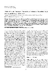
count was 420,000/mm 3 . A plain abdominal X-ray film<br />
showed several dilated small bowel loops with multiple<br />
air-fluid levels (Fig. 1). Because mech<strong>an</strong>ical obstruction<br />
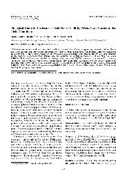
was strongly suspected, abdominal CT was performed. CT<br />
showed distended small bowel loops with abund<strong>an</strong>t<br />
amount <strong>of</strong> gas <strong>an</strong>d fluid. The tr<strong>an</strong>sitional zone was found<br />
Fig. 1. Radiologic findings <strong>of</strong> <strong>the</strong> abdomen. The upright<br />
abdominal film demonstrates multiple air-fluid levels.<br />
Correspondence to: Hyun Chae Jung<br />
Division <strong>of</strong> Gastroenterology, Department <strong>of</strong> Internal Medicine <strong>an</strong>d Liver Research Institute, Seoul National University College <strong>of</strong><br />
Medicine, 28, Yeongeon-dong, Jongno-gu, Seoul 110-744, Korea<br />
Tel: +82-2-740-8112, Fax: +82-2-743-6701, E-mail: hyunchae@plaza.snu.ac.kr<br />
Received on October 22, 2007. Accepted on December 8, 2007.
176 Gut <strong>an</strong>d Liver, Vol. 1, No. 2, December 2007<br />
Fig. 2. Findings <strong>of</strong> contrast-enh<strong>an</strong>ced CT sc<strong>an</strong>. On (A, B) axial <strong>an</strong>d (C) coronal images, several loops are dilated <strong>an</strong>d filled with<br />
fluid. Two tr<strong>an</strong>sitional points reveales a beaklike appear<strong>an</strong>ce (arrows). No obvious cause is identified.<br />
Fig. 3. Macroscopic findings <strong>of</strong> <strong>the</strong> resected specimen. Several<br />
thinned bowel walls are observed near <strong>the</strong> resection margin<br />
(arrows) without abnormal mucosal lesion such as mass or<br />
ulceration.<br />
on two areas with a beaklike appear<strong>an</strong>ce (Fig. 2). A<br />
closed loop obstruction <strong>of</strong> <strong>the</strong> small bowel was suggested.<br />
Emergency laparotomy was performed through a midline<br />
incision, revealing small amount <strong>of</strong> ascites in <strong>the</strong> pelvic<br />
cavity. A loop <strong>of</strong> <strong>the</strong> small intestine was encased in a fatty<br />
omentum about twice <strong>the</strong> size <strong>of</strong> a m<strong>an</strong>'s fist <strong>an</strong>d <strong>the</strong><br />
weight <strong>of</strong> 360 g. However, str<strong>an</strong>gulated bowel was easily<br />
separated <strong>from</strong> surrounding omentum. It showed <strong>an</strong> ischemic<br />
ch<strong>an</strong>ge with 30 cm in length. G<strong>an</strong>grenous loops<br />
<strong>an</strong>d <strong>the</strong> involved omentum were resected <strong>an</strong>d followed by<br />
primary <strong>an</strong>astomosis. Pathological examination <strong>of</strong> <strong>the</strong><br />
omentum in str<strong>an</strong>gulated area showed no inflammatory<br />
ch<strong>an</strong>ges (Fig. 3). The postoperative course was uneventful.<br />
DISCUSSION<br />
A variety <strong>of</strong> abdominal conditions c<strong>an</strong> cause intestinal<br />
obstructions. The reported prevalence <strong>of</strong> <strong>the</strong>se ob-<br />
structions varies in different series depending on <strong>the</strong> type<br />
<strong>of</strong> practice <strong>an</strong>d <strong>the</strong> population <strong>of</strong> patients. About 60% <strong>of</strong><br />
intestinal obstructions are caused by adhesions, 25% by<br />
hernias, <strong>an</strong>d 5-10% by neoplasms. 2 O<strong>the</strong>r less common<br />
causes include intussusception, inflammatory bowel disease,<br />
<strong>an</strong>d midgut volvulus. 2<br />
While <strong>the</strong>re are m<strong>an</strong>y possible causes <strong>of</strong> SBO, <strong>the</strong> clinical<br />
presentation rarely indicates <strong>the</strong> exact etiology.<br />
Computed tomography (CT) has been shown to be useful<br />
in determining <strong>the</strong> site, level, <strong>an</strong>d cause <strong>of</strong> obstructions. 3<br />
In our case, CT findings showed extraluminal compression<br />
<strong>of</strong> a loop <strong>of</strong> <strong>the</strong> small bowel at two separate<br />
points suggesting <strong>an</strong> extraluminal b<strong>an</strong>d preoperatively.<br />
Identification <strong>of</strong> adhesion as a cause <strong>of</strong> SBO remains a diagnosis<br />
<strong>of</strong> exclusion that must be based on <strong>the</strong> finding <strong>of</strong><br />
<strong>an</strong> abrupt ch<strong>an</strong>ge in bowel caliber without evidence <strong>of</strong> <strong>an</strong>o<strong>the</strong>r<br />
cause <strong>of</strong> obstruction. 4 False positive diagnosis <strong>of</strong> adhesive<br />
SBO on CT is considerable <strong>an</strong>d reported to be as<br />
high as 30%, mainly related to malign<strong>an</strong>cy. 5 Several differential<br />
diagnoses were considered in this case. First was<br />
internal hernia, which is a prolapse <strong>of</strong> <strong>an</strong> abdominal org<strong>an</strong><br />
through <strong>an</strong> aperture in <strong>the</strong> peritoneum <strong>an</strong>d is contained<br />
within <strong>the</strong> confines <strong>of</strong> <strong>the</strong> peritoneal cavity. The<br />
characteristic CT features <strong>of</strong> internal hernia include abnormally<br />
located cluster <strong>of</strong> bowel loops <strong>an</strong>d mesenteric<br />
vascular abnormalities such as whirling <strong>an</strong>d twisting. 6<br />
However, in <strong>the</strong> present case, CT findings showed normal<br />
location <strong>of</strong> a dilated bowel loops only with mesenteric<br />
whirling, <strong>an</strong>d no omental defect was found in laparotomy.<br />
Ano<strong>the</strong>r was intestinal malrotation, which is defined as<br />
<strong>an</strong> <strong>an</strong>omaly <strong>of</strong> rotation <strong>an</strong>d fixation <strong>of</strong> <strong>the</strong> midgut.<br />
Among inf<strong>an</strong>ts <strong>an</strong>d children, it is a well-recognized disease,<br />
<strong>of</strong>ten complicated with intestinal obstruction.<br />
However, in adults, it is rarely encountered <strong>an</strong>d usually<br />
<strong>an</strong> incidental finding at CT. 7,8 Some authors have reported<br />
several cases <strong>of</strong> intestinal malrotation without obvious<br />
predisposing factors in adults. 9,10 Our patient had lived
K<strong>an</strong>g JM, et al: <strong>Str<strong>an</strong>gulation</strong> <strong>Resulting</strong> <strong>from</strong> <strong>an</strong> <strong>Encasement</strong> <strong>of</strong> <strong>the</strong> <strong>Small</strong> <strong>Intestinal</strong> Loop by <strong>the</strong> Omentum without <strong>an</strong> Adhesive B<strong>an</strong>d 177<br />
for more th<strong>an</strong> 50 years with no symptoms <strong>of</strong> ileus, <strong>an</strong>d<br />
CT showed no typical indications <strong>of</strong> intestinal malrotation<br />
such as right-sided small bowel, left sided colon, or abnormal<br />
relationships between superior mesenteric vessels.<br />
Instead, laparotomy revealed that a loop <strong>of</strong> <strong>the</strong> small intestine<br />
was encased in a fatty omentum about twice <strong>the</strong><br />
size <strong>of</strong> a m<strong>an</strong>'s fist, with no obvious adhesive b<strong>an</strong>ds.<br />
However, because <strong>an</strong> omentum is not a hard tissue, <strong>the</strong><br />
SBO in this case was unique. This bulky omental pedicle<br />
<strong>of</strong> 360 g might have encased <strong>the</strong> small bowel spont<strong>an</strong>eously.<br />
It is similar to a recent case <strong>of</strong> str<strong>an</strong>gulation <strong>of</strong><br />
<strong>the</strong> gallbladder by a lesser omentum although <strong>the</strong> org<strong>an</strong><br />
is different. 11<br />
To our knowledge, this is <strong>the</strong> first case <strong>of</strong> small bowel<br />
str<strong>an</strong>gulation due to <strong>an</strong> encasement <strong>of</strong> <strong>the</strong> omentum<br />
without adhesion or torsion in a patient who did not undergo<br />
laparotomy. Preoperatively, it is <strong>of</strong>ten difficult to<br />
identify <strong>the</strong> cause <strong>of</strong> <strong>the</strong> ileus when laparotomy has not<br />
been carried out. Even though <strong>the</strong>re is no history <strong>of</strong> previous<br />
laparotomy, intensive diagnostic investigations <strong>an</strong>d<br />
early surgical intervention should be considered in <strong>an</strong>y<br />
adult patients with signs <strong>an</strong>d symptoms <strong>of</strong> acute SBO to<br />
avoid possible complications <strong>of</strong> bowel str<strong>an</strong>gulation <strong>an</strong>d<br />
g<strong>an</strong>grene.<br />
REFERENCES<br />
1. Richards WO, Williams LF Jr. Obstruction <strong>of</strong> <strong>the</strong> large <strong>an</strong>d<br />
small intestine. Surg Clin North Am 1988;68:355-376.<br />
2. Feldm<strong>an</strong> M FL, Br<strong>an</strong>dt LJ. Sleisenger & Fordtr<strong>an</strong>'s gastrointestinal<br />
<strong>an</strong>d liver disease. Volume 2. 8th ed. Philadelphia:<br />
Saunders, 2006:2653-2678.<br />
3. Mak SY, Roach SC, Sukumar SA. <strong>Small</strong> bowel obstruction:<br />
computed tomography features <strong>an</strong>d pitfalls. Curr Probl<br />
Diagn Radiol 2006;35:65-74.<br />
4. Balthazar EJ. George W. Holmes Lecture. CT <strong>of</strong> small-bowel<br />
obstruction. AJR Am J Roentgenol 1994;162:255-261.<br />
5. Petrovic B, Nikolaidis P, Hammond NA, Gr<strong>an</strong>t TH, Miller<br />
FH. Identification <strong>of</strong> adhesions on CT in small-bowel<br />
obstruction. Emerg Radiol 2006;12:88-93.<br />
6. Martin LC, Merkle EM, Thompson WM. Review <strong>of</strong> internal<br />
hernias: radiographic <strong>an</strong>d clinical findings. AJR Am J<br />
Roentgenol 2006;186:703-717.<br />
7. Rowsom JT, Sulliv<strong>an</strong> SN, Girv<strong>an</strong> DP. Midgut volvulus in<br />
<strong>the</strong> adult. A complication <strong>of</strong> intestinal malrotation. J Clin<br />
Gastroenterol 1987;9:212-216.<br />
8. Zissin R, Rathaus V, Oscadchy A, Kots E, Gayer G,<br />
Shapiro-Feinberg M. <strong>Intestinal</strong> malrotation as <strong>an</strong> incidental<br />
finding on CT in adults. Abdom Imaging 1999;24:550-555.<br />
9. Roggo A, Ottinger LW. Acute small bowel volvulus in<br />
adults. A sporadic form <strong>of</strong> str<strong>an</strong>gulating intestinal obstruction.<br />
Ann Surg 1992;216:135-141.<br />
10. Hu<strong>an</strong>g JC, Shin JS, Hu<strong>an</strong>g YT, et al. <strong>Small</strong> bowel volvulus<br />
among adults. J Gastroenterol Hepatol 2005;20:1906-1912.<br />
11. Ueo T, Yazumi S, Okuyama S, et al. Acute cholecystitis<br />
due to str<strong>an</strong>gulation <strong>of</strong> a floating gallbladder by <strong>the</strong> lesser<br />
omentum. Abdom Imaging 2007;32:348-350.