Embryonic and post-embryonic development of the ... - BioMed Central
Embryonic and post-embryonic development of the ... - BioMed Central
Embryonic and post-embryonic development of the ... - BioMed Central

You also want an ePaper? Increase the reach of your titles
YUMPU automatically turns print PDFs into web optimized ePapers that Google loves.
Rawlinson Frontiers in Zoology 2010, 7:12<br />
http://www.frontiersinzoology.com/content/7/1/12<br />
RESEARCH<br />
<strong>BioMed</strong> <strong>Central</strong><br />
Open Access<br />
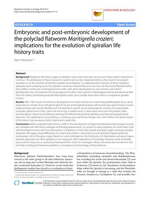
Research <strong>Embryonic</strong> <strong>and</strong> <strong>post</strong>-<strong>embryonic</strong> <strong>development</strong> <strong>of</strong><br />
<strong>the</strong> polyclad flatworm Maritigrella crozieri;<br />
implications for <strong>the</strong> evolution <strong>of</strong> spiralian life<br />
history traits<br />
Kate A Rawlinson 1,2<br />
Abstract<br />
Background: Planktonic life history stages <strong>of</strong> spiralians share some muscular, nervous <strong>and</strong> ciliary system characters in<br />
common. The distribution <strong>of</strong> <strong>the</strong>se characters is patchy <strong>and</strong> can be interpreted ei<strong>the</strong>r as <strong>the</strong> result <strong>of</strong> convergent<br />
evolution, or as <strong>the</strong> retention <strong>of</strong> primitive spiralian larval features. To underst<strong>and</strong> <strong>the</strong> evolution <strong>of</strong> <strong>the</strong>se characters<br />
adequate taxon sampling across <strong>the</strong> Spiralia is necessary. Polyclad flatworms are <strong>the</strong> only free-living Platyhelmin<strong>the</strong>s<br />
that exhibit a continuum <strong>of</strong> <strong>development</strong>al modes, with direct <strong>development</strong> at one extreme, <strong>and</strong> indirect<br />
<strong>development</strong> via a trochophore-like larval stage at <strong>the</strong> o<strong>the</strong>r. Here I present embryological <strong>and</strong> larval anatomical data<br />
from <strong>the</strong> indirect developing polyclad Maritrigrella crozieri, <strong>and</strong> consider <strong>the</strong>se data within a comparative spiralian<br />
context.<br />
Results: After 196 h hours <strong>of</strong> <strong>embryonic</strong> <strong>development</strong>, M. crozieri hatches as a swimming, planktotrophic larva. Larval<br />
myoanatomy consists <strong>of</strong> an orthogonal grid <strong>of</strong> circular <strong>and</strong> longitudinal body wall muscles plus parenchymal muscles.<br />
Diagonal body wall muscles develop over <strong>the</strong> planktonic period. Larval neuroanatomy consists <strong>of</strong> an apical plate,<br />
neuropile, paired nerve cords, a peri-oral nerve ring, a medial nerve, a ciliary b<strong>and</strong> nerve net <strong>and</strong> putative ciliary<br />
photoreceptors. Apical neural elements develop first followed by <strong>post</strong>erior perikarya <strong>and</strong> later pharyngeal neural<br />
elements. The ciliated larva is encircled by a continuous, pre-oral b<strong>and</strong> <strong>of</strong> longer cilia, which follows <strong>the</strong> distal margins<br />
<strong>of</strong> <strong>the</strong> lobes; it also possesses distinct apical <strong>and</strong> caudal cilia.<br />
Conclusions: Within polyclads heterochronic shifts in <strong>the</strong> <strong>development</strong> <strong>of</strong> diagonal bodywall <strong>and</strong> pharyngeal muscles<br />
are correlated with life history strategies <strong>and</strong> feeding requirements. In contrast to many spiralians, M. crozieri hatch with<br />
well developed nervous <strong>and</strong> muscular systems. Comparisons <strong>of</strong> <strong>the</strong> ciliary b<strong>and</strong>s <strong>and</strong> apical organs amongst spiralian<br />
planktonic life-stages reveal differences; M. crozieri lack a distinct ciliary b<strong>and</strong> muscle <strong>and</strong> flask-shaped epidermal<br />
serotonergic cells <strong>of</strong> <strong>the</strong> apical organ. Based on current phylogenies, <strong>the</strong> distribution <strong>of</strong> ciliary b<strong>and</strong>s <strong>and</strong> apical organs<br />
between polyclads <strong>and</strong> o<strong>the</strong>r spiralians is not congruent with a hypo<strong>the</strong>sis <strong>of</strong> homology. However, some similarities<br />
exist, <strong>and</strong> this study sets an anatomical framework from which to investigate cellular <strong>and</strong> molecular mechanisms that<br />
will help to distinguish between parallelism, convergence <strong>and</strong> homology <strong>of</strong> <strong>the</strong>se features.<br />
Background<br />
Flatworms (phylum Platyhelmin<strong>the</strong>s) have long been<br />
viewed as <strong>the</strong> sister group to all o<strong>the</strong>r bilaterian metazoans,<br />
due in large part to <strong>the</strong>ir blind gut <strong>and</strong> relatively simple,<br />
acoelomate body plan [1]. However, recent molecular<br />
phylogenetic studies have forced a re-evaluation <strong>of</strong> classi-<br />
* Correspondence: k.rawlinson@ucl.ac.uk<br />
1 Smithsonian Marine Station, 701 Seaway Drive, Fort Pierce, Florida, 34949 USA<br />
Full list <strong>of</strong> author information is available at <strong>the</strong> end <strong>of</strong> <strong>the</strong> article<br />
cal hypo<strong>the</strong>ses <strong>of</strong> metazoan intrarelationships. The Platyhelmin<strong>the</strong>s<br />
(including catenulids <strong>and</strong> rhabditophorans,<br />
but excluding <strong>the</strong> acoels <strong>and</strong> nemertodermatids [2]) now<br />
nest within <strong>the</strong> Spiralia, <strong>the</strong> protostomian sister clade to<br />
Ecdysozoa [[3] based on [4]]. Resolution <strong>of</strong> intrarelationships<br />
within <strong>the</strong> Spiralia is increasing, <strong>and</strong> <strong>the</strong> Platyhelminths<br />
are thought to belong to a clade that includes <strong>the</strong><br />
Bryozoa, Entoprocta, Cycliophora [5], <strong>and</strong> possibly Gas-<br />
© 2010 Rawlinson; licensee <strong>BioMed</strong> <strong>Central</strong> Ltd. This is an Open Access article distributed under <strong>the</strong> terms <strong>of</strong> <strong>the</strong> Creative Commons<br />
Attribution License (http://creativecommons.org/licenses/by/2.0), which permits unrestricted use, distribution, <strong>and</strong> reproduction in<br />
any medium, provided <strong>the</strong> original work is properly cited.
Rawlinson Frontiers in Zoology 2010, 7:12<br />
http://www.frontiersinzoology.com/content/7/1/12<br />
trotricha <strong>and</strong> Gnathostomulida [6]. This clade, in turn, is<br />
<strong>the</strong> sister group to a clade that includes annelids, mollusks,<br />
nemerteans <strong>and</strong> brachiopods [4,6]. This new phylogenic<br />
hypo<strong>the</strong>sis has a number <strong>of</strong> pr<strong>of</strong>ound implications<br />
for scenarios <strong>of</strong> metazoan morphological <strong>and</strong> life history<br />
evolution. This is largely due to <strong>the</strong> patchy distribution <strong>of</strong><br />
ciliated planktonic ("larval") stages across spiralians <strong>and</strong><br />
deuterostomes.<br />
A cursory examination <strong>of</strong> <strong>the</strong> phylogenetic distribution<br />
<strong>of</strong> larval stages within <strong>the</strong> mollusk/annelid/nemertean/<br />
branchiopod clade suggests that a ciliated "trochophore"<br />
larva could have been a primitive feature <strong>of</strong> this group.<br />
However, within its sister clade (Platyhelmin<strong>the</strong>s, Gastrotricha,<br />
Gnathostomulida, Bryozoa, Entoprocta,<br />
Cycliophora) <strong>the</strong> distribution <strong>of</strong> larval forms is scarcer,<br />
<strong>and</strong> absent in <strong>the</strong> gastrotrichs <strong>and</strong> gnathostomulids. This<br />
distribution <strong>of</strong> larval characters within <strong>the</strong> Spiralia could<br />
be interpreted in two ways: ei<strong>the</strong>r as <strong>the</strong> result <strong>of</strong><br />
repeated convergent evolution <strong>of</strong> intermediate planktonic<br />
life stages [7], or, perhaps more controversially, as <strong>the</strong><br />
retention <strong>of</strong> a primitive spiralian larval form in certain<br />
groups, with multiple instances <strong>of</strong> independent loss in<br />
o<strong>the</strong>r lineages [8]. To distinguish between <strong>the</strong>se alternative<br />
hypo<strong>the</strong>ses <strong>of</strong> character evolution, detailed morphological<br />
<strong>and</strong> <strong>development</strong>al data are needed from a wide<br />
diversity <strong>of</strong> taxa within <strong>the</strong> Spiralia - <strong>and</strong> in particular,<br />
from <strong>the</strong> relatively understudied indirect developing<br />
members <strong>of</strong> <strong>the</strong> Platyhelmin<strong>the</strong>s, Bryozoa, Entoprocta,<br />
Cycliophora. Polyclad flatworms are unique among <strong>the</strong><br />
free-living marine platyhelminths in having a gradient <strong>of</strong><br />
<strong>development</strong>al modes. These modes <strong>of</strong> <strong>development</strong> can<br />
be roughly categorized as direct <strong>development</strong> (embryos<br />
hatching as a benthic juvenile), intermediate <strong>development</strong><br />
(larva with lobes <strong>and</strong> ciliary b<strong>and</strong> retained within<br />
an egg case, <strong>and</strong> hatching as a benthic juvenile), <strong>and</strong> indirect<br />
<strong>development</strong> (with a planktonic form with lobes <strong>and</strong><br />
ciliary b<strong>and</strong>) [9]. Polyclad planktonic larval forms are,<br />
<strong>the</strong>mselves, morphologically diverse, <strong>and</strong> include <strong>the</strong><br />
four-lobed Götte's larva, <strong>the</strong> eight-lobed Müller's larva, as<br />
well as additional six- <strong>and</strong> ten-lobed forms [[10,11], pers<br />
obs]. To date, Müller's larvae have been described in both<br />
suborders <strong>of</strong> polyclads - <strong>the</strong> Cotylea <strong>and</strong> Acotylea - while<br />
Götte's larva, intracapsular larva <strong>and</strong> direct <strong>development</strong><br />
are all found exclusively with <strong>the</strong> Acotylea [12]. However,<br />
a dearth <strong>of</strong> data on <strong>the</strong> intrarelationships <strong>of</strong> polyclads <strong>and</strong><br />
insufficient taxonomic sampling <strong>of</strong> <strong>the</strong>ir life history strategies<br />
precludes inference <strong>of</strong> <strong>the</strong> primitive mode <strong>of</strong> <strong>development</strong><br />
for this clade.<br />
The ciliated larvae <strong>of</strong> polyclads have been thought to<br />
superficially resemble <strong>the</strong> pilidium <strong>of</strong> nemertines [10],<br />
<strong>and</strong> it has been suggested - based on morphological <strong>and</strong><br />
topological similarities between <strong>the</strong>ir ciliary b<strong>and</strong>s <strong>and</strong><br />
cephalic ganglia - that polyclad larvae are homologous<br />
with <strong>the</strong> trochophore larva <strong>of</strong> o<strong>the</strong>r spiralians [13]. How-<br />
Page 2 <strong>of</strong> 25<br />
ever, current hypo<strong>the</strong>ses <strong>of</strong> platyhelminth phylogeny support<br />
direct <strong>development</strong> as <strong>the</strong> primitive condition for<br />
<strong>the</strong> clade [2,14,15], <strong>and</strong> <strong>the</strong> appearance <strong>of</strong> ciliated larval<br />
forms within a single order <strong>of</strong> free-living platyhelminths<br />
may, instead, point to <strong>the</strong> convergent evolution <strong>of</strong> a<br />
planktonic life stage in polyclads. Never<strong>the</strong>less, due to <strong>the</strong><br />
variable position <strong>of</strong> <strong>the</strong> Platyhelmin<strong>the</strong>s within <strong>the</strong> Spiralia<br />
[e.g. [5] <strong>and</strong> [6]] <strong>and</strong> <strong>of</strong> <strong>the</strong> Polycladida within <strong>the</strong><br />
Platyhelmin<strong>the</strong>s [see [16]] <strong>the</strong> question <strong>of</strong> polyclad-trochophore<br />
larval homology or convergence remains open,<br />
<strong>and</strong> comparative analyses <strong>of</strong> <strong>the</strong> <strong>development</strong> <strong>of</strong> putatively<br />
homologous larval structures are needed to distinguish<br />
between <strong>the</strong>se alternative scenarios.<br />
Larval characters that are widespread amongst spiralian<br />
larvae include an apical tuft, paired ventral nerve<br />
cords, paired lateral nerve cords, cerebral ganglia, circumoral<br />
nerve loops, perikarya associated with <strong>the</strong> pedal<br />
nerve cord <strong>and</strong> <strong>the</strong> establishment <strong>of</strong> an apical muscle grid<br />
during myogenesis [17,18]. To date, descriptive studies on<br />
indirect developing polyclads using modern microscopy<br />
<strong>and</strong> fluorescent labeling techniques have characterized<br />
two anatomical similarities shared between spiralian <strong>and</strong><br />
polyclad larvae - <strong>the</strong> early establishment <strong>of</strong> an apical muscle<br />
grid in <strong>the</strong> acotylean Hoploplana inquilina [19] <strong>and</strong><br />
cotylean Maritigrella crozieri [20], <strong>and</strong> paired ventral <strong>and</strong><br />
lateral nerve cords in <strong>the</strong> acotylean Imogine mcgrathi<br />
[21]. Ultrastructural studies <strong>of</strong> polyclad larvae have<br />
greatly increased our underst<strong>and</strong>ing <strong>of</strong> <strong>the</strong> peripheral <strong>and</strong><br />
central nervous system (including <strong>the</strong> brain <strong>and</strong> ciliary<br />
b<strong>and</strong> - [21-24]). However, many aspects <strong>of</strong> polyclad<br />
<strong>embryonic</strong> <strong>and</strong> <strong>post</strong>-<strong>embryonic</strong> <strong>development</strong> remain<br />
unknown. For instance, <strong>the</strong> fate <strong>of</strong> <strong>the</strong> primary longitudinal<br />
muscles <strong>and</strong> <strong>development</strong> <strong>of</strong> pharyngeal muscles is<br />
undescribed, <strong>and</strong> data on <strong>the</strong> <strong>development</strong> <strong>and</strong> distribution<br />
<strong>of</strong> <strong>embryonic</strong> <strong>and</strong> larval neurons, <strong>the</strong>ir spatial relationship<br />
to <strong>the</strong> <strong>embryonic</strong> <strong>and</strong> larval musculature, <strong>and</strong><br />
<strong>the</strong> <strong>development</strong> <strong>of</strong> <strong>the</strong> ciliary b<strong>and</strong> <strong>and</strong> apical organs are<br />
lacking.<br />
Here, I provide a detailed account <strong>of</strong> <strong>embryonic</strong> <strong>and</strong><br />
larval <strong>development</strong> in <strong>the</strong> cotylean polyclad Maritigrella<br />
crozieri. Using a nuclear stain, phalloidin staining for Factin,<br />
<strong>and</strong> immunohistochemical staining <strong>of</strong> <strong>the</strong> nervous<br />
system, I show in detail: early cleavage, gastrulation <strong>and</strong><br />
<strong>development</strong> <strong>of</strong> <strong>the</strong> ciliary b<strong>and</strong>; myogenesis <strong>and</strong> neurogenesis.<br />
I discuss how different temporal patterns <strong>of</strong><br />
bodywall muscle <strong>development</strong> relate to different life history<br />
strategies within polyclads. This first analysis <strong>of</strong> neurotransmittor<br />
distribution shows a well-developed<br />
nervous system in <strong>the</strong> hatchling <strong>of</strong> M. crozieri. Myogenic<br />
<strong>and</strong> neurogenic elements <strong>of</strong> <strong>the</strong> ciliary b<strong>and</strong> <strong>and</strong> apical<br />
plate in M. crozieri are compared to those in o<strong>the</strong>r spiralian<br />
pelagic life history stages. Evidence for <strong>and</strong> against<br />
homology <strong>of</strong> <strong>the</strong>se organs is given, however, <strong>the</strong>se data<br />
are not sufficient to make <strong>the</strong> non-arbitrary distinction
Rawlinson Frontiers in Zoology 2010, 7:12<br />
http://www.frontiersinzoology.com/content/7/1/12<br />
between homology, parallelism <strong>and</strong> convergence. Efforts<br />
needed to decipher between <strong>the</strong>se three scenarios are discussed<br />
<strong>and</strong> this study provides a morphogenetic <strong>and</strong><br />
embryological foundation in polyclads for future comparative<br />
studies <strong>of</strong> spiralian <strong>development</strong>al gene expression<br />
<strong>and</strong> function.<br />
Results<br />
Overview <strong>of</strong> Maritigrella crozieri embryogenesis<br />
In a quiescent state mature Maritigrella crozieri measure<br />
31.3 (± 2.7) mm in length <strong>and</strong> 18.1 (± 1.9) mm in width<br />
(mean ± SD; n = 20). Individuals lay multiple egg batches,<br />
each containing anywhere from 50 to over 1000 individually<br />
encapsulated eggs. The eggs are nearly spherical <strong>and</strong><br />
measure 220 (± 15.6) μm (mean ± SD, n = 20) in diameter.<br />
At 22°C <strong>embryonic</strong> <strong>development</strong> took 196 (± 16) h (mean<br />
+ SD, n = 200) to hatching. <strong>Embryonic</strong> stages are<br />
described as hours <strong>post</strong> oviposition (hpo) <strong>and</strong>, in order to<br />
make <strong>the</strong>m comparable between taxa, as a percentage <strong>of</strong><br />
<strong>embryonic</strong> <strong>development</strong> time (% d.t.). There was no difference<br />
in <strong>development</strong> time between encapsulated<br />
embryos <strong>and</strong> those reared outside <strong>of</strong> <strong>the</strong> egg capsule.<br />
Cleavage in Maritigrella crozieri is quartet, spiral <strong>and</strong><br />
equal. The first cleavage takes place 8 hpo (4% d.t., Fig.<br />
1A), followed by <strong>the</strong> second cleavage at 11 hpo (6% d.t.,<br />
Fig. 1B). The four blastomeres are <strong>the</strong> same size making<br />
quadrant identification difficult. Following <strong>the</strong> cleavage<br />
nomenclature <strong>of</strong> Surface [25], <strong>the</strong> first quartet micromeres<br />
develop at 14 hpo (7% d.t., Fig. 1C), <strong>the</strong> second quartet<br />
micromeres at 18 hpo (9% d.t., Fig. 1D), <strong>the</strong> very small<br />
third quartet micromeres at 21 hpo (11% d.t.), <strong>and</strong> <strong>the</strong><br />
large fourth quartet micromeres (~90 μm) <strong>and</strong> very small<br />
macromeres (10 μm) at 23 hpo (12% d.t., Fig. 1E). The<br />
number <strong>of</strong> micromeres from <strong>the</strong> first three quartets<br />
increases to more than 83 in <strong>the</strong> next 10 hours, <strong>the</strong>se<br />
form an irregular double layer at <strong>the</strong> animal pole <strong>and</strong><br />
form a cap over <strong>the</strong> 4th quartet micromeres (Fig. 1F). At<br />
<strong>the</strong> onset <strong>of</strong> gastrulation a 4 th quartet micromere divides<br />
(33 hpo, 17% d.t., Fig. 1G) - this, according to Surface<br />
[25], is 4d. Gastrulation proceeds by epiboly - <strong>the</strong><br />
micromeres at <strong>the</strong> animal pole divide <strong>and</strong> migrate vegetally<br />
as a sheet, enveloping <strong>the</strong> 4 th quartet micromeres<br />
<strong>and</strong> macromeres (Fig. 1H, I, J). It appears as though <strong>the</strong><br />
micromeres 4a-c do not divide, but coalesce into <strong>the</strong> yolk<br />
filled interior <strong>of</strong> <strong>the</strong> embryo. The four small fourth quartet<br />
macromeres are visible <strong>and</strong> remain undivided until<br />
<strong>the</strong> ectoderm is established as a layer <strong>and</strong> just before it<br />
begins to invaginate at <strong>the</strong> lower pole to form <strong>the</strong> pharynx.<br />
By 95 hpo (48% d.t.) epiboly is almost complete <strong>and</strong><br />
<strong>the</strong> animal-vegetal axis turns into <strong>the</strong> antero-<strong>post</strong>erior<br />
axis.<br />
Following gastrulation, <strong>development</strong> may be considered<br />
in three main stages; i) <strong>development</strong> <strong>of</strong> <strong>the</strong> epidermis<br />
<strong>and</strong> organ primordia, ii) organ differentiation, iii)<br />
Page 3 <strong>of</strong> 25<br />
<strong>development</strong> <strong>of</strong> larval lobes. Development <strong>of</strong> <strong>the</strong> epidermis<br />
<strong>and</strong> organ primordia takes place between 69 <strong>and</strong> 125<br />
hpo (35% <strong>and</strong> 63% d.t.). During this time <strong>the</strong> embryo is<br />
round in shape <strong>and</strong> a small outpocketing develops at <strong>the</strong><br />
<strong>post</strong>erior end, in <strong>the</strong> centre <strong>of</strong> which lies <strong>the</strong> stomodaeum.<br />
Smooth epi<strong>the</strong>lial ectodermal cells are differentiated<br />
from more rounded deep cells. At 69 hpo (35% d.t.),<br />
epidermal cilia stain positive for acetylated tubulin, <strong>and</strong><br />
rotation <strong>of</strong> <strong>the</strong> embryos starts at 75 hpo (38% d.t.). At 109<br />
hpo (56% d.t.) a pre-oral ciliary b<strong>and</strong> forms a circle subequatorially<br />
around <strong>the</strong> <strong>post</strong>erior end <strong>of</strong> <strong>the</strong> embryo <strong>and</strong><br />
a ring <strong>of</strong> longer cilia forms around <strong>the</strong> developing stomodaeum<br />
(Fig. 2A). Sixteen hours later, <strong>the</strong> ciliary b<strong>and</strong> has<br />
four <strong>of</strong>fshoots which branch upwards towards <strong>the</strong> anterior<br />
<strong>of</strong> <strong>the</strong> embryo (Fig. 2C). The epidermal eyespot<br />
forms at 115 hpo (58% d.t.), in an anterio-dorsal position.<br />
From 126 hpo (64% d.t.) to 169 hpo (86% d.t.) organ differentiation<br />
takes place <strong>and</strong> <strong>the</strong> embryo becomes obovate<br />
by elongating slightly along <strong>the</strong> antero-<strong>post</strong>erior axis.<br />
During this period two acetylated tubulin (acTub)<br />
expressing structures are visible, <strong>the</strong>se are <strong>the</strong> two developing<br />
protonephridial canal cells (129 hpo, Fig. 2D). They<br />
extend from ei<strong>the</strong>r side <strong>of</strong> <strong>the</strong> stomodeum, across <strong>the</strong> epi<strong>the</strong>lium<br />
<strong>and</strong> basement membrane <strong>and</strong> proceed anteriorly.<br />
The two putative terminal cells <strong>of</strong> <strong>the</strong> protonephridia are<br />
visible in acTub stained wholemounts later (160 hpo) as<br />
two bilaterally symmetric, unbranched, elongated <strong>and</strong><br />
heavy ciliated vesicles, located in <strong>the</strong> upper third <strong>of</strong> <strong>the</strong><br />
embryo ei<strong>the</strong>r side <strong>of</strong> <strong>the</strong> oral hood (Fig. 2F). The left<br />
cerebral eye develops first (139 hpo [70% d.t.]), followed<br />
shortly by <strong>the</strong> right cerebral eye, <strong>the</strong>y are 20 μm from<br />
each o<strong>the</strong>r <strong>and</strong> are positioned more ventrally relative to<br />
<strong>the</strong> epidermal eye. The stomodaeum migrates from a<br />
<strong>post</strong>erior position to a mid-ventral position at 150 hpo<br />
(Fig. 2E), <strong>the</strong> pharynx invaginates later at 160 hpo (Fig.<br />
2F), while <strong>the</strong> ciliary b<strong>and</strong> migrates equatorially <strong>and</strong><br />
demarcates <strong>the</strong> position where <strong>the</strong> eight larval lobes will<br />
develop (Fig. 2E). The F-actin containing microtubular<br />
sheaths <strong>of</strong> rhabdite cells are visible at <strong>the</strong> anterior <strong>and</strong><br />
<strong>post</strong>erior tip <strong>of</strong> <strong>the</strong> embryo <strong>and</strong> become more numerous.<br />
From 170 hpo (87% d.t.) to 196 hpo (100% d.t.) <strong>the</strong><br />
embryo's shape changes from obovate to a complex morphology<br />
with eight protruding lobes. Lobe formation<br />
starts with <strong>the</strong> protrusion <strong>of</strong> <strong>the</strong> oral hood (170 hpo [87%<br />
d.t.]) followed by <strong>the</strong> dorsal lobe. The paired lobes follow<br />
sequentially with <strong>the</strong> ventro-lateral lobes (175 hpo, 89%<br />
d.t.), followed by <strong>the</strong> lateral lobes <strong>and</strong> shortly after, <strong>the</strong><br />
dorso-lateral lobes (which elongate fur<strong>the</strong>r after hatching).<br />
The lateral lobes protrude higher up <strong>the</strong> embryo<br />
than <strong>the</strong> ventolateral <strong>and</strong> dorsolateral lobes. The pharynx<br />
<strong>and</strong> gut lumen meet at 175 hpo (89% d.t.). The gut is<br />
blind, ciliated throughout, un-branched (Fig. 2G) <strong>and</strong><br />
reaches almost to <strong>the</strong> latitude <strong>of</strong> <strong>the</strong> brain in <strong>the</strong> developed<br />
larva. Hatching occurs on day 8 after 196 hours <strong>of</strong>
Rawlinson Frontiers in Zoology 2010, 7:12<br />
http://www.frontiersinzoology.com/content/7/1/12<br />
Page 4 <strong>of</strong> 25<br />
Figure 1 Confocal laser scanning micrographs (CLSM) <strong>of</strong> <strong>the</strong> blastula <strong>and</strong> gastrula stages <strong>of</strong> Maritigrella crozieri. (F-actin labeled with phalloidin<br />
564 [red], nucleic acids stained with Sytox Green [green]). (A-D) - animal views <strong>of</strong> cleavage cycles 1-4; showing <strong>the</strong> first quartet micromeres (arrowheads<br />
in C) <strong>and</strong> second quartet micromeres (arrowheads in D). (E) Animal view showing <strong>the</strong> third quartet micromeres (arrowheads), vegetal view<br />
showing large fourth quartet micromeres (4a-d, double arrowheads) <strong>and</strong> mini-macromeres (mm)(4A-D). (F) Lateral view shows irregular double layer<br />
<strong>of</strong> dividing 1 st to 3 rd quartet micromeres at <strong>the</strong> animal pole over <strong>the</strong> 4a-d quartet. (G) Animal view <strong>of</strong> micromere cap <strong>and</strong> vegetal view <strong>of</strong> 4d division.<br />
(H-J) Animal, lateral <strong>and</strong> vegetal views respectively <strong>of</strong> gastrulation by epiboly; micromere cap migrates vegetally enveloping <strong>the</strong> fourth quartet micromeres<br />
<strong>and</strong> macromeres, which remain visible until <strong>the</strong> ectoderm invaginates at <strong>the</strong> lower pole to form <strong>the</strong> stomodeum. Scale bars 100 μm, hpo =<br />
hours <strong>post</strong> oviposition, dt = <strong>development</strong> time.
Rawlinson Frontiers in Zoology 2010, 7:12<br />
http://www.frontiersinzoology.com/content/7/1/12<br />
<strong>embryonic</strong> <strong>development</strong>. The larva is positively phototactic<br />
<strong>and</strong> swims in a right-h<strong>and</strong>ed helix - led by epidermal<br />
eye (ie anterio-dorsal side leading). It is planktotrophic<br />
[26] <strong>and</strong> was observed to feed on cultures <strong>of</strong> unicellular<br />
algae introduced into <strong>the</strong> seawater (pers obs). Rhabdites,<br />
<strong>and</strong> pigmentation do not extend onto <strong>the</strong> lobes. The protonephridia<br />
extend anteriorly from below <strong>the</strong> mouth<br />
through <strong>the</strong> basement membrane, branching mid-larvae<br />
at <strong>the</strong> level <strong>of</strong> <strong>the</strong> lateral lobes, <strong>and</strong> re-enter <strong>the</strong> epidermis<br />
ventro-anteriorly just above <strong>the</strong> oral hood at <strong>the</strong> same latitude<br />
as <strong>the</strong> brain. They run interior to, but in close proximity<br />
with, <strong>the</strong> lateral nerve connectives. The average size<br />
<strong>of</strong> a hatchling is 250 (± 12, n = 10) μm in length <strong>and</strong> 200<br />
(± 8, n = 10) μm wide. The gross anatomy is illustrated at<br />
two-day <strong>post</strong> hatching (Fig. 3).<br />
Larval muscle anatomy<br />
The body wall musculature at two-days <strong>post</strong>-hatching (n<br />
= 30)(Fig. 4) is composed <strong>of</strong> an apical complex on top <strong>of</strong><br />
circular <strong>and</strong> longitudinal fibers organized in a lattice-like<br />
arrangement. Parenchymal muscles include dorso-ventral<br />
muscles <strong>and</strong> those associated with <strong>the</strong> mouth <strong>and</strong><br />
pharynx.<br />
The apical complex (Fig 4A), at <strong>the</strong> anterior pole <strong>of</strong> <strong>the</strong><br />
larva, was made up <strong>of</strong> 5 spirally arranged coils or closed<br />
circular muscles. The apical coil had a diameter <strong>of</strong> 15 μm.<br />
Page 5 <strong>of</strong> 25<br />
Figure 2 Ciliary b<strong>and</strong> migration in Maritigrella crozieri embryos. CLSM micrographs <strong>of</strong> <strong>post</strong>-gastrulation acetylated tubulin stained wholemounts.<br />
(A) Posterior view showing ciliary ring around stomodeum (st) at <strong>post</strong>erior pole <strong>and</strong> <strong>the</strong> early ciliary b<strong>and</strong> (cb). (B-C) Lateral view - anterior up - (B)<br />
ciliated epidermis <strong>and</strong> sub-equatorial ciliary b<strong>and</strong>, (C) four branches <strong>of</strong> ciliary b<strong>and</strong> migrate anteriorly, (D) Ventral view at focal plane <strong>of</strong> developing<br />
protonephridial canal cells (pnp). (E) Ventral view - stomodeum migrates ventrally, (F) Ventral <strong>and</strong> left lateral view - ciliary b<strong>and</strong> demarcates position<br />
where <strong>the</strong> eight larval lobes will form (oral hood, oh, ventro-lateral lobe, vll, lateral lobe, ll, dorsal lobe, dl), cilia in <strong>the</strong> protonephridia lumen (pnp) <strong>and</strong><br />
pharynx (ph) are visible. (G) Left lateral view-pre-hatchling showing continual b<strong>and</strong> <strong>of</strong> longer cilia round <strong>the</strong> lobes <strong>and</strong> fused intestine (in) <strong>and</strong> pharynx.<br />
Scale bars 100 μm<br />
These muscles serve as attachment sites for most <strong>of</strong> <strong>the</strong><br />
longitudinal muscles. Approximately 6 longitudinal muscles<br />
met in <strong>the</strong> centre <strong>of</strong> <strong>the</strong> complex at <strong>the</strong> anterior pole.<br />
Below, an orthogonal grid <strong>of</strong> 12-22 outer circular <strong>and</strong> 12-<br />
16 inner longitudinal muscles constitutes <strong>the</strong> framework<br />
Figure 3 Gross anatomy <strong>of</strong> Maritigrella crozieri two-day <strong>post</strong>hatching<br />
pelagic stage. A) ventral view, B) left lateral view. (ac) apical<br />
cilium, (ee) epidermal eye, (ce) cerebral eye, (in) intestine, (oh) oral<br />
hood, (pnp) protonephridia, (ll) lateral lobe, (vll) ventro-lateral lobe, (ph)<br />
pharynx, (m) mouth, (sop) sub-oral plate, (cci) caudal cilium.
Rawlinson Frontiers in Zoology 2010, 7:12<br />
http://www.frontiersinzoology.com/content/7/1/12<br />
Page 6 <strong>of</strong> 25<br />
Figure 4 Confocal projections <strong>of</strong> <strong>the</strong> phalloidin-labelled musculature <strong>of</strong> Maritigrella crozieri two-day <strong>post</strong>-hatching pelagic stage. Apical<br />
views (ventral up) <strong>of</strong> A) Apical complex (ac) <strong>of</strong> circular <strong>and</strong> coiled muscles above <strong>the</strong> orthogonal grid <strong>of</strong> body wall muscles, <strong>and</strong> B) optical sections<br />
below <strong>the</strong> apical complex showing mouth retractor muscle (mrm) <strong>and</strong> a pair <strong>of</strong> anterior-<strong>post</strong>erior parenchymal muscles (asterisk) extending from <strong>the</strong><br />
apical complex to <strong>the</strong> mouth (m in C). A pair <strong>of</strong> dorso-ventral parenchymal muscles (arrow heads in B) extends from <strong>the</strong> dorsal lobe (dl) to <strong>the</strong> oral<br />
hood (oh). C) Ventral, D) lateral <strong>and</strong> E) dorsal view <strong>of</strong> orthogonal grid <strong>of</strong> body wall muscles, mouth retractor muscle (mrm) <strong>and</strong> oral hood muscles;<br />
longitudinal (arrowheads in C), oblique (double arrowhead in C) <strong>and</strong> circular (arrowheads in D). Prominent circular muscles include <strong>the</strong> anterior primary<br />
circular muscle (apcm), <strong>the</strong> <strong>post</strong>erior primary circular muscle (ppcm), <strong>and</strong> <strong>the</strong> lateral lobe circular muscle (llcm). Two thick muscle b<strong>and</strong>s extend<br />
between <strong>the</strong> oral hood <strong>and</strong> dorsal lobe (double arrowheads in D & E), <strong>the</strong> endings branch obliquely across <strong>the</strong> lobes (double arrowhead in C) <strong>and</strong><br />
interdigitate with endings from <strong>the</strong> opposing fiber. Primary longitudinal muscles (arrow heads in E), epidermal eye (ee). Roots <strong>of</strong> <strong>the</strong> apical <strong>and</strong> caudal<br />
cilia are visible in <strong>the</strong> epidermis (asterisks in C, D & E). (F) Dorsal view, optical sections inside <strong>of</strong> body wall musculature at focal plane <strong>of</strong> dorsoventral<br />
parenchymal muscles (arrowheads), three pairs in bilateral organisation lateral to <strong>the</strong> intestine (in). (G) Ventral view, single optical section showing<br />
many parenchymal muscles radiating from <strong>the</strong> <strong>post</strong>erior primary circular muscle (ppcm) laterally (arrow heads) <strong>and</strong> anteriorly (asterisk), concentric<br />
rings <strong>of</strong> mouth muscles (double arrow heads). (H) More ventral still, an optical section behind <strong>the</strong> ventral lobes shows <strong>the</strong> mouth retractor muscle<br />
(mrm) extending from <strong>the</strong> apical complex <strong>and</strong> a ring <strong>of</strong> muscle (double arrowheads) around <strong>the</strong> pharynx (ph). Scale bars 50 μm.
Rawlinson Frontiers in Zoology 2010, 7:12<br />
http://www.frontiersinzoology.com/content/7/1/12<br />
<strong>of</strong> <strong>the</strong> body wall musculature. Of <strong>the</strong>se, two circular <strong>and</strong><br />
two longitudinal muscles are considered primary muscle<br />
fibres, as <strong>the</strong>y were <strong>the</strong> earliest distinguishable l<strong>and</strong>marks<br />
during myogenesis (see below). These muscles were more<br />
deeply set than <strong>the</strong> rest <strong>of</strong> <strong>the</strong> body wall muscle grid. The<br />
primary circular muscles (Fig. 4D) consist <strong>of</strong> an anterior<br />
primary circular muscle <strong>and</strong> <strong>the</strong> <strong>post</strong>erior primary circular<br />
muscle. The former follows <strong>the</strong> lower contour <strong>of</strong> <strong>the</strong><br />
oral <strong>and</strong> dorsal lobes. The <strong>post</strong>erior primary circular<br />
muscle forms <strong>the</strong> lower muscle b<strong>and</strong> <strong>of</strong> <strong>the</strong> ventro-lateral<br />
<strong>and</strong> dorso-lateral lobes, but is not connected to <strong>the</strong> lateral<br />
lobe, which protrudes higher up <strong>the</strong> larvae. However, it<br />
runs close to <strong>the</strong> lateral lobe circular muscle ventrally<br />
above <strong>the</strong> mouth. On <strong>the</strong> dorsal side two primary longitudinal<br />
muscles (Fig. 4E) run parallel to each o<strong>the</strong>r down<br />
from <strong>the</strong> apical complex to <strong>the</strong> caudal pole, under <strong>the</strong><br />
sub-oral plate to join <strong>the</strong> mouth muscles on <strong>the</strong> ventral<br />
surface.<br />
Bilateral symmetry <strong>of</strong> <strong>the</strong> longitudinal muscles is<br />
apparent from <strong>the</strong> apical view (Fig. 4A). 2-3 major longitudinal<br />
muscle fibres extend <strong>post</strong>eriorly from <strong>the</strong> apical<br />
complex into each lateral lobe. At <strong>the</strong> end <strong>of</strong> <strong>the</strong> lobes<br />
<strong>the</strong>se muscles digitate into fine muscle fibres. Of <strong>the</strong>se<br />
longitudinal muscles, <strong>the</strong> two that form <strong>the</strong> lateral ridge<br />
<strong>of</strong> <strong>the</strong> lateral lobes are particularly thick <strong>and</strong> prominent.<br />
Four longitudinal muscles on <strong>the</strong> dorsal side, including<br />
<strong>the</strong> two primary longitudinal muscles, pass under <strong>the</strong><br />
sub-oral plate <strong>and</strong> are joined by many fine muscles leading<br />
to <strong>the</strong> mouth. The lateral lobe circular muscle (Fig.<br />
4D) runs around <strong>the</strong> larvae at <strong>the</strong> level <strong>of</strong> <strong>the</strong> two lateral<br />
lobes <strong>and</strong> over <strong>the</strong> anterior opening <strong>of</strong> <strong>the</strong> mouth. Many<br />
<strong>of</strong> <strong>the</strong> prominent circular <strong>and</strong> longitudinal muscles<br />
appear to be double str<strong>and</strong>ed. Many fine circular muscles<br />
converge at <strong>the</strong> mouth. A first diagonal body wall muscle<br />
(Fig. 4D, E) runs from a <strong>post</strong>erior medial position on <strong>the</strong><br />
dorsal side around to <strong>the</strong> right lateral side, arcing up<br />
behind <strong>the</strong> lateral lobe <strong>and</strong> curves down towards <strong>the</strong> ventro-lateral<br />
lobe. This is named <strong>the</strong> dorsal diagonal arc in<br />
Hoploplana inquilina [19], <strong>and</strong> in M. crozieri it becomes<br />
more pronounced, extending from <strong>the</strong> left caudal side to<br />
<strong>the</strong> right side <strong>of</strong> <strong>the</strong> larvae as it develops. By 10 days <strong>post</strong>hatching<br />
o<strong>the</strong>r diagonal fibres are visible including two<br />
short diagonal muscles that cross each o<strong>the</strong>r above <strong>the</strong><br />
dorsal lobe (see additional file 1 figure S1a).<br />
The musculature <strong>of</strong> <strong>the</strong> concave oral hood consists <strong>of</strong> 5-<br />
7 circular muscles (Fig. 4D) <strong>and</strong> 3-8 oblique muscle fibres<br />
(Fig. 4C) radiating from paired thick b<strong>and</strong>s <strong>of</strong> muscle on<br />
ei<strong>the</strong>r side <strong>of</strong> <strong>the</strong> hood (Fig. 4D). These paired muscles<br />
b<strong>and</strong>s run from <strong>the</strong> upper surface <strong>of</strong> <strong>the</strong> dorsal lobe to <strong>the</strong><br />
oral hood. A second pair extends (Fig. 4D) along <strong>the</strong><br />
lower margin <strong>of</strong> <strong>the</strong> dorsal <strong>and</strong> oral lobes, <strong>and</strong> <strong>the</strong>y converge<br />
ventrally with <strong>the</strong> anterior primary circular muscle<br />
<strong>and</strong> a lower circular muscle to form <strong>the</strong> thick, prominent<br />
muscle at <strong>the</strong> front <strong>of</strong> <strong>the</strong> lobe. There are 9 fine longitudi-<br />
Page 7 <strong>of</strong> 25<br />
nal muscles (Fig. 4C) in <strong>the</strong> oral hood that are positioned<br />
inside <strong>of</strong> <strong>the</strong> circular <strong>and</strong> oblique muscles. These muscles<br />
connect to <strong>the</strong> upper margin <strong>of</strong> <strong>the</strong> mouth muscles. The<br />
dorsal lobe is made up <strong>of</strong> four circular muscles (including<br />
<strong>the</strong> anterior primary circular muscle) <strong>and</strong> <strong>the</strong> two pairs <strong>of</strong><br />
thick muscle b<strong>and</strong>s that wrap around <strong>the</strong> larva from <strong>the</strong><br />
oral hood to <strong>the</strong> dorsal lobe. The endings branch <strong>and</strong><br />
interdigitate on <strong>the</strong> dorsal lobe protrusion (Fig 4E). The<br />
three pairs <strong>of</strong> lateral lobes that protrude from <strong>the</strong> body<br />
wall each have 2-3 longitudinal muscles <strong>and</strong> several circular<br />
muscles. Numerous fine circular <strong>and</strong> diagonal muscles<br />
are found along <strong>the</strong> length <strong>of</strong> <strong>the</strong> lobes <strong>and</strong> at <strong>the</strong> tips.<br />
There are five pairs <strong>of</strong> bilaterally symmetrical parenchymal<br />
dorsoventral muscles; three run lateral to <strong>the</strong><br />
intestine, between <strong>the</strong> mouth <strong>and</strong> anterior primary circular<br />
muscle (Fig. 4F). The fourth pair lies inside <strong>of</strong> <strong>the</strong><br />
anterior primary circular muscle <strong>and</strong> joins <strong>the</strong> dorsal lobe<br />
to <strong>the</strong> oral hood (Fig. 4F). The fifth pair is more anterior<br />
<strong>and</strong> is at <strong>the</strong> same latitude as <strong>the</strong> 8 th circular muscle from<br />
<strong>the</strong> apex (Fig. 4B). Several more dorsoventral parenchymal<br />
muscles are found in <strong>the</strong> sub-oral plate. Six - twelve<br />
diagonal parenchymal muscle fibres run anteriorly from<br />
<strong>the</strong> <strong>post</strong>erior primary circular muscle just above <strong>the</strong><br />
mouth margin, two connect below <strong>the</strong> apical complex,<br />
several run to <strong>the</strong> frontal muscle <strong>of</strong> <strong>the</strong> oral hood <strong>and</strong> <strong>the</strong><br />
o<strong>the</strong>rs connect to circular muscles laterally below <strong>the</strong> latitude<br />
<strong>of</strong> <strong>the</strong> cerebral eyes (Fig. 4G). These diagonal muscles<br />
probably act as mouth dilator muscles. The mouth<br />
retractor muscle (Fig 4B, C, H) bifurcates at <strong>the</strong> anterior<br />
pole <strong>of</strong> <strong>the</strong> larva, each branch attaches to ei<strong>the</strong>r side <strong>of</strong><br />
<strong>the</strong> apical complex. Just below <strong>the</strong> cerebral eyes it forks<br />
into several branches, which divide into many fingers.<br />
Some <strong>of</strong> <strong>the</strong>se fingers reach down to <strong>the</strong> mouth circular<br />
muscles, while o<strong>the</strong>rs attach to <strong>the</strong> lower muscles <strong>of</strong> <strong>the</strong><br />
oral hood. Three ring-shaped fibres form a sphincter at<br />
<strong>the</strong> mouth (Fig. 4G <strong>and</strong> 4H). F-actin in <strong>the</strong> cilial b<strong>and</strong> was<br />
stained with phalloidin <strong>and</strong> two rows <strong>of</strong> cilia were<br />
observed on <strong>the</strong> oral <strong>and</strong> ventro-lateral lobes, while only<br />
one is observed on <strong>the</strong> lateral, dorso-lateral <strong>and</strong> dorsal<br />
lobes (additional file 1 figure S1 b).<br />
Larval neuroanatomy<br />
The ground pattern <strong>of</strong> nerves at two-days <strong>post</strong>-hatching<br />
(n = 50) is described based on acetylated tubulin (acTub)<br />
stained wholemounts (Fig. 5A &5B). Acetylated tubulin<br />
marks stabilized microtubules that form in, for example,<br />
axon fibers <strong>and</strong> gl<strong>and</strong> cell necks. In order to begin to distinguish<br />
neural from non-neural elements, <strong>the</strong> distribution<br />
<strong>of</strong> <strong>the</strong> neurotransmittors serotonin (5HT) <strong>and</strong><br />
FMRF-amide was mapped onto <strong>the</strong> ground pattern. Neurons<br />
that showed immunoreactivity to <strong>the</strong> neurotransmittors<br />
were located in <strong>the</strong> apical plate, <strong>the</strong> neuropile,<br />
paired dorso-lateral <strong>and</strong> ventro-lateral nerve cords, a<br />
pharyngeal nerve ring, a medial nerve, a ciliary b<strong>and</strong>
Rawlinson Frontiers in Zoology 2010, 7:12<br />
http://www.frontiersinzoology.com/content/7/1/12<br />
Figure 5 The neural groundplan <strong>of</strong> <strong>the</strong> Maritigrella crozieri twoday<br />
<strong>post</strong>-hatching pelagic stage. (A & B) based on acetylated tubulin<br />
wholemounts, left lateral <strong>and</strong> ventral view respectively (bright<br />
green = neurons that show serotonin <strong>and</strong>/or FMRFamide immunoreactivity,<br />
dark green = structures not immunoreactive to serotonin <strong>and</strong><br />
FMRF-amide antibodies). (C) Serotonin expression - ventral view. (D)<br />
FMRF-amide expression - ventral view. (aa) apical plate, (apl) apical<br />
plexus, (cbnr) ciliary b<strong>and</strong> ring, (cc) cerebral commissure, (ccb) commissural<br />
cell bodies (ccip) caudal cilium perikarya, (cp) caudal perikarya,<br />
(dlc) dorso-lateral connective, (een) epidermal eye nerve, (gcn) gl<strong>and</strong><br />
cell necks, (iec) intra-epidermal cells, (ien) intra-epidermal nerves, (ienp)<br />
intra-epidermal nerve plexi, (mc) medial cluster, (mn) medial nerve,<br />
(on) oellar nerve, (phnr) pharyngeal nerve ring, (rc) ring connectives,<br />
(sor) supra-oral ring, (vlc) ventro-lateral connective, (vp) ventral perikarya.<br />
nerve ring, <strong>and</strong> two intra-epidermal nerve plexi (Fig. 5C<br />
&5D). Many acTub labeled structures did not show 5HT<br />
or FMRFamide immunoreactivity, indicating that o<strong>the</strong>r<br />
neurotransmittors might be active or that <strong>the</strong>y are nonneural<br />
structures (see below, Fig. 5).<br />
The apical plate is located above <strong>the</strong> apical spiral complex<br />
<strong>of</strong> <strong>the</strong> body wall muscles (Fig. 6A) <strong>and</strong> consists <strong>of</strong>: 1)<br />
a single apical cilium (Fig. 6A, 7A), 2) a ring <strong>of</strong> longer cilia<br />
surrounding <strong>the</strong> apical cilium (Fig. 6A) <strong>and</strong> 3) a ring <strong>of</strong><br />
5HT (Fig. 6E) <strong>and</strong> FMRFamide expression beneath <strong>the</strong><br />
ring <strong>of</strong> long cilia. Whe<strong>the</strong>r <strong>the</strong> ring <strong>of</strong> long cilia are from<br />
cells that are 5HT <strong>and</strong> FMRFamide positive or whe<strong>the</strong>r<br />
<strong>the</strong>se epidermal ciliated cells are non-neuronal but are in<br />
contact with dendrites <strong>of</strong> 5HT <strong>and</strong> FMRFamide positive<br />
cells is not discernable from this data <strong>and</strong> requires fur<strong>the</strong>r<br />
Page 8 <strong>of</strong> 25<br />
investigation. Indeed how <strong>the</strong> apical plate is connected to<br />
<strong>the</strong> neuropile is not evident from <strong>the</strong>se data. The acTub<br />
structures leading from <strong>the</strong> apical plate to <strong>the</strong> cerebral<br />
commissure are not 5HT or FMRFamide-positive <strong>and</strong> it<br />
is possible that <strong>the</strong>y could be <strong>the</strong> microtubules in <strong>the</strong><br />
gl<strong>and</strong> cell necks (Fig 7A).<br />
Intraepi<strong>the</strong>lial neurons surrounding <strong>the</strong> apical plate<br />
extend <strong>post</strong>eriorly between <strong>the</strong> body wall muscles to a<br />
cluster <strong>of</strong> axons - <strong>the</strong> apical plexus, which is situated in<br />
<strong>the</strong> midline, anterior to, but in contact with, <strong>the</strong> cerebral<br />
commissure <strong>and</strong> forms part <strong>of</strong> <strong>the</strong> neuropile (see<br />
below)(Fig. 7A, D, E, F). The brain <strong>of</strong> <strong>the</strong> larvae consists<br />
<strong>of</strong> a bilaterally symmetric central neuropile made up <strong>of</strong><br />
two distinct regions; <strong>the</strong> apical plexus <strong>and</strong> <strong>the</strong> cerebral<br />
commissure. The apical plexus consists <strong>of</strong> a rectangular<br />
shaped network <strong>of</strong> FMRF-amide expressing cell processes<br />
(Fig. 7B) <strong>and</strong> <strong>the</strong> spherical median cluster expressing<br />
5HT <strong>and</strong> FMRFamide - this may be a synaptic glomeruli<br />
where <strong>the</strong> sensory afferents innervating <strong>the</strong> apical plate,<br />
intra-epidermal cells <strong>and</strong> o<strong>the</strong>r intra-epidermal neurites<br />
converge (Fig. 7C). Nerves expressing both neurotransmittors<br />
proceed from <strong>the</strong> apical plexus to <strong>the</strong> epidermal<br />
eye (Fig. 7F) (5HT data not shown). The cerebral commissure<br />
consists <strong>of</strong> an orderly array <strong>of</strong> neurons, with subsets<br />
showing immunoreactivity to FMRF-amide <strong>and</strong> 5HT<br />
(Fig. 7A, B). Two very distinct 5HT-expressing cell bodies<br />
sit ei<strong>the</strong>r side <strong>of</strong> <strong>the</strong> cerebral commisure (Fig. 6A, C, E, F<br />
<strong>and</strong> 7A, C). Neurons from <strong>the</strong> commissure can be traced<br />
into all four main body nerve cords; <strong>the</strong> paired ventro-lateral<br />
<strong>and</strong> dorso-lateral nerves (Fig. 6A, B, D, E F &7A, B,<br />
C). The two cerebral eyes sit ei<strong>the</strong>r side <strong>of</strong> <strong>the</strong> apical<br />
plexus <strong>and</strong> acTub visible fibres lead from <strong>the</strong> cerebral<br />
commissure to <strong>the</strong> eyes (possible ocellar nerves) (Fig. 7E).<br />
The dorso-lateral <strong>and</strong> ventro-lateral nerves (5HT, FMR-<br />
Famide IR) diverge at <strong>the</strong> junction <strong>of</strong> <strong>the</strong> prominent 5HTexpressing<br />
commissural cell bodies. The ventro-lateral<br />
nerves extend ventrally, <strong>and</strong> branch at <strong>the</strong> level <strong>of</strong> <strong>the</strong><br />
anterior primary circular muscle. One branch aligns with<br />
this muscle around <strong>the</strong> oral hood, while <strong>the</strong> second<br />
branch follows <strong>the</strong> inner margin <strong>of</strong> <strong>the</strong> ventro-lateral<br />
lobes (Fig. 6D). The dorso-lateral nerve cord extends <strong>post</strong>erio-laterally<br />
(Fig. 6A, E, 7A, B) until it reaches <strong>the</strong> body<br />
wall muscles where it aligns with <strong>the</strong> prominent lateral<br />
longitudinal muscle. Projections branch <strong>of</strong>f <strong>the</strong> dorso-lateral<br />
nerve cord dorsally with many following <strong>the</strong> contours<br />
<strong>of</strong> circular muscle b<strong>and</strong>s. Below <strong>the</strong> lateral lobe <strong>the</strong> main<br />
branch <strong>of</strong> <strong>the</strong> dorso-lateral nerve sits external to <strong>the</strong><br />
bodywall musculature <strong>and</strong> meets its pair at <strong>the</strong> caudal cilium<br />
(Fig. 6B). The caudal cilium is slightly dorsal to <strong>the</strong><br />
<strong>post</strong>erior pole, <strong>and</strong> is located beneath <strong>the</strong> two primary<br />
longitudinal muscles (Fig. 6D). One neuronal cell body<br />
(5HT <strong>and</strong> FMRFamide IR) innervates <strong>the</strong> caudal cilium,<br />
<strong>and</strong> two more perikarya are located on both sides <strong>of</strong> <strong>the</strong><br />
caudal tuft on <strong>the</strong> dorso-lateral nerve cord (Fig. 6D).
Rawlinson Frontiers in Zoology 2010, 7:12<br />
http://www.frontiersinzoology.com/content/7/1/12<br />
Page 9 <strong>of</strong> 25<br />
Figure 6 CLSM micrographs <strong>of</strong> <strong>the</strong> nervous system in two-day <strong>post</strong>-hatching Maritigrella crozieri. (phalloidin-white, acTub-green, 5HT-magenta,<br />
FMRFamide-blue [except in C where 5HT <strong>and</strong> FMRFamide-magenta]). (A) Mid optical sections <strong>of</strong> triple labeled larva (ventral view), showing neural<br />
components; commissural cell bodies [ccb], dorso-lateral connective [dlc], pharyngeal nerve ring [phnr], intra-epidermal cells [iec], epidermal nerve<br />
plexi [enp], apical plate [ap] <strong>and</strong> apical cilium [ac], in relation to <strong>the</strong> apical complex muscles (ac) <strong>and</strong> <strong>post</strong>erior primary circular muscle (ppcm). (B) FM-<br />
RFamide expression in <strong>the</strong> apical plexus (apl), dorso-lateral coonective, pharyngeal nerve ring <strong>and</strong> supra-oral nerve ring (arrowhead) Ventral view.<br />
Rhabdites (rb). (C) Dorso-lateral view showing <strong>the</strong> neural activity in <strong>the</strong> apical plexus, <strong>the</strong> cerebral commissure (cc) <strong>and</strong> commissural cell bodies, <strong>and</strong><br />
<strong>the</strong> spatial relationships between <strong>the</strong> intra-epidermal cells, epidermal nerve plexi, pharyngeal nerve ring, protonephridia (pnp), <strong>and</strong> intestine (in). (D<br />
& E) Ventral <strong>and</strong> lateral views <strong>of</strong> <strong>the</strong> nerve <strong>and</strong> muscle orthogons showing <strong>the</strong> ventro-lateral (vlc) <strong>and</strong> dorso-lateral connectives, <strong>the</strong> innervation <strong>of</strong> <strong>the</strong><br />
sub-oral plate (caudal perikarya (cp) <strong>and</strong> caudal cilia perikaryon (ccip) on <strong>the</strong> dorso-lateral connective <strong>and</strong> ventral perikarya (vp) on <strong>the</strong> medial nerve),<br />
<strong>and</strong> <strong>the</strong> intra-epidermal 5HT expression at <strong>the</strong> apical plate <strong>and</strong> <strong>the</strong> ciliary b<strong>and</strong> (cb). (F) Dorsal view showing <strong>the</strong> spatial relations <strong>of</strong> <strong>the</strong> protonephridia<br />
(pnp) <strong>and</strong> dorso-lateral connectives, <strong>the</strong> pharyngeal nerve ring <strong>and</strong> <strong>the</strong> pharynx (ph) <strong>and</strong> intestine, <strong>and</strong> <strong>the</strong> intra-epidermal cells <strong>and</strong> <strong>the</strong> neuropile <strong>and</strong><br />
commissural cell bodies. (G) Right lateral view <strong>of</strong> head, 5HT expression in epidermal nerve plexus external to <strong>the</strong> apical complex <strong>of</strong> body wall muscles<br />
<strong>and</strong> acTub IR in intra-epidermal cells. (H) Neuromusculature <strong>of</strong> left lateral lobe (lll) showing nerves external to body wall muscles. (I) Ventral view <strong>of</strong><br />
FMRFamide expression in <strong>the</strong> pharyngeal nervous system shows connections between <strong>the</strong> pharyngeal nerve ring <strong>and</strong> <strong>the</strong> dorso-lateral connectives<br />
(arrowheads) <strong>and</strong> cerebral commisure (double arrowheads). Scale bars 50 μm.
Rawlinson Frontiers in Zoology 2010, 7:12<br />
http://www.frontiersinzoology.com/content/7/1/12<br />
Page 10 <strong>of</strong> 25<br />
Figure 7 The larval brain <strong>of</strong> Maritigrella crozieri - CSLM micrographs <strong>of</strong> neural components. (A-E) Ventral views <strong>of</strong> <strong>the</strong> anterior end <strong>of</strong> <strong>the</strong> larvae.<br />
(A) AcTub (green) <strong>and</strong> 5HT (magenta) expression showing <strong>the</strong> <strong>post</strong>erio-lateral extension <strong>of</strong> <strong>the</strong> dorso-lateral nerve cords (dlc) <strong>and</strong> anterior <strong>and</strong> anterior-lateral<br />
extension <strong>of</strong> intra-epi<strong>the</strong>lial neurites (ien) to <strong>the</strong> apical plate (ap) <strong>and</strong> surface from <strong>the</strong> cerebral commissures (cc). 5HT expression is pronounced<br />
in <strong>the</strong> commissural cell bodies (ccb) <strong>and</strong> median cluster (mc). The protonephridia (pnp), intra-epidermal cells (iec) <strong>and</strong> cilia in <strong>the</strong> intestine<br />
(in) show acTub IR <strong>and</strong> rhabdites (rb) are visible in <strong>the</strong> epidermis. Inset shows neural connection (arrow heads) between <strong>the</strong> intra-epidermal cells <strong>and</strong><br />
<strong>the</strong> cerebral commisure. (B) FMRFamide (blue) <strong>and</strong> acTub (green) expression, note lack <strong>of</strong> FMRFamide expression in commissural cell bodies but immunoreactivity<br />
in <strong>the</strong> apical plexus (apl) above <strong>the</strong> cerebral commissure <strong>and</strong> in two nerves connecting <strong>the</strong> brain to <strong>the</strong> pharynx (double arrowheads).<br />
Inset shows FMRFamide <strong>and</strong> acTub expression in cerebral commissure. (C, D, E) Dual labeling <strong>of</strong> acTub (green) <strong>and</strong> FMRFamide <strong>and</strong> 5HT simultaneously<br />
(magenta) showing; (C) ventral section <strong>of</strong> brain with commissural cell bodies, cerebral commissure, apical plexus <strong>and</strong> intraepi<strong>the</strong>lial neurites,<br />
(D) dorsal section <strong>of</strong> brain consisting <strong>of</strong> median cluster <strong>and</strong> intraepi<strong>the</strong>lial neurites in close proximity to intra-epidermal cells (iec) <strong>and</strong> apical cilia (ac).<br />
(E) Optical section at <strong>the</strong> focus plane <strong>of</strong> <strong>the</strong> cerebral eyes (ce) ei<strong>the</strong>r side <strong>of</strong> median cluster, acTub expressing optic nerves (on) visible. (F) Left lateral<br />
view <strong>of</strong> anterior end <strong>of</strong> larva showing FMRFamide (blue) expression in <strong>the</strong> median cluster dorsal to <strong>the</strong> apical plexus. FMRFamide <strong>and</strong> acTub IR is seen<br />
in epidermal eye nerve (een) extending dorsally to <strong>the</strong> epidermal eye (asterisk) Inset shows neuron (arrowhead) from intra-epidermal cell passing<br />
through <strong>the</strong> basement membrane. Scale bars 50 μm, inset in B 25 μm.
Rawlinson Frontiers in Zoology 2010, 7:12<br />
http://www.frontiersinzoology.com/content/7/1/12<br />
From <strong>the</strong> caudal cilium, two medial FMRFamide <strong>and</strong><br />
5HT expressing nerves extend to a ventral perikarya<br />
before diverging <strong>and</strong> connecting to <strong>the</strong> pharyngeal nerve<br />
ring (Fig. 6B, D). Several 5HT <strong>and</strong> FMRFamide expressing<br />
nerves (Fig. 6B, C, F, I) form a net around <strong>the</strong> concentric<br />
rings <strong>of</strong> <strong>the</strong> pharyngeal musculature (Fig. 6A, D).<br />
Numerous (2-5) bilaterally symmetric perikarya are visible<br />
on this nerve ring (Fig. 6A, C, F). Many nerves (2-4)<br />
radiate laterally from each side <strong>of</strong> <strong>the</strong> pharyngeal ring to<br />
join <strong>the</strong> dorso-lateral nerves <strong>and</strong> two nerves join <strong>the</strong> cerebral<br />
commissure at <strong>the</strong> commissural cell bodies (FMRFamide<br />
IR only) (Fig. 4I). No nerves or plexi were observed<br />
around <strong>the</strong> intestine. Anterior to <strong>the</strong> pharyngeal nerve<br />
ring sits a ring <strong>of</strong> FMRF-amide expressing cells (Fig. 6B).<br />
This is located intra-epidermally between <strong>the</strong> ventro-lateral<br />
lobes.<br />
A grid <strong>of</strong> nerves that express FMRFamide <strong>and</strong> 5HT<br />
innervate <strong>the</strong> musculature <strong>of</strong> <strong>the</strong> larval lobes. The nerves<br />
are external to <strong>the</strong> musculature <strong>and</strong> circular (ring or<br />
transverse) nerves form commissures connecting <strong>the</strong> four<br />
main nerve cords (Fig. 6D <strong>and</strong> 6H). 5HT <strong>and</strong> FMRFamide<br />
expression reveal two putative intra-epidermal nerve<br />
plexi; <strong>the</strong> first beneath <strong>the</strong> cilia <strong>and</strong> <strong>the</strong> second external to<br />
<strong>the</strong> body wall musculature (Fig. 6A, E, G <strong>and</strong> 7F), though<br />
where this second plexus sits in relation to <strong>the</strong> basement<br />
membrane needs verification at <strong>the</strong> ultrastructural level.<br />
5HT <strong>and</strong> FMRFamide expression is particulary dense<br />
below <strong>the</strong> ciliary b<strong>and</strong> with many transmittor release sites<br />
between <strong>the</strong> lobe muscles <strong>and</strong> ciliated cells <strong>of</strong> <strong>the</strong> ciliary<br />
b<strong>and</strong> (Fig. 6E, additional file 1 figure S1c. FMRFamide<br />
data not shown).<br />
A comparison <strong>of</strong> <strong>the</strong> aminergic (5HT) <strong>and</strong> peptidergic<br />
(FMRFamide) components <strong>of</strong> M. crozieri larval nervous<br />
system reveals <strong>the</strong>re is significant overlap in immunoreactivity<br />
in <strong>the</strong> neuropil, dorso-lateral <strong>and</strong> ventro-lateral<br />
connectives, pharyngeal nerve ring <strong>and</strong> peripheral nerve<br />
plexus (especially under <strong>the</strong> ciliary b<strong>and</strong>), though colocalisation<br />
was not tested. However only FMRFamide immunoreactivity<br />
was detected in <strong>the</strong> supra-oral ring <strong>and</strong> in<br />
portions <strong>of</strong> <strong>the</strong> pharyngeal nervous system, such as<br />
nerves leading to <strong>the</strong> dorso-lateral connectives <strong>and</strong> brain<br />
(Fig. 5D). Only 5HT immunoreactivity was detected in<br />
<strong>the</strong> two cell bodies at each side <strong>of</strong> <strong>the</strong> cerebral commissures<br />
(Fig. 5C).<br />
Many acTub labeled structures did not show 5HT or<br />
FMRFamide immunoreactivity; <strong>the</strong> fibres leading to <strong>the</strong><br />
cerebral eyes <strong>and</strong> structures leading from <strong>the</strong> neuropile to<br />
<strong>the</strong> apical plate (<strong>the</strong>se have been discussed above). A<br />
prominent third feature identified by acTub staining was<br />
two clusters <strong>of</strong> 2 - 8 intra-epidermal cells at <strong>the</strong> anterior<br />
pole <strong>of</strong> <strong>the</strong> larvae (Fig. 6G). These clusters are located<br />
dorso-laterally to each cerebral eye, <strong>and</strong> a thin process<br />
extends from each cell through <strong>the</strong> basement membrane<br />
to <strong>the</strong> brain (Fig. 7A &7F). These cells are similar in <strong>the</strong>ir<br />
Page 11 <strong>of</strong> 25<br />
acTub expression to <strong>the</strong> 'apical gl<strong>and</strong> cells' described in<br />
Imogine mcgrathi [21] <strong>and</strong> may correspond to <strong>the</strong> intraepi<strong>the</strong>lial<br />
multiciliated cells described in Pseudoceros<br />
canadensis [24]. They are hypo<strong>the</strong>sized to be ciliary photoreceptors<br />
(see discussion).<br />
The ontogeny <strong>of</strong> <strong>the</strong> nervous <strong>and</strong> muscle systems<br />
1) 0-125 hpo (0-63% d.t.)<br />
During <strong>the</strong> first 54% <strong>of</strong> <strong>development</strong> time, F-actin was<br />
only visible in zonulae adhaerentes <strong>of</strong> epidermal cells.<br />
Approximately 12 hours after gastrulation, between 107 -<br />
119 hpo (55- 60% d.t.), <strong>the</strong> first muscles developed; 2-3<br />
spiral coiled muscles at <strong>the</strong> anterior pole, plus a ring <strong>of</strong><br />
interweaved circular muscles at <strong>the</strong> <strong>post</strong>erior pole surrounding<br />
<strong>the</strong> stomodeum (Fig. 8A). The first immunoreactive<br />
elements <strong>of</strong> <strong>the</strong> nervous system were a 5HT<br />
expressing ectodermal cell at <strong>the</strong> site <strong>of</strong> <strong>the</strong> developing<br />
epidermal eye, <strong>and</strong> a single neuron that connected this<br />
cell to fur<strong>the</strong>r 5HT immunoreactive cells in <strong>the</strong> apical<br />
epidermis (Fig. 8A &10A). The two primary longitudinal<br />
muscles formed subsequently (121 hpo, 62% d.t.), 40 μm<br />
apart from each o<strong>the</strong>r <strong>and</strong> running parallel from <strong>the</strong> <strong>post</strong>erior<br />
pole to <strong>the</strong> apical circular muscles. They ran close<br />
to <strong>the</strong> epidermal eye indicating a dorsal position (Fig. 8B).<br />
Oblique muscles branching <strong>of</strong>f from <strong>the</strong> longitudinal<br />
muscles developed into circular muscles, including <strong>the</strong><br />
anterior primary circular muscle (Fig. 8B). Fur<strong>the</strong>r circular<br />
muscles developed at <strong>the</strong> <strong>post</strong>erior pole <strong>and</strong> equatorially<br />
(Fig. 8C). Myogenesis appears to proceed by<br />
branching or fission <strong>of</strong> <strong>the</strong> pioneer my<strong>of</strong>ibrils <strong>of</strong> <strong>the</strong> apical<br />
complex <strong>and</strong> primary circular <strong>and</strong> longitudinal muscles,<br />
many <strong>of</strong> which had forked ends (Fig. 8C). The two<br />
5HT-expressing cell bodies <strong>of</strong> <strong>the</strong> cerebral commissure<br />
appear at 62% d.t. in close proximity to <strong>the</strong> epidermal eye,<br />
<strong>and</strong> 5HT expression is detected in a caudal perikaryon<br />
(Fig. 8C).<br />
2) 126-169 hpo (64-86% d.t.)<br />
The first cerebral commissure labeled with acTub was<br />
observed at 126 hpo (64% d.t.) <strong>and</strong> from this extended<br />
one intra-epi<strong>the</strong>lial neuron to <strong>the</strong> apical plate (not<br />
shown). At this stage 5HT expression was also visible in<br />
1-2 caudal perikarya in <strong>the</strong> epidermis (Fig. 8C, D). By 139<br />
hpo (70% d.t.) <strong>the</strong> cerebral eyes in <strong>the</strong> epidermis showed<br />
5HT immunoreactivity. A ring-shaped cluster <strong>of</strong> 5HT<br />
expressing cells (12-14) formed in <strong>the</strong> apical epidermis<br />
between <strong>the</strong> cerebral eyes above <strong>the</strong> apical spiral complex<br />
<strong>of</strong> muscles (Fig. 8E). This signaled <strong>the</strong> start <strong>of</strong> brain condensation<br />
as fur<strong>the</strong>r <strong>development</strong> <strong>of</strong> <strong>the</strong> commissural cell<br />
bodies <strong>and</strong> axons ensued below <strong>the</strong> body wall muscles.<br />
Migrations <strong>of</strong> <strong>the</strong> dorso-lateral <strong>and</strong> ventro-lateral connectives<br />
from <strong>the</strong> commissural cell bodies commenced.<br />
The former was thicker <strong>and</strong> more prominent than <strong>the</strong><br />
later, <strong>and</strong> <strong>the</strong>y grew out from <strong>the</strong> brain before <strong>the</strong> main<br />
period <strong>of</strong> longitudinal muscle growth. Early traces <strong>of</strong> dor-
Rawlinson Frontiers in Zoology 2010, 7:12<br />
http://www.frontiersinzoology.com/content/7/1/12<br />
Page 12 <strong>of</strong> 25<br />
Figure 8 Early <strong>development</strong> <strong>of</strong> <strong>the</strong> nervous <strong>and</strong> muscle systems in Maritigrella crozieri. CLSM micrographs <strong>of</strong> fluorescently labeled wholemounts<br />
from 55-70% <strong>development</strong> time (phalloidin - white, 5HT - magenta). (A) Left - anterior view, right - <strong>post</strong>erior view. Early <strong>development</strong> <strong>of</strong> apical<br />
circular my<strong>of</strong>ibrils (acm) <strong>and</strong> <strong>post</strong>erior circular muscles (pcm) around <strong>the</strong> stomodeum (st). 5HT immunoreactivity in <strong>the</strong> developing epidermal eye (ee)<br />
<strong>and</strong> it's associated nerve (een). (B) Dorsal view showing <strong>the</strong> primary longitudinal muscles (plm) <strong>and</strong> <strong>the</strong>ir position relative to <strong>the</strong> epidermal eye, apical<br />
circular muscles <strong>and</strong> anterior primary circular muscle (apcm). Rudiments <strong>of</strong> <strong>the</strong> commissural cell bodies (ccb) are developing. (C) Left lateral view showing<br />
fur<strong>the</strong>r <strong>development</strong> <strong>of</strong> circular muscles (cm) <strong>and</strong> 5HT expression in caudal perikarya (cp). (D) Anterior <strong>and</strong> left lateral view show neuropil condensation.<br />
An apical ring (ar) <strong>of</strong> 5HT expressing cells in <strong>the</strong> epidermis sits between <strong>the</strong> cerebral eyes (asterisks) <strong>and</strong> above <strong>the</strong> apical circular muscles, <strong>the</strong><br />
commissural cell bodies <strong>and</strong> nerves are below <strong>the</strong> body wall muscles. The dorso-lateral (dlc) <strong>and</strong> ventro-lateral (vlc) connectives migrate <strong>post</strong>eriorly<br />
<strong>and</strong> <strong>the</strong> ring commisures extend dorsally (double arrowheads). Several longitudinal muscles (arrowheads) form <strong>and</strong> early mouth retractor muscles<br />
(mrm) grow from <strong>the</strong> apical complex <strong>post</strong>erior-ventrally to <strong>the</strong> mouth. (E) Ventral view shows predominance <strong>of</strong> circular muscles <strong>and</strong> <strong>the</strong> first signs <strong>of</strong><br />
5HT expression under <strong>the</strong> ciliary b<strong>and</strong> (cb). (F) Anterio-lateral view shows <strong>the</strong> establishment <strong>of</strong> <strong>the</strong> orthogonal body wall musculature with <strong>the</strong> <strong>development</strong><br />
<strong>of</strong> up to 12 longitudinal muscles. The mouth retractor muscles (mrm) are visible on <strong>the</strong> ventral side <strong>and</strong> increasing numbers <strong>of</strong> rhabdites (rb)<br />
develop in <strong>the</strong> epidermis Scale bars 100 μm.
Rawlinson Frontiers in Zoology 2010, 7:12<br />
http://www.frontiersinzoology.com/content/7/1/12<br />
Page 13 <strong>of</strong> 25<br />
Figure 9 Late <strong>embryonic</strong> <strong>development</strong> <strong>of</strong> <strong>the</strong> nervous <strong>and</strong> muscle systems in Maritigrella crozieri. CLSM micrographs <strong>of</strong> fluorescently labeled<br />
wholemounts from 71-100% <strong>development</strong> time (phalloidin - white, 5HT - magenta, acTub - green, FMRFamide - blue). (A) Ventro-left lateral view<br />
showing acTub <strong>and</strong> FMRFamide immunoreactivity in <strong>the</strong> cerebral commissure (cc), intraepi<strong>the</strong>lial nerves (ien) <strong>and</strong> dorso-lateral connective (dlc). Ac-<br />
Tub expression is also in <strong>the</strong> protonephridia (pnp) <strong>and</strong> in cilia in <strong>the</strong> pharynx (ph). (B) Ventral view showing circular muscle formation around <strong>the</strong><br />
mouth (double arrowhead) <strong>and</strong> 5HT expression in <strong>the</strong> pharyngeal nerve ring (phnr) with its bilaterally symmetric perikarya (php). Two nerves extend<br />
<strong>post</strong>eriorly from <strong>the</strong> pharyngeal nerve ring <strong>and</strong> converge at a ventral perikarya (vp) before joining <strong>the</strong> dorso-lateral connective with it's caudal perikaya<br />
(cp). My<strong>of</strong>ibrils diverge from <strong>the</strong> orthogonal muscle grid as lobe formation commences, a circular muscle loops over <strong>the</strong> oral hood (arrowhead). (C)<br />
Left lateral view 10 hours before hatching showing areas <strong>of</strong> double muscle fibers (arrowhead), flattened circular muscles (double arrowheads) <strong>and</strong><br />
circular nerves <strong>of</strong> <strong>the</strong> dorsal plexus (asterisks) following <strong>the</strong> anterior primary circular muscles (apcm). Scale bars 100 μm.
Rawlinson Frontiers in Zoology 2010, 7:12<br />
http://www.frontiersinzoology.com/content/7/1/12<br />
sal <strong>and</strong> ventral circular commissures were visible. Faint<br />
5HT immunoreactivity under <strong>the</strong> cilial b<strong>and</strong> was<br />
detected (Fig. 8D). By 74% <strong>development</strong> time a third caudal<br />
perikarya had formed along <strong>the</strong> dorso-lateral connective<br />
in <strong>the</strong> suboral plate <strong>and</strong> 5HT expression was strong<br />
in <strong>the</strong> epidermis under <strong>the</strong> developing ciliary b<strong>and</strong> (Fig.<br />
8E). The first acTub labeled intra-epidermal cell formed<br />
at 160 hpo (81% d.t.).<br />
Fur<strong>the</strong>r circular muscles developed during this period<br />
between <strong>the</strong> anterior primary circular muscle <strong>and</strong> <strong>the</strong><br />
apical spiral complex. These single fibres appear to form<br />
<strong>the</strong> lower coils <strong>of</strong> <strong>the</strong> apical spiral complex closing to<br />
completely encircle <strong>the</strong> embryo. During this period <strong>the</strong><br />
number <strong>of</strong> longitudinal my<strong>of</strong>ibrils also grew within <strong>the</strong><br />
body wall. The first parenchymal muscles, early mouth<br />
retractor muscles, were visible at 139 hpo (71% d.t.) <strong>and</strong><br />
grew from <strong>the</strong> apical complex <strong>post</strong>erior-ventrally to <strong>the</strong><br />
forming mouth (Fig. 8D). By 160 hpo (81% d.t.) up to 14<br />
circular muscles <strong>and</strong> 12 longitudinal muscles formed<br />
around <strong>the</strong> embryo establishing an orthogonal muscle<br />
grid (Fig. 8F). A dense b<strong>and</strong> <strong>of</strong> circular muscles developed<br />
at <strong>the</strong> latitude <strong>of</strong> <strong>the</strong> developing dorsal lobe <strong>and</strong> oral<br />
hood. One prominent equatorial circular muscle was evident.<br />
3) 170 hpo (87% d.t.) - hatching<br />
From 170 hpo (87%), muscles began to deviate from <strong>the</strong><br />
orthogonal grid. On <strong>the</strong> ventral side circular muscles<br />
arched from one developing ventrolateral lobe up over<br />
<strong>the</strong> oral hood <strong>and</strong> down to <strong>the</strong> opposite ventrolateral lobe<br />
(Fig. 9B). A series <strong>of</strong> concentric muscle rings formed<br />
around <strong>the</strong> pharynx (Fig. 9B), <strong>and</strong> parenchymal mouth<br />
dilator muscles developed connecting <strong>the</strong> anterior margin<br />
<strong>of</strong> <strong>the</strong> mouth to <strong>the</strong> apical complex <strong>and</strong> circular body<br />
wall muscles. From 176 hpo (90%) onwards <strong>the</strong> roots <strong>of</strong><br />
<strong>the</strong> apical tuft cilium was visible above <strong>the</strong> apical complex<br />
Page 14 <strong>of</strong> 25<br />
Figure 10 <strong>Embryonic</strong> <strong>development</strong> <strong>of</strong> <strong>the</strong> serotonergic nervous system in Maritigrella crozieri. (A) 111 hours <strong>post</strong>-oviposition (hpo) - ventral<br />
view, (B) 125 hpo - ventral view, (C) 139 hpo - i) ventral view, ii) left lateral view, (D) 175 hpo - ventral view. (ar) apical ring, (cb) ciliary b<strong>and</strong>, (cc) cerebral<br />
commissure, (ccb) commissural cell bodies, (ce) cerebral eye, (cp) caudal perikaryon, (dlc) dorso-lateral connective, (ee) epidermal eye, (mn) medial<br />
nerve, (phnr) pharyngeal nerve ring, (rc) ring commissures, (st) stomodaeum, (vlc) ventro-lateral connective, (vp) ventral perikarya.<br />
(not shown). Serotonin expression was detected in <strong>the</strong><br />
pharyngeal nerve ring (Fig. 10D), which forms in close<br />
association with <strong>the</strong> pharyngeal circular muscles. Bilaterally<br />
symmetric pharyngeal perikarya (up to 4 in number)<br />
develop at this stage. From <strong>the</strong> two lower perikarya on<br />
<strong>the</strong> nerve ring two axons extend caudally <strong>and</strong> converge at<br />
a ventro-caudal perikaryon, <strong>the</strong>se nerves <strong>the</strong>n run adjacent<br />
to <strong>the</strong> primary longitudinal muscles <strong>and</strong> join <strong>the</strong><br />
dorso-lateral connective at <strong>the</strong> caudal cilium.<br />
Between 180-196 hpo (92-100% d.t.) many <strong>of</strong> <strong>the</strong> spiral<br />
muscle fibres <strong>of</strong> <strong>the</strong> apical complex are transformed into a<br />
more or less circular muscle system. Secondary longitudinal<br />
<strong>and</strong> circular muscles were observed to line up with<br />
<strong>the</strong> primary muscle fibres <strong>of</strong> <strong>the</strong> orthogonal muscle grid<br />
to give areas <strong>of</strong> double fibers (Fig. 9C). Many circular<br />
muscles formed thin flattened laminar sheets from two<br />
fibers (Fig. 9C). The number <strong>of</strong> acTub expressing intraepidermal<br />
cells increased <strong>and</strong> <strong>the</strong> commissural cell bodies<br />
migrated fur<strong>the</strong>r from <strong>the</strong> epidermis <strong>and</strong> closer toge<strong>the</strong>r.<br />
The dorso-lateral connectives ran adjacent to <strong>the</strong> prominent<br />
double-str<strong>and</strong>ed lateral longitudinal muscles that<br />
form part <strong>of</strong> <strong>the</strong> lateral lobes <strong>and</strong> weave ei<strong>the</strong>r side <strong>of</strong> <strong>the</strong><br />
circular body wall muscles (Fig. 9C). Below <strong>the</strong> lateral<br />
lobe this nerve crosses <strong>the</strong> body wall muscles <strong>and</strong> proceeds<br />
in <strong>the</strong> epidermis to <strong>the</strong> caudal tuft. A nerve plexus<br />
extends dorsally from <strong>the</strong> dorso-lateral nerve cord with<br />
two circular nerves running external to <strong>the</strong> circular muscles<br />
<strong>of</strong> <strong>the</strong> oral hood <strong>and</strong> dorsal lobe (Fig. 9C).<br />
Discussion<br />
In this study, confocal laser scanning microscopy combined<br />
with fluorescent staining <strong>and</strong> immunohistochemistry<br />
has enabled <strong>the</strong> detailed description <strong>of</strong> polyclad<br />
cleavage, gastrulation, ciliation, myo- <strong>and</strong> neurogenesis,<br />
<strong>and</strong> larval myo- <strong>and</strong> neuroanatomy. This is <strong>the</strong> first study
Rawlinson Frontiers in Zoology 2010, 7:12<br />
http://www.frontiersinzoology.com/content/7/1/12<br />
to detail <strong>the</strong> <strong>development</strong> <strong>and</strong> topology <strong>of</strong> <strong>the</strong> larval nervous<br />
system using antibodies against neurotransmittors,<br />
<strong>and</strong> to demonstrate <strong>the</strong> position <strong>of</strong> neural elements relative<br />
to musculature. It is now possible to compare <strong>the</strong><br />
results found in Maritigrella crozieri amongst o<strong>the</strong>r polyclads<br />
<strong>and</strong> platyhelminths. The anatomy <strong>and</strong> <strong>development</strong><br />
<strong>of</strong> o<strong>the</strong>r spiralian larvae have received considerable attention<br />
using <strong>the</strong>se techniques (see [27] <strong>and</strong> references<br />
<strong>the</strong>rein). The addition <strong>of</strong> embryological <strong>and</strong> larval data<br />
from a polyclad widens <strong>the</strong> taxon sampling within <strong>the</strong><br />
Spiralia, giving a more comprehensive comparative<br />
framework within which to discuss <strong>the</strong> evolution <strong>of</strong> larval<br />
anatomies.<br />
Polyclads are <strong>the</strong> only free-living platyhelminths where<br />
embryos can be reared in vitro outside <strong>of</strong> <strong>the</strong>ir impermeable<br />
egg capsule. As such <strong>the</strong>y are an important taxon<br />
with which to address platyhelminth <strong>development</strong> <strong>and</strong><br />
investigate <strong>the</strong> spiral pattern <strong>of</strong> early cleavage. In Maritigrella<br />
crozieri cleavage is <strong>of</strong> equal quartet spiral type <strong>and</strong><br />
identification <strong>of</strong> <strong>the</strong> D quadrant occurs late, at <strong>the</strong> onset<br />
<strong>of</strong> epiboly. There appears to be no difference in blastomere<br />
size at <strong>the</strong> two or four-cell stage. In polyclads <strong>the</strong> macromeres<br />
are much larger than <strong>the</strong> first to third quartet<br />
micromeres. However, <strong>the</strong> sixth cleavage cycle gives rise<br />
to significantly larger micromeres than macromeres at<br />
<strong>the</strong> 4 th quartet stage. This may be a derived feature <strong>of</strong><br />
polyclads, although verification in o<strong>the</strong>r archoophoran<br />
Rhabditophora such as macrostomids has not been possible<br />
as cleavage patterns beyond <strong>the</strong> third division are<br />
obscured by yolk [28]. However, an observation <strong>of</strong> a similar<br />
pattern has been recorded in <strong>the</strong> neoophoran Lecithoepi<strong>the</strong>liates<br />
[29].<br />
Anti-tubulin immunohistochemistry on Maritigrella<br />
crozieri embryos showed that <strong>the</strong> ciliary b<strong>and</strong> is <strong>the</strong> first<br />
ciliary feature to appear, well before <strong>the</strong> apical <strong>and</strong> caudal<br />
cilia. The ciliary b<strong>and</strong> forms <strong>post</strong>-gastrulation as a complete<br />
continuous b<strong>and</strong> around <strong>the</strong> stomodaeum, which<br />
<strong>the</strong>n migrates to <strong>the</strong> position <strong>of</strong> <strong>the</strong> developing larval<br />
lobes. Ruppert [22] describes <strong>the</strong> distribution <strong>of</strong> prototrochal<br />
cells in ano<strong>the</strong>r Muller's larva as a circular pre-oral<br />
b<strong>and</strong> interrupted by <strong>the</strong> eight arm-like outgrowths <strong>of</strong> <strong>the</strong><br />
body wall. However, <strong>the</strong>re is variation in cilial b<strong>and</strong> morphology<br />
in 4 <strong>and</strong> 6-lobed polyclad larva. In Imogine<br />
mcgrathi one ciliary b<strong>and</strong> forms a complete circle around<br />
<strong>the</strong> larva at <strong>the</strong> latitiude <strong>of</strong> <strong>the</strong> oral hood, while a second<br />
ciliary b<strong>and</strong> encircles <strong>the</strong> two lateral, peri-oral lobes [21].<br />
In Pseudoceros canadensis, <strong>the</strong> ciliary b<strong>and</strong> consists <strong>of</strong><br />
discontinuous ventral <strong>and</strong> dorsal loops <strong>and</strong> ciliated features<br />
on <strong>the</strong> suboral plate [23]. This variation could be<br />
related to feeding strategies. The larvae <strong>of</strong> M. crozieri are<br />
actively planktotrophic [26] <strong>and</strong> <strong>the</strong> mechanism for<br />
planktivory is assumed to be <strong>the</strong> use <strong>of</strong> <strong>the</strong> ciliary b<strong>and</strong><br />
for down-stream feeding. I. mcgrathi larvae have been<br />
Page 15 <strong>of</strong> 25<br />
reported to feed passively on microplankton [21]. There<br />
is no evidence on whe<strong>the</strong>r P. canadensis is planktotrophic,<br />
however, given <strong>the</strong> accessory ciliary features<br />
that it possesses [23] it is highly likely that <strong>the</strong>se are<br />
involved in feeding. Tubulin staining also highlights <strong>the</strong><br />
apical <strong>and</strong> caudal cilia, <strong>the</strong> cilia in <strong>the</strong> developing pharynx,<br />
gut lumen, <strong>and</strong> paired protonephridia. It has also<br />
been used to visualize axon fiber <strong>development</strong> [21] (discussed<br />
below). A collar <strong>of</strong> cilia at <strong>the</strong> junction <strong>of</strong> <strong>the</strong> stomodaeum<br />
<strong>and</strong> larval gut is present in M. crozieri as in<br />
o<strong>the</strong>r polyclad larvae investigated by Ruppert [22].<br />
Myogenesis <strong>and</strong> larval myoanatomy<br />
Adult musculature <strong>of</strong> Platyhelmin<strong>the</strong>s consists <strong>of</strong> body<br />
wall muscles made up <strong>of</strong> circular, longitudinal <strong>and</strong> diagonal<br />
my<strong>of</strong>ibrils, <strong>and</strong> parenchymal muscles [30]. Compared<br />
to o<strong>the</strong>r Platyhelmin<strong>the</strong>s, <strong>the</strong>re is a paucity <strong>of</strong> data on <strong>the</strong><br />
musculature <strong>of</strong> adult polyclads, with data for only three<br />
species: Planocera gilchristi [31], Notoplana acticola<br />
[32,33] <strong>and</strong> Discocelides langi [34]. However, <strong>the</strong>re are<br />
ontogenetic data on muscle <strong>development</strong> for three species:<br />
Melloplana ferruginea [20], Hoploplana inquilina<br />
[19] <strong>and</strong> Maritigrella crozieri [[20], this study]. The later<br />
two are indirect developers.<br />
Comparisons between Maritigrella crozieri <strong>and</strong> Hoploplana<br />
inquilina highlight <strong>the</strong> similarities in muscle <strong>development</strong><br />
<strong>and</strong> topology between six <strong>and</strong> eight-lobed larvae<br />
from two different species in different sub-orders (Cotylea<br />
<strong>and</strong> Acotylea respectively). Similarities in myogenesis<br />
include: 1) <strong>the</strong> early formation <strong>of</strong> a primary muscle grid,<br />
composed <strong>of</strong> two longitudinal muscles, an anterior <strong>and</strong> a<br />
<strong>post</strong>erior circular muscle. 2) An initial period <strong>of</strong> circular<br />
muscle <strong>development</strong> followed by longitudinal muscle<br />
growth. 3) Similarities in mouth retractor muscle position<br />
<strong>and</strong> number, <strong>and</strong> rings <strong>of</strong> concentric circular muscles<br />
around <strong>the</strong> mouth. The radial pharynx muscles described<br />
in H. inquilina probably equate to <strong>the</strong> parenchymal<br />
mouth dilator muscles described in M. crozieri. 4) Similar<br />
chronology <strong>and</strong> timing <strong>of</strong> larval lobe <strong>development</strong>, with<br />
<strong>the</strong> oral hood <strong>and</strong> ventro-lateral lobe anlages visible at<br />
92% <strong>development</strong> time in H. inquilina <strong>and</strong> by 89% in M.<br />
crozieri.<br />
However, <strong>the</strong>re are several notable differences in muscles<br />
<strong>development</strong> <strong>and</strong> topology between M. crozieri <strong>and</strong><br />
H. inquilina. The first primary longitudinal muscles <strong>of</strong> H.<br />
inquilina were located in lateral positions compared to<br />
dorsal positions in M. crozieri. Two transverse muscles<br />
extend over <strong>the</strong> apex <strong>of</strong> H. inquilina embryos at 92%<br />
<strong>development</strong> time <strong>and</strong> <strong>the</strong>se were not observed in M. crozieri<br />
embryos. Pharyngeal longitudinal muscles described<br />
in H. inquilina were not observed in M. crozieri. A prominent<br />
diagonal muscle runs around <strong>the</strong> dorsal side <strong>of</strong> <strong>the</strong><br />
embryo in H. inquilina. This is not in M. crozieri<br />
embryos, though one diagonal muscle was counted in
Rawlinson Frontiers in Zoology 2010, 7:12<br />
http://www.frontiersinzoology.com/content/7/1/12<br />
two-day old larvae. This muscle becomes thicker over <strong>the</strong><br />
following 8 days, by which time fur<strong>the</strong>r diagonal muscles<br />
have formed. This suggests that in indirect developing<br />
polyclads diagonal muscles are added to <strong>the</strong> body wall<br />
musculature during <strong>the</strong> planktonic period. The observed<br />
acceleration <strong>of</strong> diagonal muscle <strong>development</strong> in H.<br />
inquilina relative to M. crozieri correlates with length <strong>of</strong><br />
time in <strong>the</strong> plankton. H. inquilina larvae have been<br />
observed to settle at 16 days <strong>post</strong>-hatching [11] while M.<br />
crozieri larvae settle after approximately 3.5 weeks [35].<br />
At hatching, <strong>the</strong> direct developing Melloplana ferruginea<br />
have approximately 10 pairs <strong>of</strong> diagonal body wall<br />
muscles on <strong>the</strong> dorsal side <strong>and</strong> 5 on <strong>the</strong> ventral side, many<br />
parenchymal dorso-ventral muscles, <strong>and</strong> a more elaborate<br />
musculature <strong>of</strong> <strong>the</strong> pharynx [20] compared to Maritigrella<br />
crozieri larvae. M. crozieri larvae develop diagonal<br />
muscles throughout <strong>the</strong> pelagic period <strong>and</strong> it is predicted<br />
that dorso-ventral muscles develop in later larval stages,<br />
<strong>the</strong>reby giving M. crozieri pelagic forms a similar body<br />
wall musculature to that <strong>of</strong> direct developing hatchlings<br />
at time <strong>of</strong> settlement. As such a comparable sequence <strong>of</strong><br />
body wall musculature differentiation is seen between <strong>the</strong><br />
different <strong>development</strong>al modes; with <strong>the</strong> initial establishment<br />
<strong>of</strong> <strong>the</strong> orthogonal grid <strong>of</strong> circular <strong>and</strong> longitudinal<br />
muscles, followed by <strong>the</strong> formation <strong>of</strong> diagonal <strong>and</strong><br />
dorso-ventral muscles. Comparisons between M. crozieri,<br />
H. inquilina <strong>and</strong> M. ferruginea reveal heterochrony in <strong>the</strong><br />
<strong>development</strong> <strong>of</strong> two muscle types that may correlate with<br />
<strong>the</strong> different life history strategies. Both diagonal body<br />
wall muscles <strong>and</strong> complex pharyngeal muscles develop<br />
earlier in species that spend no (Melloplana ferruginea)<br />
to little (Hoploplana inquilina) time in <strong>the</strong> water column<br />
compared to species that spend longer periods feeding in<br />
<strong>the</strong> plankton (Maritigrella crozieri).<br />
Features <strong>of</strong> <strong>the</strong> larval anatomy may be retained in <strong>the</strong><br />
adult body plan as metamorphosis consists <strong>of</strong> a series <strong>of</strong><br />
gradual changes involving <strong>the</strong> dorso-ventral flattening <strong>of</strong><br />
<strong>the</strong> body <strong>and</strong> <strong>the</strong> reabsorption <strong>of</strong> <strong>the</strong> larval lobes [22,36].<br />
This is quite unlike <strong>the</strong> catastrophic movements that<br />
effect metamorphosis in some spiralian larvae (e.g. nemerteans<br />
[37] <strong>and</strong> bryozoans [38]). Data from late stage<br />
polyclad larvae <strong>of</strong> many different species collected from<br />
<strong>the</strong> plankton show that growth <strong>of</strong> diagonal body wall<br />
muscles <strong>and</strong> dorsoventral parenchymal muscles dorsoventrally<br />
flatten <strong>the</strong> larva whilst it is still actively swimming<br />
in <strong>the</strong> water column (Rawlinson, unpubl data). It is<br />
highly likely that <strong>the</strong> body wall musculature that develops<br />
in <strong>the</strong> Maritigrella crozieri larva persists through to<br />
adulthood <strong>and</strong> this is likely to be a common feature <strong>of</strong><br />
polyclad larval <strong>development</strong>. Development <strong>of</strong> <strong>the</strong> musculature<br />
associated with a ventral sucker has been observed<br />
in late-stage larvae <strong>of</strong> unidentified species (presumably<br />
belonging to <strong>the</strong> sub-order Cotylea) (additional file 1 fig-<br />
Page 16 <strong>of</strong> 25<br />
ure S1d), though whe<strong>the</strong>r this occurs in M. crozieri is<br />
unknown. Post-settlement myogenesis must continue in<br />
order to develop <strong>the</strong> high density <strong>of</strong> circular, longitudinal<br />
<strong>and</strong> diagonal body wall muscles <strong>and</strong> <strong>the</strong> complex muscle<br />
systems associated with <strong>the</strong> powerful tubular pharynx,<br />
<strong>the</strong> tentacles <strong>and</strong> reproductive structures <strong>of</strong> <strong>the</strong> adult<br />
worm.<br />
"Intermediate-type" is <strong>the</strong> term used by Wanninger [27]<br />
to differentiate <strong>the</strong> mode <strong>of</strong> <strong>development</strong> <strong>of</strong> circular muscles<br />
in indirect developing polyclads from that in o<strong>the</strong>r<br />
worm-like spiralians. Based on myogenetic descriptions<br />
from Hoploplana inquilina [19] <strong>and</strong> Pseudoceros<br />
canadensis (Semmler <strong>and</strong> Wanninger, unpubl as cited in<br />
[27]), he summarises early myogenesis as <strong>the</strong> formation<br />
<strong>of</strong> pairs <strong>of</strong> secondary circular muscles in between <strong>the</strong><br />
original two primary circular founder muscles, which<br />
develop synchronously at opposite ends <strong>of</strong> <strong>the</strong> embryo.<br />
Fission or duplication <strong>of</strong> existing muscle fibres is considered<br />
as <strong>the</strong> driving force in ring muscle growth in P.<br />
canadensis (Semmler <strong>and</strong> Wanninger, unpubl data as<br />
cited in [27]). This pattern <strong>of</strong> circular muscle distribution<br />
is also found in Maritigrella crozieri. However, <strong>the</strong> mechanisms<br />
that lead to <strong>the</strong> regular distribution <strong>of</strong> <strong>the</strong>se circular<br />
muscles are not clear. Early myogenesis in M. crozieri<br />
shows my<strong>of</strong>ibrils developing from <strong>the</strong> apical complex <strong>and</strong><br />
primary circular muscles by means <strong>of</strong> fission (as seen in<br />
fibres with forked ends), <strong>and</strong> duplication (as seen in fibres<br />
lying in very close proximity to each o<strong>the</strong>r). Fur<strong>the</strong>rmore,<br />
circular muscles were observed to branch <strong>of</strong>f from <strong>the</strong><br />
primary longitudinal muscles.<br />
While fission <strong>and</strong> duplication <strong>of</strong> pioneer muscle fibers<br />
appear to be <strong>the</strong> mode <strong>of</strong> myogenesis in Maritigrella crozieri,<br />
it has been suggested that nerve cords play a central<br />
guiding function during <strong>the</strong> <strong>development</strong> <strong>of</strong> circular<br />
muscles in <strong>the</strong> closely related rhabditophoran Macrostomum<br />
hystricinum marinum [39]. In M. hystricinum marinum<br />
migration <strong>and</strong> differentiation <strong>of</strong> pre-myocytes <strong>of</strong><br />
circular muscles appear to be spatially restricted to <strong>the</strong><br />
nerve cord. The cell bodies <strong>of</strong> <strong>the</strong>se muscle fibers stay<br />
close to <strong>the</strong> main longitudinal nerve cords sending out<br />
<strong>the</strong> contractile part <strong>of</strong> <strong>the</strong>ir cells in a dorsal <strong>and</strong> ventral<br />
direction. In F-actin <strong>and</strong> serotonin labelled M. crozieri<br />
embryos, circular muscles form before <strong>the</strong> dorso-ventral<br />
<strong>and</strong> dorso-lateral connectives are visible (Fig. 8B <strong>and</strong> 8C)<br />
<strong>and</strong> early my<strong>of</strong>ibrils are not spatially restricted to <strong>the</strong><br />
developing dorso-lateral connective but are evenly distributed<br />
around <strong>the</strong> circumference <strong>of</strong> <strong>the</strong> embryo. Communication<br />
between <strong>the</strong>se cell types is highly probable, as<br />
later in <strong>development</strong> <strong>the</strong> prominent lateral longitudinal<br />
muscles in M. crozieri do form in alignment with <strong>the</strong><br />
already developed dorso-lateral connectives (Fig. 9C).<br />
Fur<strong>the</strong>rmore, <strong>the</strong>re is close association between <strong>the</strong> cell<br />
types, particularly around <strong>the</strong> mouth <strong>and</strong> pharynx.
Rawlinson Frontiers in Zoology 2010, 7:12<br />
http://www.frontiersinzoology.com/content/7/1/12<br />
Neurogenesis <strong>and</strong> larval neuroanatomy<br />
Polyclad larval neuroanatomy has been described at <strong>the</strong><br />
ultrastructural level in unknown species <strong>of</strong> Göttes <strong>and</strong><br />
Müllers larvae [22] <strong>and</strong> in detail in Pseudoceros canadensis<br />
[23,24,40]. A cursory examination <strong>of</strong> <strong>the</strong> serotonergic<br />
nervous system has been carried out in Stylostomum sanjuana<br />
[41]. Development <strong>of</strong> <strong>the</strong> larval nervous system in<br />
Imogine mcgrathi was demonstrated at <strong>the</strong> ultrastructural<br />
level <strong>and</strong> using an antibody against acetylated tubulin<br />
[21].<br />
An apical or frontal organ has been identified by ultrastructural<br />
studies in polyclad larvae [22-24] <strong>and</strong> a gl<strong>and</strong>ulo-sensory<br />
function has been suggested [22].<br />
According to <strong>the</strong>se studies, <strong>the</strong> apical organ consists <strong>of</strong> a<br />
cluster <strong>of</strong> monociliated sensory cells surrounded by a circle<br />
<strong>of</strong> gl<strong>and</strong> cell necks supported peripherally by microtubules.<br />
The gl<strong>and</strong> cell necks pass through <strong>the</strong> basement<br />
membrane extending <strong>post</strong>eriorly, dorsal <strong>and</strong> ventral to<br />
<strong>the</strong> neuropile to secretory cell bodies above, below <strong>and</strong><br />
behind <strong>the</strong> brain [22,24]. The bases <strong>of</strong> <strong>the</strong> monociliated<br />
sensory cells were located on top <strong>of</strong> <strong>the</strong> brain (called apical<br />
cells, Lacalli [24]), but no direct connection to <strong>the</strong><br />
brain was observed, Lacalli [24] <strong>the</strong>refore determined<br />
that <strong>the</strong>se apical cells were probably not nerve cells.<br />
Immunohistochemical studies [[21], this study] show<br />
immunoreactivity in <strong>the</strong> form <strong>of</strong> an apical plate. 5HT <strong>and</strong><br />
FMRFamide are expressed in a ring around <strong>the</strong> apical<br />
cilia <strong>and</strong> <strong>the</strong>se expression patterns may be located in <strong>the</strong><br />
monociliated sensory cells described by Lacalli [23,24]. If<br />
<strong>the</strong> apical organ described in Pseudoceros canadensis is in<br />
<strong>the</strong> same position in Maritigrella crozieri <strong>the</strong>n <strong>the</strong>re<br />
appears to be little 5HT or FMRFamide immunoreactivity<br />
in <strong>the</strong> organ. It is possible that some <strong>of</strong> <strong>the</strong> acetylated<br />
tubulin staining visible intraepidermally under <strong>the</strong> apical<br />
plate is associated with <strong>the</strong> microtubules that give peripheral<br />
support to <strong>the</strong> gl<strong>and</strong> cell necks (Fig. 7A). Fur<strong>the</strong>rmore<br />
<strong>the</strong> lack <strong>of</strong> immunoreactivity in <strong>the</strong> apical cells<br />
described by Lacalli [24], may confirm his suspicions that<br />
<strong>the</strong>se cells lying on top <strong>of</strong> <strong>the</strong> brain (that give rise to<br />
numerous microvilli <strong>and</strong> to one cilium) are not nerve<br />
cells, although this requires fur<strong>the</strong>r analysis, as many<br />
additional neuropeptides <strong>and</strong> neurotransmittors are<br />
found in larval nervous systems [42,43]. These findings<br />
may suggest that <strong>the</strong> frontal/apical organ (sensu Ruppert<br />
<strong>and</strong> Lacalli) may be more gl<strong>and</strong>ular than sensory, but that<br />
it is surrounded by neural activity in <strong>the</strong> neuropile <strong>and</strong><br />
apical plate. Hay-Schmidt [41] described two serotonergic<br />
cells in <strong>the</strong> 'apical ganglion' <strong>of</strong> Stylostomum sanjuana<br />
larvae. These serotonin-expressing cells are equivalent to<br />
<strong>the</strong> commissural cell bodies described here in M. crozieri<br />
<strong>and</strong> <strong>the</strong> lateral cell masses described in P. canadensis [23].<br />
They are not homologous to <strong>the</strong> serotonergic epidermal<br />
cells found in <strong>the</strong> apical organ <strong>of</strong> some o<strong>the</strong>r spiralian larvae<br />
(see below). Comparative studies on direct develop-<br />
Page 17 <strong>of</strong> 25<br />
ing polyclad species would determine whe<strong>the</strong>r <strong>the</strong>se<br />
species have a similar apical sensory plate or whe<strong>the</strong>r it is<br />
a unique character <strong>of</strong> <strong>the</strong> pelagic life history stage. Cilia at<br />
apical <strong>and</strong> caudal ends, with a presumed sensory function,<br />
have been identified in direct developing hatchlings<br />
[44].<br />
Acetylated tubulin-expressing intra-epidermal cells at<br />
<strong>the</strong> anterior end <strong>of</strong> larvae have been recognized in Imogine<br />
mcgrathi ('apical gl<strong>and</strong> cells' in [21]) <strong>and</strong> Maritigrella<br />
crozieri (this study). In Imogine mcgrathi 14-17 <strong>of</strong> <strong>the</strong>se<br />
cells were found in later stage embryos, similarly in Maritigrella<br />
crozieri larvae, 2-8 acetylated tubulin-expressing<br />
cells formed bilaterally symmetric clusters in <strong>the</strong> dorsal<br />
epidermis at <strong>the</strong> latitude <strong>of</strong> <strong>the</strong> cerebral eyes. It is possible<br />
that <strong>the</strong>se acTub expressing cells are homologous to <strong>the</strong><br />
intra-epi<strong>the</strong>lial multiciliated cells buried entirely within<br />
<strong>the</strong> epi<strong>the</strong>lium described in Pseudoceros canadensis by<br />
Lacalli [24]. He concludes that due to <strong>the</strong>ir multiciliated<br />
organization that <strong>the</strong>re should be some involvement with<br />
sensory reception <strong>and</strong> <strong>the</strong> nervous system. Findings in M.<br />
crozieri corroborate this hypo<strong>the</strong>sis as fine connections<br />
were observed leading from <strong>the</strong>se cells to <strong>the</strong> neuropile<br />
(Fig. 5A, F). Fur<strong>the</strong>rmore <strong>the</strong>ir strong acTub immunoreactivity<br />
could be consistent with dense ciliation. There<br />
are two differences observed between <strong>the</strong> acTub reactive<br />
intra-epidermal cells in Imogine mcgrathi <strong>and</strong> Maritigrella<br />
crozieri. Firstly, <strong>the</strong> processes leading from <strong>the</strong>se<br />
cells are fine (much thinner than intra-epi<strong>the</strong>lial nerves)<br />
in M. crozieri but thick <strong>and</strong> swollen in I. mcgrathi. Secondly,<br />
<strong>the</strong> processes from <strong>the</strong>se cells extend to <strong>the</strong> brain<br />
in M. crozieri, whereas in I. mcgrathi <strong>the</strong>y end at <strong>the</strong> apical<br />
plate. These differences may be due to inter-specific<br />
variation, or differences between larval types (Müller's vs<br />
Götte's). It is possible that <strong>the</strong> intra-epidermal acTub<br />
reactive cells observed in M. crozieri <strong>and</strong> I. mcgrathi <strong>and</strong><br />
<strong>the</strong> intra-epi<strong>the</strong>lial multiciliated cells in P. canadensis are<br />
ciliary photoreceptors <strong>and</strong> that <strong>the</strong>se may persist through<br />
ontogeny to <strong>the</strong> adult body plan. In adult macrostomid<br />
species <strong>and</strong> o<strong>the</strong>r rhabditophorans putative light-perceiving<br />
organs are common, <strong>and</strong> <strong>the</strong>se consist <strong>of</strong> pericerebral<br />
aggregations <strong>of</strong> cells with an internal cavity into which<br />
axonemata <strong>of</strong> modified cilia project [45]. Ciliated cells<br />
<strong>and</strong> 'sensory cups' (showing acTub immunoreactivity)<br />
associated with apical sensory organs have been<br />
described in <strong>the</strong> larvae <strong>of</strong> <strong>the</strong> polychaete Platynereis<br />
dumerilii [46] <strong>and</strong> <strong>the</strong> gastropod Nerita melanotragus<br />
[47]. In P. dumerilii <strong>the</strong>se cells have been shown to<br />
express opsins [46], suggesting that <strong>the</strong>y are ciliary photoreceptors<br />
<strong>and</strong> might function in non-directional photoresponse<br />
such as <strong>the</strong> control <strong>of</strong> photoperiodic behaviour.<br />
Indeed extraocular photoreceptors are found in many<br />
marine larvae [48]. To determine whe<strong>the</strong>r <strong>the</strong>se intra-epidermal<br />
cells in M. crozieri are ciliary photoreceptors, <strong>the</strong><br />
expression <strong>of</strong> opsins could be investigated <strong>and</strong> ablation
Rawlinson Frontiers in Zoology 2010, 7:12<br />
http://www.frontiersinzoology.com/content/7/1/12<br />
experiments used to demonstrate <strong>the</strong>ir role, plus future<br />
<strong>development</strong>al studies on later stage larvae <strong>and</strong> juveniles<br />
are needed to determine whe<strong>the</strong>r <strong>the</strong>se cells persist in <strong>the</strong><br />
adult body plan.<br />
The anatomy <strong>of</strong> <strong>the</strong> larval neuropile is generally consistent<br />
between Pseudoceros canadensis, Imogine mcgrathi<br />
<strong>and</strong> Maritigrella crozieri. It consists <strong>of</strong> a spherical tangle<br />
<strong>of</strong> cell processes called <strong>the</strong> apical plexus (or median cluster<br />
in I. mcgrathi [21]), which lies anterio-dorsal to <strong>the</strong><br />
orderly array <strong>of</strong> neurites that form <strong>the</strong> cerebral commissure.<br />
The neurites associated with <strong>the</strong> receptor cells <strong>of</strong><br />
<strong>the</strong> cerebral eyes are distinctive for <strong>the</strong>ir large diameter in<br />
P. canadensis <strong>and</strong> ra<strong>the</strong>r than entering <strong>the</strong> brain directly,<br />
<strong>the</strong>se pass down <strong>the</strong> side <strong>of</strong> <strong>the</strong> brain on ei<strong>the</strong>r side to <strong>the</strong><br />
ends <strong>of</strong> <strong>the</strong> commissure. The same pattern is seen in <strong>the</strong><br />
ocellar nerves <strong>of</strong> M. crozieri. The lateral cell masses<br />
described in P. canadensis [23] probably correspond to<br />
<strong>the</strong> commissural cell bodies in M. crozieri.<br />
Lacalli's [23,24] work on <strong>the</strong> neuroanatomy <strong>of</strong> <strong>the</strong><br />
Muller's larva <strong>of</strong> Pseudoceros canadensis revealed <strong>the</strong><br />
organization <strong>of</strong> <strong>the</strong> central <strong>and</strong> peripheral nervous systems.<br />
He described <strong>the</strong> central nervous system as comprising<br />
<strong>of</strong> a brain, apical organ <strong>and</strong> four nerve cords, all <strong>of</strong><br />
which lie beneath <strong>the</strong> basement membrane. The peripheral<br />
nervous system is intra-epi<strong>the</strong>lial <strong>and</strong> lies entirely<br />
outside <strong>the</strong> basement membrane consisting <strong>of</strong> nerves in<br />
association with <strong>the</strong> ciliary b<strong>and</strong>. The only direct contact<br />
between <strong>the</strong> central <strong>and</strong> peripheral systems occurs at <strong>the</strong><br />
ends <strong>of</strong> <strong>the</strong> dorsolateral cords, where a few neurites cross<br />
<strong>the</strong> basement membrane to <strong>the</strong> adjacent ciliary nerves<br />
[23]. A similar topology <strong>of</strong> nerves is seen in <strong>the</strong> larva <strong>of</strong><br />
Maritigrella crozieri using immunohistochemical techniques,<br />
although some differences are recorded. In M.<br />
crozieri <strong>the</strong> distinction between a central <strong>and</strong> peripheral<br />
nervous system is not as distinct, as nerves appear to<br />
cross <strong>the</strong> basement membrane in at least two places.<br />
First, contrary to <strong>the</strong> findings in P. canadensis where <strong>the</strong><br />
dorso-lateral connectives terminate at <strong>the</strong> ciliary b<strong>and</strong><br />
[24], <strong>the</strong> dorso-lateral connectives in M. crozieri extend<br />
to <strong>the</strong> lateral lobes, where <strong>the</strong>y become external to <strong>the</strong><br />
body wall musculature <strong>and</strong> continue in <strong>the</strong> sub-oral plate<br />
to <strong>the</strong> caudal tuft where <strong>the</strong>y meet <strong>the</strong>ir co-lateral pair.<br />
Secondly, nerves associated with <strong>the</strong> pharyngeal nervous<br />
system may cross <strong>the</strong> basement membrane, especially<br />
FMRFamide expressing nerves. These nerves connect to<br />
<strong>the</strong> intra-epidermal caudal tuft perikaryon via <strong>the</strong> medial<br />
nerve <strong>and</strong> to <strong>the</strong> supra-oral nerve ring, while also connecting,<br />
sub-epidermally, to <strong>the</strong> dorso-lateral nerves <strong>and</strong><br />
brain. Several o<strong>the</strong>r notable differences are observed in<br />
neuroanatomy <strong>of</strong> Pseudoceros canadensis <strong>and</strong> Maritigrella<br />
crozieri larvae; 1) <strong>the</strong> ventro-lateral cords terminate<br />
at <strong>the</strong> sides <strong>of</strong> <strong>the</strong> oral hood in P. canadensis, whereas in<br />
M. crozieri <strong>the</strong> two ventral connectives bifurcate, with<br />
one branch meeting it's co-lateral pair at <strong>the</strong> distal por-<br />
Page 18 <strong>of</strong> 25<br />
tion <strong>of</strong> <strong>the</strong> oral hood <strong>and</strong> <strong>the</strong> second branch extending<br />
<strong>post</strong>eriorly. 2) In P. canadensis suboral plate innervation<br />
consists <strong>of</strong> three nerve cells below <strong>the</strong> rejectory cell (a ciliated<br />
cell that functions as a food rejection system)[41].<br />
However, in M. crozieri <strong>the</strong>re is considerable sub-oral<br />
plate innervation as intra-epidermal innervation surrounding<br />
<strong>the</strong> caudal tuft <strong>and</strong> leading to <strong>the</strong> pharyngeal<br />
nerve ring.<br />
In Pseudoceros canadensis Lacalli [23] found that most<br />
neural elements outside <strong>of</strong> <strong>the</strong> basement membrane<br />
belong to a separate intraepi<strong>the</strong>lial system <strong>of</strong> nerves associated<br />
with <strong>the</strong> ciliary b<strong>and</strong>. These ciliary nerves ran<br />
beneath <strong>the</strong> b<strong>and</strong> along most <strong>of</strong> its length, <strong>the</strong> largest<br />
being those in <strong>the</strong> ventrolateral lobes where <strong>the</strong> sensory<br />
cells are <strong>the</strong> most numerous. A large nerve passed above<br />
<strong>the</strong> mouth to connect <strong>the</strong> ciliary nerves <strong>of</strong> <strong>the</strong> ventrolateral<br />
lobes. In Maritigrella crozieri 5HT <strong>and</strong> FMRFamide<br />
immunoreactive cells beneath <strong>the</strong> epidermal cilia indicate<br />
a sub-cilial nerve plexus, <strong>the</strong>se increase in density<br />
beneath <strong>the</strong> ciliary b<strong>and</strong>, forming a prominent network<br />
following its entire length. No distinct nerve tracts were<br />
identified with <strong>the</strong> three neural markers used. There<br />
appeared to be a second nerve plexus beneath <strong>the</strong> sub-cilial<br />
plexus but above <strong>the</strong> body wall musculature, which<br />
was situated between <strong>and</strong> in contact with both <strong>the</strong> ciliary<br />
nerve b<strong>and</strong> <strong>and</strong> <strong>the</strong> body wall musculature. The differences<br />
in neural anatomy described between M. crozieri<br />
<strong>and</strong> P. canadensis may be variation between species, artifacts<br />
<strong>of</strong> different types <strong>of</strong> analysis (immunohistochemistry<br />
vs electron microscopy) or ontogenetic differences<br />
whereby <strong>the</strong> smaller (150 μm anterior-<strong>post</strong>erior axis), less<br />
elaborate (6 lobes) hatchling <strong>of</strong> P. canadensis is slightly<br />
less developed.<br />
A supra-oral ring <strong>of</strong> FMRFamide expressing cells was<br />
found in Maritigrella crozieri larvae (Fig. 4B). This has<br />
not been described from o<strong>the</strong>r Müller's larvae but Ruppert<br />
[22] observed an anastomosis <strong>of</strong> intraepi<strong>the</strong>lial neurons<br />
between <strong>the</strong> two ventral clusters <strong>of</strong> neurons just<br />
anterior to <strong>the</strong> mouth in a Götte's larva. Perhaps this zone<br />
<strong>of</strong> neural activity is associated with a food groove as<br />
found in o<strong>the</strong>r spiralian larvae, where heavily innervated<br />
cilia convey particles towards <strong>the</strong> mouth or control<br />
mucous secretion.<br />
A comparison <strong>of</strong> <strong>the</strong> larval serotonergic nervous system<br />
is possible between <strong>the</strong> 8-lobed larvae <strong>of</strong> two closely<br />
related euryleptids; Stylostomum sanjuana [41] <strong>and</strong> Maritigrella<br />
crozieri (this study). Hay-Schmidt [41] describes<br />
an apical ganglion housing a pair <strong>of</strong> serotonergic cell bodies<br />
from which an axon extends laterally towards <strong>the</strong> <strong>post</strong>erior<br />
end where <strong>the</strong>y join a ring <strong>of</strong> serotonergic<br />
processes coming from six serotonergic cell bodies (one<br />
for each lateral lobe). Projections from <strong>the</strong> serotonergic<br />
neurons enter <strong>the</strong> ciliary b<strong>and</strong> <strong>and</strong> <strong>the</strong> serotonergic larval<br />
nervous system initially stops at <strong>the</strong> prototroch. However,
Rawlinson Frontiers in Zoology 2010, 7:12<br />
http://www.frontiersinzoology.com/content/7/1/12<br />
<strong>the</strong> serotonergic nervous system in S. sanjuana looks<br />
remarkably similar to that <strong>of</strong> M. crozieri <strong>and</strong> on close<br />
examination <strong>of</strong> <strong>the</strong> images I would disagree with Hay-<br />
Schmidt's interpretation that <strong>the</strong> six serotonergic cell<br />
bodies represent one for each lateral lobe <strong>and</strong> that <strong>the</strong><br />
nervous system does not extend caudally beyond <strong>the</strong> ciliary<br />
b<strong>and</strong>. The six serotonergic cell bodies he has indicated<br />
(his Fig 1A) look equivalent to those <strong>of</strong> <strong>the</strong> dorsolateral<br />
connective in M. crozieri, which are found in <strong>the</strong><br />
sub-oral plate.<br />
From this study it is evident that early neurogenesis in<br />
M. crozieri starts with neurotransmittor (5HT <strong>and</strong> FMR-<br />
Famide) immunoreactivity in <strong>the</strong> epidermis with <strong>the</strong><br />
establishment <strong>of</strong> brain <strong>and</strong> apical plate primordia, <strong>and</strong><br />
expression in caudal perikarya. The 5HT <strong>and</strong> FMRFamide<br />
expressing dorso-lateral <strong>and</strong> ventro-lateral connectives<br />
migrate <strong>post</strong>eriorly. The dorso-lateral<br />
connective connects <strong>the</strong> cerebral commisure to <strong>the</strong> caudal<br />
perikarya <strong>and</strong> <strong>the</strong> thinner, less prominent ventro-lateral<br />
connective bifurcates with one extension following<br />
<strong>the</strong> oral hood <strong>and</strong> <strong>the</strong> second following <strong>the</strong> inner margin<br />
<strong>of</strong> <strong>the</strong> future ventro-lateral lobes. Neurogenesis has only<br />
been studied in one o<strong>the</strong>r polyclad, Imogine mcgrathi,<br />
this was carried out using immunohistochemistry against<br />
acTub [21]. I. mcgrathi shows asymmetric <strong>development</strong> <strong>of</strong><br />
brain - with <strong>the</strong> neuropile being thicker <strong>and</strong> extending<br />
fur<strong>the</strong>r on <strong>the</strong> right side, with <strong>the</strong> right cerebral eye<br />
developing first. At <strong>the</strong> two eye stage in I. mcgrathi (<strong>the</strong>ir<br />
stage 7) a small dorso-medial cluster <strong>of</strong> neurons forms in<br />
<strong>the</strong> brain that sends branched processes toward <strong>the</strong> apical<br />
tuft. This is interpreted as a forerunner <strong>of</strong> <strong>the</strong> apical<br />
plexus. Antibody staining <strong>of</strong> I. mcgrathi was not achieved<br />
after this stage, however, this was possible in Maritigrella<br />
crozieri <strong>and</strong> it can be confirmed that this medial cluster is<br />
most likely <strong>the</strong> anlage <strong>of</strong> <strong>the</strong> apical plexus ('tangle') <strong>of</strong> <strong>the</strong><br />
larval neuropile.<br />
Lacalli [24] considered that <strong>the</strong> adult <strong>and</strong> larval<br />
orthogons cannot be considered equivalent or directly<br />
comparable without fur<strong>the</strong>r information on how <strong>the</strong> former<br />
develops from <strong>the</strong> later. It is unlikely that <strong>the</strong> adult<br />
nervous system develops entirely separately from <strong>the</strong> larval<br />
nervous system due to <strong>the</strong> gradual nature <strong>of</strong> polyclad<br />
metamorphosis [22,36]. The neural anatomy <strong>of</strong> <strong>the</strong> benthic<br />
stage Maritigrella crozieri is unknown. However,<br />
from <strong>the</strong> acotylean, Notoplana acticola, Koopowitz [49]<br />
described <strong>the</strong> adult nervous system consisting <strong>of</strong> two<br />
nerve networks that radiate outwards from an anterior<br />
brain: 1) a ventral network <strong>of</strong> coarse nerves with a meshwork<br />
<strong>of</strong> finer fibres between <strong>the</strong> large nerves, 2) a dorsal<br />
network <strong>of</strong> fine fibres. It is hypo<strong>the</strong>sised that <strong>the</strong> transition<br />
in M. crozieri from pelagic multi-lobed larvae to<br />
dorso-ventrally flattened benthic adult may include <strong>the</strong><br />
following rearrangement to <strong>the</strong> nervous system topology:<br />
<strong>the</strong> prominent dorso-lateral connectives become <strong>the</strong><br />
Page 19 <strong>of</strong> 25<br />
main ventral nerve cords in <strong>the</strong> flattened adult; <strong>the</strong> dorsal<br />
ring nerves become commissural nerves connecting <strong>the</strong><br />
lateral nerves dorsally <strong>and</strong> forming part <strong>of</strong> <strong>the</strong> dorsal network;<br />
<strong>the</strong> 5HT expressing ventral perikaryon on <strong>the</strong><br />
nerve cords that extend between <strong>the</strong> caudal tuft <strong>and</strong> pharyngeal<br />
nerve ring become associated with <strong>the</strong> ventral<br />
sucker.<br />
If indeed <strong>the</strong> rearrangement <strong>of</strong> muscles <strong>and</strong> nerves<br />
involved with settlement to <strong>the</strong> benthos is gradual <strong>and</strong><br />
slight, <strong>the</strong>n perhaps <strong>the</strong> polyclad gastrula embarks on<br />
<strong>development</strong> towards a juvenile, but <strong>the</strong> early stages <strong>of</strong><br />
this process co-exist with transient larval structures (e.g.<br />
lobes <strong>and</strong> ciliary b<strong>and</strong>) to allow a temporary pelagic life<br />
<strong>and</strong> <strong>the</strong> ability to capture food. A fur<strong>the</strong>r study on late<br />
stage larval <strong>development</strong> <strong>and</strong> settlement <strong>of</strong> Maritigrella<br />
crozieri would allow <strong>the</strong> morphogenetic change involved<br />
in settlement to be assessed. Fur<strong>the</strong>rmore, a working phylogeny<br />
<strong>of</strong> polyclads is needed to determine <strong>the</strong> polarity <strong>of</strong><br />
change for life-history evolution within this clade. If an<br />
indirect life history was plesiomorphic <strong>the</strong>n one might<br />
ask <strong>the</strong> role <strong>of</strong> <strong>the</strong> larval muscles <strong>and</strong> nerves in <strong>the</strong> <strong>development</strong><br />
<strong>of</strong> <strong>the</strong> juvenile/adult body plan. If, however, <strong>the</strong><br />
intercalation <strong>of</strong> pelagic stages occurred many times independently<br />
<strong>the</strong>n one might consider how elements <strong>of</strong> <strong>the</strong><br />
juvenile/adult musculature <strong>and</strong> nervous system have<br />
been co-opted or embellished to serve in mechanic <strong>and</strong><br />
sensory roles in planktonic larvae.<br />
Spiralian larval ciliary b<strong>and</strong>s <strong>and</strong> apical sensory organs<br />
Nei<strong>the</strong>r spiral cleavage nor planktonic life stages are<br />
found universally amongst <strong>the</strong> Platyhelmin<strong>the</strong>s or <strong>the</strong><br />
Spiralia. Yet, it has been proposed that <strong>the</strong> trochophore<br />
larva represents <strong>the</strong> ancestral feeding larva <strong>of</strong> <strong>the</strong> Spiralia<br />
<strong>and</strong> possibly <strong>of</strong> <strong>the</strong> protostomes [50-53]. The name trochophore<br />
has been used in a fairly wide sense <strong>and</strong> has<br />
been redefined by Rouse [54] to include larval forms that<br />
have a prototroch originating from trochoblasts, an apical<br />
plate (<strong>and</strong> usually an apical tuft) <strong>and</strong> a pair <strong>of</strong> protonephridia.<br />
According to Rouse [54] <strong>the</strong> taxa that have a<br />
trochophore are <strong>the</strong> Annelida, Echiura, Entoprocta, Mollusca,<br />
<strong>and</strong> Sipuncula. Nielsen [13] added <strong>the</strong> Nemerteans<br />
<strong>and</strong> Platyhelmin<strong>the</strong>s to this list. In accordance with<br />
Rouse's [54] definition, <strong>the</strong> polyclad larva could come<br />
under <strong>the</strong> term trochophore, <strong>and</strong> <strong>the</strong>re is a long history<br />
defending a phylogenetic link between polyclad <strong>and</strong> trochophore<br />
larvae [10,13,22,53,55-58]. However, Elher's<br />
[59] platyhelminth phylogeny <strong>and</strong> recent molecular studies<br />
[[4-6], see [16]] have established a new framework for<br />
interpreting <strong>the</strong> distribution <strong>of</strong> larval forms within Platyhelmin<strong>the</strong>s<br />
<strong>and</strong> <strong>the</strong> broader Spiralia. As polyclads are <strong>the</strong><br />
only free-living Platyhelminth lineage to have planktonic<br />
larval forms, <strong>the</strong> likelihood <strong>of</strong> planktonic larvae being a<br />
primitive feature <strong>of</strong> this clade is low. Indeed, <strong>the</strong> primitive<br />
life history condition for polyclads remains unclear, due
Rawlinson Frontiers in Zoology 2010, 7:12<br />
http://www.frontiersinzoology.com/content/7/1/12<br />
to <strong>the</strong> patchy distribution <strong>of</strong> larval characters within <strong>the</strong><br />
group. Fur<strong>the</strong>rmore, <strong>the</strong> platyhelminths, <strong>the</strong>mselves, are<br />
nested within a clade whose members do not universally<br />
exhibit planktonic larval stages. As such, it seems unparsimonious<br />
to suggest that <strong>the</strong> ciliary b<strong>and</strong> <strong>and</strong> apical plate<br />
<strong>of</strong> polyclad larvae are homologous with those <strong>of</strong> o<strong>the</strong>r<br />
spiralian larvae. Never<strong>the</strong>less arguments <strong>of</strong> homology <strong>of</strong><br />
larval traits have arisen due to morphological similarities<br />
in larval features, <strong>and</strong> from assumptions that <strong>the</strong> convergent<br />
evolution <strong>of</strong> <strong>the</strong>se features is highly unlikely. The<br />
<strong>development</strong>al data presented here from Martigrella crozieri<br />
enables a more detailed comparison <strong>of</strong> <strong>the</strong> ciliation,<br />
myo- <strong>and</strong> neurogenesis associated with <strong>the</strong> ciliary b<strong>and</strong><br />
<strong>and</strong> apical organ amongst spiralian larvae.<br />
The prototroch is defined as a ring <strong>of</strong> (usually compound)<br />
cilia on multiciliate cells that is derived from trochoblasts,<br />
which exhibit a constant cell lineage <strong>and</strong><br />
pattern <strong>of</strong> organisation [54,56,60,61]. In trochophore larvae,<br />
<strong>the</strong> prototroch is a pre-oral b<strong>and</strong> <strong>of</strong> ciliary cells that<br />
function as <strong>the</strong> principal locomotory cilial b<strong>and</strong> <strong>and</strong> as a<br />
motor generating a feeding current towards <strong>the</strong> mouth.<br />
In Maritigrella crozieri larvae <strong>the</strong> pre-oral ciliary b<strong>and</strong><br />
serves <strong>the</strong> same functions <strong>and</strong> homology <strong>of</strong> <strong>the</strong> polyclad<br />
ciliary b<strong>and</strong> with <strong>the</strong> prototroch <strong>of</strong> o<strong>the</strong>r spiralian larvae<br />
has been proposed based on shared spiral cleavage [10],<br />
positional similarities [22], <strong>and</strong> <strong>the</strong> position <strong>of</strong> <strong>the</strong> cilial<br />
b<strong>and</strong> cells along <strong>the</strong> borderline <strong>of</strong> first <strong>and</strong> second quartet<br />
micromere derivatives [[13] based on <strong>the</strong> interpretation<br />
<strong>of</strong> cell lineage data <strong>of</strong> [62]]. However, convergent evolution<br />
is suggested by Ax [1] <strong>and</strong> Jenner [63] based on <strong>the</strong><br />
phylogenetic distribution <strong>of</strong> larval forms <strong>and</strong> characters.<br />
Rouse [54] suggests that a prototroch is absent in Platyhelmin<strong>the</strong>s,<br />
based on <strong>the</strong> statement by van den Biggelaar<br />
et al. [64,65] that polyclad larvae are uniformly ciliated<br />
<strong>and</strong> do not posses a discrete ciliary b<strong>and</strong>.<br />
Data from Maritigrella crozieri reveal a number <strong>of</strong> similarities<br />
<strong>and</strong> differences between <strong>the</strong>se ciliary b<strong>and</strong>s <strong>and</strong><br />
<strong>the</strong> associated muscles <strong>and</strong> nerves. Contrary to <strong>the</strong> findings<br />
<strong>of</strong> van den Biggelaar et al. [64,65] a single pre-oral<br />
b<strong>and</strong> <strong>of</strong> cells with long cilia is present in M. crozieri (<strong>and</strong><br />
in all o<strong>the</strong>r polyclad larvae examined by <strong>the</strong> author,<br />
unpub data). It develops following gastrulation <strong>and</strong> ciliation<br />
<strong>of</strong> <strong>the</strong> epidermis, <strong>and</strong> prior to <strong>the</strong> <strong>development</strong> <strong>of</strong> <strong>the</strong><br />
apical or caudal cilia. The prototroch is also <strong>the</strong> first ciliary<br />
feature to appear in <strong>the</strong> trochophore larvae <strong>of</strong> some<br />
polychaetes [66,67] <strong>and</strong> mollusks [60]. There are two<br />
rows <strong>of</strong> cilia on <strong>the</strong> peri-oral lobes in M. crozieri ie. <strong>the</strong><br />
oral hood <strong>and</strong> both ventro-lateral lobes. Two rows <strong>of</strong> cilia<br />
are also present in <strong>the</strong> prototroch <strong>of</strong> <strong>the</strong> trochophore <strong>of</strong><br />
<strong>the</strong> polychaete Pomatoceros lamarckii [66].<br />
Differences between <strong>the</strong> musculature <strong>and</strong> innervation<br />
<strong>of</strong> <strong>the</strong> ciliary b<strong>and</strong> <strong>of</strong> M. crozieri larvae <strong>and</strong> <strong>the</strong> prototrochs<br />
<strong>of</strong> various spiralian larvae are noted. M. crozieri<br />
Page 20 <strong>of</strong> 25<br />
lacks a distinct prototroch muscle directly beneath <strong>the</strong><br />
ciliated prototrochal cells. Prototroch muscle rings <strong>of</strong> this<br />
nature are found in some mollusk <strong>and</strong> annelid larvae but<br />
are lost at metamorphosis [17,66,68]. These muscle rings<br />
are lacking in some taxa with a very short larval phase<br />
such as <strong>the</strong> Scaphopod, Antalis entalis (metamorphic<br />
competence reached 94 hours <strong>post</strong>fertilisation)[69] <strong>and</strong><br />
<strong>the</strong> Sipuncula, Phascolion strombus (metamorphic competence<br />
72 hpf)[70]. In M. crozieri <strong>the</strong> ciliary b<strong>and</strong> cells<br />
form a convoluted continuous b<strong>and</strong> around <strong>the</strong> eight larval<br />
lobes, multiple longitudinal <strong>and</strong> circular body wall<br />
muscles are involved in <strong>the</strong> movement <strong>and</strong> orientation <strong>of</strong><br />
<strong>the</strong> b<strong>and</strong> during <strong>the</strong> comparatively long planktonic phase.<br />
As <strong>the</strong>se muscles are part <strong>of</strong> <strong>the</strong> orthogonal grid <strong>of</strong> body<br />
wall musculature, it seems unlikely that <strong>the</strong>y are truly larval<br />
muscles <strong>and</strong> probably become part <strong>of</strong> <strong>the</strong> juvenile<br />
musculature.<br />
The serotonin immunoreactivity, in <strong>the</strong> form <strong>of</strong> a dense<br />
nerve net between <strong>the</strong> ciliated cells <strong>of</strong> <strong>the</strong> ciliary b<strong>and</strong><br />
<strong>and</strong> <strong>the</strong> muscles <strong>of</strong> <strong>the</strong> lobes <strong>of</strong> M. crozieri, bares more<br />
resemblance to <strong>the</strong> extensive nerve net in polyplacophoran<br />
[71] <strong>and</strong> ectoproct larvae [72] than to <strong>the</strong> distinct<br />
serotonin-labelled axons <strong>of</strong> <strong>the</strong> prototroch nerve rings <strong>of</strong><br />
<strong>the</strong> larvae <strong>of</strong> some polychaetes [66], bivalves [73] <strong>and</strong> gastropod<br />
mollusks [74]. In early Platynereis dumerilii trochophores,<br />
ciliary b<strong>and</strong> beating <strong>and</strong> phototactic<br />
movement is under <strong>the</strong> control <strong>of</strong> larval photoreceptors<br />
that are in direct contact with <strong>the</strong> prototroch; <strong>the</strong> two larval<br />
photoreceptors work as sensory-motor neurons [43].<br />
In M. crozieri larvae <strong>the</strong>re is no direct coupling <strong>of</strong> light<br />
sensing <strong>and</strong> ciliary motor control as <strong>the</strong> eyespots are not<br />
located near <strong>the</strong> ciliary b<strong>and</strong>. Instead neurons from <strong>the</strong><br />
eyes are centralized through <strong>the</strong> brain <strong>and</strong> integrated<br />
neuronal processes probably coordinate ciliary b<strong>and</strong><br />
beating <strong>and</strong> <strong>the</strong> activity <strong>of</strong> longitudinal, circular <strong>and</strong> diagonal<br />
muscles to steer <strong>the</strong> larvae. Sensory <strong>and</strong> motor<br />
nerves may already have differentiated before hatching in<br />
M. crozieri. Future investigations at <strong>the</strong> molecular level<br />
will help determine whe<strong>the</strong>r <strong>the</strong>re are similar <strong>development</strong>al<br />
mechanisms involved in ciliary b<strong>and</strong> morphogenesis<br />
between larvae <strong>of</strong> different taxa, <strong>and</strong> <strong>the</strong> early<br />
appearance <strong>of</strong> <strong>the</strong> ciliary b<strong>and</strong> in polyclad embryos may<br />
serve as a reference for <strong>embryonic</strong> gene expression patterns.<br />
Many marine protostome larvae have an apical cluster<br />
<strong>of</strong> neurons positioned below an apical cilium or patch <strong>of</strong><br />
cilia. Neurons within this apical ganglion are among <strong>the</strong><br />
first cells to express a neuronal phenotype during<br />
embryogenesis [75-78], <strong>and</strong> it has been suggested that<br />
<strong>the</strong>se cells induce <strong>the</strong> <strong>development</strong> <strong>of</strong> <strong>the</strong> cerebral system<br />
[79,80]. During <strong>the</strong> pelagic stage <strong>of</strong> <strong>the</strong> life-history it is<br />
generally considered that this apical ganglion functions in<br />
a sensorimotor capacity to transduce environmental
Rawlinson Frontiers in Zoology 2010, 7:12<br />
http://www.frontiersinzoology.com/content/7/1/12<br />
stimuli into motor signals that modulate muscular <strong>and</strong><br />
ciliary activities <strong>of</strong> <strong>the</strong> larva [74], eventually triggering<br />
larval settlement <strong>and</strong> metamorphosis [81,82].<br />
It is becoming evident that many neurotransmittors are<br />
expressed in <strong>the</strong>se apical neurons [43,83]. However, due<br />
to <strong>the</strong> commonly available antibody to serotonin <strong>and</strong> its<br />
conservation across <strong>the</strong> Metazoa [41], this neurotransmittor<br />
<strong>and</strong> its spatial <strong>and</strong> temporal distribution in <strong>embryonic</strong><br />
<strong>and</strong> larval nerves has received most attention to<br />
date. Apical clusters <strong>of</strong> serotonin expressing larval neurons<br />
have been called a variety <strong>of</strong> names including 'apical<br />
sensory organ', 'cephalic sensory organ', apical disc', 'apical<br />
organ' <strong>and</strong> 'apical ganglion' <strong>and</strong> <strong>the</strong>y have been<br />
described from species in <strong>the</strong> following spiralian clades:<br />
Mollusca [42,71,74-76,80,84,85], Annelida [66,86,87]<br />
Sipuncula [88], Phoronida [89,90], Bryozoa [91], Brachiopoda<br />
[83], Entoprocta [72].<br />
There is great diversity in shape, number <strong>and</strong> immunoreactivity<br />
<strong>of</strong> <strong>the</strong> apical ganglion neurons amongst spiralian<br />
larvae, even within phyla, with apical ganglia missing<br />
all toge<strong>the</strong>r in Echiura [92,93], <strong>the</strong> coronate larvae <strong>of</strong> a<br />
bryozoan [[38], although see [91]] <strong>and</strong> <strong>the</strong> choroid larva<br />
<strong>of</strong> Cycliophora [94]. Some <strong>of</strong> <strong>the</strong>se serotonergic epi<strong>the</strong>lial<br />
cells have neck-like apical projections giving <strong>the</strong>m a flaskshape,<br />
as seen in many mollusk taxa [71,73,74,80,84], an<br />
entoproct [72]), <strong>and</strong> a polychaete [87]. However, o<strong>the</strong>r<br />
polychaetes [66,86], bryozoans [91] <strong>and</strong> phoronids [90]<br />
do not show such cell morphology. Wanninger [18,27]<br />
considers <strong>the</strong> plesiomorphic condition for spiralians to be<br />
a simple apical organ with about four serotonergic flaskshaped<br />
cells.<br />
Phylogenetic homologies <strong>of</strong> <strong>the</strong> apical ganglion are difficult<br />
<strong>and</strong> need to be based on comparable <strong>development</strong>al<br />
stages. Results on <strong>the</strong> ontogeny <strong>of</strong> <strong>the</strong> serotonergic nervous<br />
system in a M. crozieri show that, at hatching, a simple<br />
apical plate consists <strong>of</strong> a small ring <strong>of</strong> serotoninpositive<br />
epidermal cells under <strong>the</strong> ciliary ring surrounding<br />
<strong>the</strong> apical cilium (Fig. 4E). Therefore <strong>the</strong> intra-epidermal,<br />
flask-shaped serotonergic cells <strong>of</strong> <strong>the</strong> larval apical<br />
organ, considered plesiomorphic for <strong>the</strong> spiralians<br />
[18,27], are not found in <strong>the</strong> hatchling stage <strong>of</strong> M. crozieri.<br />
However, in order to ascertain that <strong>the</strong>se apical organ<br />
cells are not found at different <strong>development</strong>al stages a<br />
couple <strong>of</strong> fur<strong>the</strong>r investigations are necessary. Firstly, in<br />
M. crozieri <strong>the</strong> <strong>embryonic</strong> transient ring <strong>of</strong> epidermal,<br />
serotonin-expressing cells between <strong>the</strong> cerebral eyes (Fig.<br />
8D & 10C) shows similar positioning to <strong>the</strong> ring <strong>of</strong> flaskshaped<br />
cells <strong>of</strong> <strong>the</strong> larvae <strong>of</strong> a polyplacophoran [71] <strong>and</strong><br />
an entoproct [72]. The close proximity <strong>of</strong> this ring <strong>of</strong> cells<br />
to <strong>the</strong> developing cerebral commissures <strong>and</strong> associated<br />
neuronal cell bodies would suggest a role in larval brain<br />
<strong>development</strong>, as has been proposed for Molluscs [80]. If<br />
this is <strong>the</strong> case, it could be hypo<strong>the</strong>sized that <strong>the</strong> polyclad<br />
equivalent <strong>of</strong> <strong>the</strong> apical organ flask-shaped serotonergic<br />
Page 21 <strong>of</strong> 25<br />
cells is an <strong>embryonic</strong> feature <strong>and</strong> that <strong>the</strong> cerebral system<br />
is already established before <strong>the</strong> larval phase. Secondly, it<br />
is necessary to determine whe<strong>the</strong>r serotonergic flaskshaped<br />
cells develop in M. crozieri near time <strong>of</strong> settlement,<br />
as <strong>the</strong>se cells are thought to function in chemosensory<br />
detection <strong>of</strong> metamorphic cues in <strong>the</strong> veliger <strong>of</strong><br />
<strong>the</strong> gastropod Phestilla sibogae [95].<br />
In <strong>the</strong> early Maritigrella crozieri larva <strong>the</strong> serotonin<br />
expressing cells in <strong>the</strong> apical plate <strong>and</strong> <strong>the</strong>ir connection to<br />
<strong>the</strong> neuropil are simple compared to <strong>the</strong> elaborate apical<br />
organs <strong>of</strong> many o<strong>the</strong>r spiralians, which may include apically<br />
derived structures as well as lateral ganglionic components.<br />
Whe<strong>the</strong>r <strong>the</strong> morphologically simple apical<br />
plate <strong>of</strong> polyclads is a primitive condition <strong>and</strong> complex<br />
apical organs have convergently evolved multiple times in<br />
separate lineages remains to be tested.<br />
While this study has highlighted several gross morphological<br />
similarities <strong>and</strong> differences between <strong>the</strong> ciliary<br />
b<strong>and</strong>s <strong>and</strong> apical organ across taxa, <strong>the</strong> current data does<br />
not allow <strong>the</strong> non-arbitrary distinction between parallelism,<br />
convergence <strong>and</strong> homology. A more robust phylogenetic<br />
analysis would allow better distinction between<br />
homology <strong>and</strong> parallelism, while more <strong>development</strong>al<br />
data would help distinguish between convergence <strong>and</strong><br />
parallelism [96]. Considering <strong>the</strong> patchy distribution <strong>of</strong><br />
<strong>the</strong> ciliary b<strong>and</strong>/prototroch <strong>and</strong> apical organ across <strong>the</strong><br />
Spiralia, three scenarios <strong>of</strong> organ-level evolution can be<br />
considered.<br />
1) Morphological homology - with <strong>the</strong> ciliary b<strong>and</strong><br />
<strong>and</strong> an apical organ being present in <strong>the</strong> pelagic stage<br />
<strong>of</strong> last common ancestor, with repetitive loss in multiple<br />
lineages explaining <strong>the</strong> unparsimonious distribution<br />
on <strong>the</strong> tree.<br />
2) Morphological similarity because <strong>of</strong> parallelism.<br />
The <strong>development</strong>al mechanisms to make <strong>the</strong> ciliary<br />
b<strong>and</strong> <strong>and</strong> apical organ in <strong>the</strong> last common ancestor <strong>of</strong><br />
Spiralia were recruited convergently in taxa with<br />
<strong>the</strong>se features.<br />
3) Complete convergence <strong>of</strong> ciliary b<strong>and</strong>s <strong>and</strong> apical<br />
organs as similar adaptations to external conditions.<br />
Independently evolved features with different <strong>development</strong>al<br />
mechanisms.<br />
Scenario 1 is accepted by some [13,27] although, based<br />
on current spiralian <strong>and</strong> platyhelmin<strong>the</strong> phylogenies, <strong>the</strong><br />
distribution <strong>of</strong> ciliary b<strong>and</strong>s (<strong>and</strong> possibly apical organs)<br />
is not congruent with a hypo<strong>the</strong>sis <strong>of</strong> homology. In order<br />
to differentiate between scenarios 2 <strong>and</strong> 3 <strong>development</strong>al<br />
similarities can be investigated by searching for common<br />
gene expressions <strong>and</strong> by examining <strong>the</strong> regulation <strong>of</strong><br />
<strong>the</strong>se genes to assess whe<strong>the</strong>r <strong>the</strong> regulatory linkages are<br />
convergent or <strong>development</strong>ally similar. This study has<br />
provided <strong>the</strong> anatomical framework upon which to test<br />
<strong>the</strong>se scenarios.
Rawlinson Frontiers in Zoology 2010, 7:12<br />
http://www.frontiersinzoology.com/content/7/1/12<br />
Conclusion<br />
Compared to many spiralian taxa with biphasic lifecycles<br />
polyclad species with pelagic stages hatch with welldeveloped<br />
muscle <strong>and</strong> nervous systems. This study gives<br />
a fur<strong>the</strong>r example <strong>of</strong> myogenesis <strong>and</strong> <strong>the</strong> myoanatomy <strong>of</strong><br />
an indirect developing polyclad species <strong>and</strong> shows that<br />
<strong>the</strong> patterns <strong>of</strong> muscle <strong>development</strong> are similar between<br />
species with different modes <strong>of</strong> <strong>development</strong> <strong>and</strong> that differences<br />
in <strong>the</strong> timing <strong>of</strong> <strong>development</strong> <strong>of</strong> diagonal bodywall<br />
muscles <strong>and</strong> pharyngeal muscles correlate with<br />
length <strong>of</strong> pelagic stage <strong>and</strong> feeding strategies. The data<br />
also suggest that in Maritigrella crozieri, <strong>and</strong> o<strong>the</strong>r species<br />
with biphasic life cycles, that significant myogenesis<br />
continues over <strong>the</strong> pelagic stage, that <strong>the</strong>se <strong>and</strong> muscles<br />
developed during embryogenesis are likely to persist in<br />
<strong>the</strong> adult bodyplan <strong>and</strong> that settlement to <strong>the</strong> benthos<br />
probably does not involve major rearrangement <strong>of</strong> bodywall<br />
muscles. These observations have bearings on <strong>the</strong><br />
evolution <strong>of</strong> <strong>the</strong> biphasic life cycle <strong>and</strong> suggest that <strong>the</strong><br />
intercalation <strong>of</strong> a pelagic stage may only require <strong>the</strong><br />
<strong>development</strong> <strong>of</strong> a ciliary b<strong>and</strong> <strong>and</strong> <strong>the</strong> lobes on which it<br />
sits.<br />
Data on <strong>the</strong> <strong>development</strong> <strong>of</strong> <strong>the</strong> nervous system in<br />
Maritigrella crozieri show that at hatching <strong>the</strong> larva has<br />
developed sensory apparatus in <strong>the</strong> form <strong>of</strong> an apical cilium<br />
<strong>and</strong> apical plate, three pigmented photoreceptors,<br />
many putative ciliary photoreceptors (intra-epidermal<br />
cells) <strong>and</strong> a caudal cilium. These are integrated into a centralized<br />
nervous system through a simple neuropile <strong>and</strong><br />
<strong>the</strong> paired nerve cords, which connect to <strong>the</strong> body wall<br />
musculature <strong>and</strong> ciliary b<strong>and</strong>. This first study detailing<br />
<strong>the</strong> distribution <strong>of</strong> <strong>the</strong> neurotransmittors serotonin <strong>and</strong><br />
FMRFamide, <strong>and</strong> demonstrating <strong>the</strong> position <strong>of</strong> neural<br />
elements relative to <strong>the</strong> musculature, provides <strong>the</strong> basis<br />
on which to examine <strong>the</strong> diversification <strong>of</strong> polyclad <strong>and</strong><br />
spiralian larval body plans.<br />
Comparisons <strong>of</strong> <strong>the</strong> ontogeny, musculature <strong>and</strong> neural<br />
system <strong>of</strong> <strong>the</strong> ciliary b<strong>and</strong> in Maritigrella crozieri with <strong>the</strong><br />
prototroch in o<strong>the</strong>r spiralian taxa reveal morphological<br />
similarities <strong>and</strong> differences, providing evidence for <strong>and</strong><br />
against <strong>the</strong> homology <strong>of</strong> <strong>the</strong>se structures. The many<br />
underlying morphological differences <strong>and</strong> <strong>the</strong> current<br />
distribution <strong>of</strong> ciliary b<strong>and</strong>s/prototrochs across <strong>the</strong> platyhelmin<strong>the</strong><br />
<strong>and</strong> spiralian phylogenies assumes that <strong>the</strong><br />
covergent evolution <strong>of</strong> <strong>the</strong> polyclad ciliary b<strong>and</strong> is most<br />
parsimonious.<br />
Methods<br />
Animal collection <strong>and</strong> culture<br />
Maritigrella crozieri adults were collected from Zane<br />
Grey Creek on Long Key <strong>and</strong> from No Name Key in <strong>the</strong><br />
Florida Keys, U.S.A. from August 2008 to February 2009.<br />
They were kept in seawater tanks <strong>and</strong> fed <strong>the</strong> ascidian<br />
Page 22 <strong>of</strong> 25<br />
Ecteinascidia turbinata (Herdman 1880). In order to<br />
carry out phalloidin staining, immunohistochemistry <strong>and</strong><br />
nuclear staining on <strong>the</strong> developing embryos it was necessary<br />
to obtain eggs lacking <strong>the</strong> impermeable egg-shell<br />
membrane. This was achieved using <strong>the</strong> technique <strong>of</strong><br />
Boyer [97], whereby <strong>the</strong> uteri <strong>of</strong> gravid adults were<br />
pierced with a sharpened tungsten needle to release<br />
mature, fertilized eggs. Naked embryos were raised in<br />
penicillin-streptomycin (100 μg/ml penicillin; 200 μg/ml<br />
streptomycin) Millipore filtered seawater in gelatin<br />
coated Petri dishes at room temperature (22°C), with<br />
daily water changes.<br />
Sampling <strong>and</strong> fixation<br />
Embryos were collected at approximately 5 hours intervals.<br />
Embryos were fixed for 1 hour at room temperature<br />
in 4% formaldehyde in 0.1 M phosphate buffered saline<br />
(PBS), pH = 7.6. Embryos were rinsed in PBS for 1 h at<br />
room temperature. Embryos for phalloidin <strong>and</strong> nuclear<br />
staining were stored in PBS <strong>and</strong> 0.1% sodium azide at 4°C.<br />
Embryos for immunohistochemistry were dehydrated<br />
<strong>and</strong> stored in Methanol at -20°C.<br />
Phalloidin staining <strong>and</strong> immunohistochemistry<br />
To visualize filamentous actin, embryos were washed 3 ×<br />
15 min with 0.1 M PBS, permeabilised for 1 h in PBS <strong>and</strong><br />
0.1% Triton x-100 (PBST), <strong>and</strong> incubated in Alexa 488 or<br />
543 phalloidin 1:500 (Molecular Probes) for 1 h at room<br />
temperature. Embryos were washed in PBST for 1 h. Blastula<br />
<strong>and</strong> gastrula stage embryos were <strong>the</strong>n counterstained<br />
with <strong>the</strong> nuclear stain Sytox Green (Invitrogen)(0.1<br />
uM Sytox Green for 30 min in <strong>the</strong> dark, rinsed<br />
for 1 h in PBST).<br />
To visualize neurogenesis <strong>and</strong> <strong>development</strong> <strong>of</strong> cilia,<br />
three primary antibodies were used; anti-serotonin<br />
(immunostar) <strong>and</strong> anti-FMRFamide (immunostar), which<br />
label <strong>the</strong> cytoplasm <strong>of</strong> a subset <strong>of</strong> central nervous system<br />
neurons (cell body <strong>and</strong> axonal processes), <strong>and</strong> anti-acetylated<br />
tubulin (Sigma), which labels <strong>the</strong> stabilized microtubules<br />
<strong>of</strong> neurons <strong>and</strong> ciliated cells. From methanol,<br />
embryos were rinsed three times in PBS followed by 4 ×<br />
20 min washes in PBST <strong>and</strong> <strong>the</strong>n placed in Image-iT FX<br />
Signal Enhancer (Molecular Probes) for 1 h to minimize<br />
non-specific staining. Specimens were rinsed in PBST for<br />
1 h <strong>and</strong> incubated in primary antibodies (anti-rabbit<br />
FMRFamide diluted 1:500, anti-rabbit Serotonin diluted<br />
1:500, or anti-mouse Acetylated Tubulin diluted 1:500)<br />
for 18 h at 4°C. All antibodies were diluted with PBST.<br />
Specimens were <strong>the</strong>n rinsed 4 × 30 min in PBST <strong>and</strong><br />
transferred to goat anti-rabbit Alexa-Fluor 488 or goat<br />
anti-mouse Alexa-Fluor 488 (Molecular Probes), as<br />
appropriate (diluted 1:200 in PBT). Specimens were incubated<br />
in secondary antibody for 18 h at 4°C <strong>and</strong> <strong>the</strong>n<br />
rinsed in PBST for 12 h. Negative controls were obtained
Rawlinson Frontiers in Zoology 2010, 7:12<br />
http://www.frontiersinzoology.com/content/7/1/12<br />
at each <strong>development</strong>al stage by omitting <strong>the</strong> primary<br />
antibody <strong>and</strong> no fluorescence signal was detected.<br />
Microscopy <strong>and</strong> image processing<br />
Immunostained embryos were dehydrated (30%, 50%,<br />
70%, 95% <strong>and</strong> 100% isopropanol), cleared with Murray's<br />
reagent (2:1 benzyl benzoate: benzyl alcohol) <strong>and</strong><br />
mounted in Permount (Fisher). Phalloidin stained<br />
embryos/larvae were mounted in Vectashield antifade<br />
mounting medium (Vector Laboratories, Burlingame,<br />
CA). 10 or more embryos per <strong>development</strong> stage were<br />
imaged using a Zeiss LSM 510 confocal laser scanning<br />
microscope. Digital images were assembled in Adobe<br />
Photoshop CS.<br />
Additional material<br />
Additional file 1 Figure S1 - CLSM micrographs showing musculature<br />
<strong>and</strong> serotonergic nervous system <strong>of</strong> polyclad pelagic stages. (phalloidin<br />
- white, 5HT - magenta). (A) 10 day <strong>post</strong> hatching Maritigrella crozieri<br />
larva, dorsal view, showing <strong>development</strong> <strong>of</strong> diagonal body wall muscles<br />
above <strong>the</strong> dorsal lobe <strong>and</strong> <strong>the</strong> dorsal diagonal arc (arrow heads). (B) 2 day<br />
<strong>post</strong> hatching M. crozieri larvae showing two rows <strong>of</strong> cilia on <strong>the</strong> ciliary b<strong>and</strong><br />
<strong>of</strong> <strong>the</strong> peri-oral lobes (arrowheads) <strong>and</strong> one row on <strong>the</strong> lateral <strong>and</strong> dorsolateral<br />
lobes (double arrowhead). (C) Right lateral view <strong>of</strong> 2 day <strong>post</strong> hatching<br />
M. crozieri larvae showing extensive synaptic serotonin expression<br />
between receptors <strong>of</strong> <strong>the</strong> ciliary b<strong>and</strong> cells, axons <strong>and</strong> musculature <strong>of</strong> lobes<br />
(cb - ciliary b<strong>and</strong>, ap - apical plate). (D) Unidentified 8-lobed polyclad larvae<br />
showing later-stage larval muscle <strong>development</strong> including <strong>development</strong> <strong>of</strong><br />
ventral sucker (vs) muscles <strong>and</strong> fur<strong>the</strong>r <strong>development</strong> <strong>of</strong> mouth (m) <strong>and</strong><br />
pharyngeal (ph) muscles. Scale bars: A-C 50 μm, D 100 μm.<br />
Competing interests<br />
The author declares that <strong>the</strong>y have no competing interests.<br />
Acknowledgements<br />
This work was supported by a Smithsonian Marine Science Network Postdoctoral<br />
Fellowship <strong>and</strong> was carried out at <strong>the</strong> Smithsonian Marine Station in Fort<br />
Pierce, Florida.<br />
I wish to thank Mary Rice, Val Paul, Julie Piraino <strong>and</strong> all staff at SMS for <strong>the</strong>ir help<br />
<strong>and</strong> encouragement, <strong>and</strong> <strong>the</strong> friends <strong>of</strong> SMS for contributing to <strong>the</strong> new Zeiss<br />
LSM 510 confocal microscope. I wish to thank Andrew Gillis for help with animal<br />
collection <strong>and</strong> Max Telford for interesting discussion. Finally, I wish to thank<br />
two anonymous reviewers for helpful comments. Smithsonian Marine Station<br />
at Fort Pierce Contribution number 815.<br />
Author Details<br />
1Smithsonian Marine Station, 701 Seaway Drive, Fort Pierce, Florida, 34949 USA<br />
<strong>and</strong> 2Current address: Department <strong>of</strong> Genetics, Evolution <strong>and</strong> Environment,<br />
University College London, Gower Street, London WC1E 6BT, UK<br />
Received: 14 December 2009 Accepted: 28 April 2010<br />
Published: 28 April 2010<br />
This © This Frontiers 2010 is article an Rawlinson; in Open is Zoology available Access licensee 2010, from: article 7:12<br />
<strong>BioMed</strong> http://www.frontiersinzoology.com/content/7/1/12<br />
distributed <strong>Central</strong> under Ltd. <strong>the</strong> terms <strong>of</strong> <strong>the</strong> Creative Commons Attribution License (http://creativecommons.org/licenses/by/2.0), which permits unrestricted use, distribution, <strong>and</strong> reproduction in any medium, provided <strong>the</strong> original work is properly cited.<br />
References<br />
1. Ax P: The position <strong>of</strong> <strong>the</strong> Gnathostomulida <strong>and</strong> Platyhelmin<strong>the</strong>s in <strong>the</strong><br />
phylogenetic system <strong>of</strong> <strong>the</strong> Bilateria. In The origins <strong>and</strong> relationships <strong>of</strong><br />
lower invertebrates Edited by: Conway Morris S, George JD, Gibson R, Platt<br />
HM. Oxford: Clarendon Press; 1985:168-180.<br />
2. Larsson K, Jondelius U: Phylogeny <strong>of</strong> Catenulida <strong>and</strong> support for<br />
Platyhelmin<strong>the</strong>s. Orgs Divers Evol 2008, 8:378-387.<br />
3. Giribet G, Dunn CW, Edgecombe GD, Hejnol A, Martindale MQ, Rouse GW:<br />
Assembling <strong>the</strong> spiralian tree <strong>of</strong> life. In Animal Evolution: genomes, fossils<br />
<strong>and</strong> trees Edited by: Telford MJ, Littlewood DTJ. Oxford University Press;<br />
2009:52-64.<br />
Page 23 <strong>of</strong> 25<br />
4. Dunn CW, Hejnol A, Matus DQ, Pang K, Browne WE, Smith SA, Seaver E,<br />
Rouse GW, Obst M, Edgecombe GD, Sorensen MV, Haddock SHD,<br />
Schmidt-Rhaesa A, Okusu A, Mobjerg Kristensen R, Wheeler WC,<br />
Martindale MQ, Giribet G: Broad phylogenomic sampling improves<br />
resolution <strong>of</strong> <strong>the</strong> animal tree <strong>of</strong> life. Nature 2008, 452:745-749.<br />
5. Paps J, Baguna J, Riutort M: Lophotrochozoa internal phylogeny: new<br />
insights from an up-to-date analysis <strong>of</strong> nuclear ribosomal genes. Proc R<br />
Soc Lond Ser B-Biol Sci 2009, 276:1245-1254.<br />
6. Hejnol A, Obst M, Stamatakis A, Ott M, Rouse GW, Edgecombe GD,<br />
Martinez P, Baguñà J, Bailly X, Jondelius U, Wiens M, Werner E, Müller G,<br />
Seaver E, Wheeler WC, Martindale MQ, Giribet G, Dunn CW: Assessing <strong>the</strong><br />
root <strong>of</strong> bilaterian animals with scalable phylogenomic methods. Proc R<br />
Soc Lond Ser B-Biol Sci 2009. doi: 10.1098/rspb.2009<br />
7. Sly BJ, Snoke MS, Raff RA: Who came first - larvae or adults? Origins <strong>of</strong><br />
bilaterian metazoan larvae. Int J Dev Biol 2003, 47:623-632.<br />
8. Arendt D, Technau U, Wittbrodt J: Evolution <strong>of</strong> <strong>the</strong> bilaterian larval<br />
foregut. Nature 2001, 409:81-85.<br />
9. Kato K: On <strong>the</strong> <strong>development</strong> <strong>of</strong> some Japanese polyclads. Jpn J Zool<br />
1940, 8:537-573.<br />
10. Jägersten G: Evolution <strong>of</strong> <strong>the</strong> metazoan life cycle. A comprehensive <strong>the</strong>ory<br />
London: Academic Press; 1972.<br />
11. Rooney LM, Boyer BC: New aspects <strong>of</strong> <strong>development</strong> <strong>of</strong> <strong>the</strong> polyclad,<br />
Hoploplana inquilina. Biol Bull 1984, 167:519-520.<br />
12. Smith NF, Johnson KB, Young C: Phylum Platyhelmin<strong>the</strong>s. In Atlas <strong>of</strong><br />
marine invertebrate larvae Edited by: Young CM, Sewell MA, Rice ME. San<br />
Diego: Academic Press; 2002:123-148.<br />
13. Nielsen C: Trochophora larvae: cell lineages, ciliary b<strong>and</strong>s <strong>and</strong> body<br />
regions.2. O<strong>the</strong>r groups <strong>and</strong> general discussion. J Exp Zool Part B 2005,<br />
304B:401-47.<br />
14. Littlewood DTJ, Rohde K, Clough KA: The inter-relationships <strong>of</strong> all major<br />
groups <strong>of</strong> Platyhelmin<strong>the</strong>s: phylogenetic evidence from morphology<br />
<strong>and</strong> molecules. Biol J Linn Soc 1999, 66:75-114.<br />
15. Lockyer AE, Olson PD, Littlewood DTJ: Utility <strong>of</strong> complete large <strong>and</strong> small<br />
subunit rRNA genes in resolving <strong>the</strong> phylogeny <strong>of</strong> <strong>the</strong> Neodermata<br />
(Platyhelmin<strong>the</strong>s): implications <strong>and</strong> a review <strong>of</strong> <strong>the</strong> cercomer <strong>the</strong>ory.<br />
Biol J Linn Soc 2003, 78:155-171.<br />
16. Baguñà J, Riutort M: Molecular phylogeny <strong>of</strong> <strong>the</strong> Platyhelmin<strong>the</strong>s. Can J<br />
Zool 2004, 82:168-193.<br />
17. Wanninger A, Haszprunar G: Chiton myogenesis: Perspectives for <strong>the</strong><br />
<strong>development</strong> <strong>and</strong> evolution <strong>of</strong> larval <strong>and</strong> adult muscle systems in<br />
mollusks. J Morphol 2002, 251:103-113.<br />
18. Wanninger A: Comparative lophotrochozoan neurogenesis <strong>and</strong> larval<br />
neuroanatomy: recent advances from previously neglected taxa. Acta<br />
Biol Hung 2008, 59:127-136.<br />
19. Rieter D, Boyer BC, Ladurner P, Mair G, Salvenmoser W, Rieger R:<br />
Differentiation <strong>of</strong> <strong>the</strong> body wall musculature in Macrostomum<br />
hystricinum <strong>and</strong> Hoploplana inquilina (Platyhelmin<strong>the</strong>s), as models for<br />
muscle <strong>development</strong> in lower Spiralia. Roux's Arch Dev Biol 1996,<br />
205:410-423.<br />
20. Bolaños DM, Litvatis M: <strong>Embryonic</strong> muscle <strong>development</strong> in direct <strong>and</strong><br />
indirect developing marine flatworms (Platyhelmin<strong>the</strong>s, Polycladida).<br />
Evol Dev 2009, 11:290-301.<br />
21. Younossi-Hartenstein A, Hartenstein V: The <strong>embryonic</strong> <strong>development</strong> <strong>of</strong><br />
<strong>the</strong> polyclad flatworm Imogine mcgrathi. Dev Genes Evol 2000,<br />
210:383-398.<br />
22. Ruppert EE: A review <strong>of</strong> metamorphosis <strong>of</strong> turbellarian larvae. In<br />
Settlement <strong>and</strong> metamorphosis <strong>of</strong> marine invertebrate larvae Edited by: Chia<br />
F-S, Rice ME. New York: Elsevier; 1978:65-81.<br />
23. Lacalli TC: The nervous system <strong>and</strong> ciliary b<strong>and</strong> <strong>of</strong> Müller's larva. Proc R<br />
Soc Lond Ser B-Biol Sci 1982, 217:37-58.<br />
24. Lacalli TC: The brain <strong>and</strong> central nervous system <strong>of</strong> Müller's larva. Can J<br />
Zool 1983, 61:39-51.<br />
25. Surface FM: The early <strong>development</strong> <strong>of</strong> a polyclad, Planocera inquilina<br />
Wh. Proc Acad Natl Sci Phil 1907, 59:514-559.<br />
26. Scarpa J, Weis B, Ruppert E, Frick J, Ford A, Wright A: Direct evidence <strong>of</strong><br />
planktotrophy in Muller's larva <strong>of</strong> <strong>the</strong> tiger flatworm Pseudoceros<br />
crozieri [abstract]. Am Zool 1996, 36(5):107.<br />
27. Wanninger A: Shaping <strong>the</strong> things to come: Ontogeny <strong>of</strong><br />
Lophotrochozoan Neuromuscular Systems <strong>and</strong> <strong>the</strong> Tetraneuralia<br />
concept. Biol Bull 2009, 216:293-306.
Rawlinson Frontiers in Zoology 2010, 7:12<br />
http://www.frontiersinzoology.com/content/7/1/12<br />
28. Morris J, Nallur R, Ladurner P, Egger B, Rieger R, Hartenstein V: The<br />
<strong>embryonic</strong> <strong>development</strong> <strong>of</strong> <strong>the</strong> flatworm Macrostomum sp. Dev Genes<br />
Evol 2004, 214(5):220-239.<br />
29. Reisinger E, Cichocki I, Erlach R, Szyskowitz T: Ontogenetische Studien an<br />
Turbellarien: ein Beitrag zur Evolution der Dotterverarbeitung im<br />
ektolecitalen. Ei. Zeitschrtft fur zoologisches Systematik und Evolutionsforschung<br />
1974, 12:161-195. 241-278<br />
30. Tyler S, Hooge M: Comparative morphology <strong>of</strong> <strong>the</strong> body wall <strong>of</strong><br />
flatworms (Platyhelmin<strong>the</strong>s). Can J Zool 2004, 82:194-210.<br />
31. Gruber SA, Ewer DW: Observations on <strong>the</strong> myo-neural physiology <strong>of</strong> <strong>the</strong><br />
polyclad, Planocera gilchristi. J Exp Biol 1962, 39:459-477.<br />
32. MacRae EK: The fine structure <strong>of</strong> muscle in a marine turbellarian. Cell<br />
<strong>and</strong> Tissue Research 1965, 68:348-362.<br />
33. Chien P, Koopowitz H: The ultrastructure <strong>of</strong> neuromuscular systems in<br />
Notoplana acticola, a free-living polyclad flatworm. Cell <strong>and</strong> Tissue<br />
Research 1972, 133:277-288.<br />
34. Pedersen KJ: The organization <strong>of</strong> <strong>the</strong> connective tissue <strong>of</strong> Discocelides<br />
langi, (Turbellaria, polycladida). Cell <strong>and</strong> Tissue Research 1966, 71:94-117.<br />
35. Johnson KB, Forward RB: Larval photoresponses <strong>of</strong> <strong>the</strong> polyclad<br />
flatworm Maritigrella crozieri (Platyhelmin<strong>the</strong>s, Polycladida) (Hyman). J<br />
Exp Mar Biol Ecol 2003, 282:103-112.<br />
36. Lang A: Die Polycladen (Seeplanarien) des Golfes von Neapel. Fauna<br />
Flora Golfes Neapel 1884, 11:1-688. Pls. 1-39<br />
37. Norenburg JL, Stricker SA: Phylum Nemertea. In Atlas <strong>of</strong> marine<br />
invertebrate larvae Edited by: Young CM, Sewell MA, Rice ME. San Diego:<br />
Academic Press; 2002:163-177.<br />
38. Wanninger A, Koop D, Degnan BM: Immunocytochemistry <strong>and</strong><br />
metamorphic fae <strong>of</strong> <strong>the</strong> larval nervous system <strong>of</strong> Triphyllozoon<br />
mucronatum (Ectoprocta: Gymnolaemata: Cheilostomata).<br />
Zoomorphology 2005, 124:161-170.<br />
39. Rieger RM, Salvenmoser W, Legniti A, Tyler S: Phalloidin-rhodamine<br />
preparations <strong>of</strong> Macrostomum hystricinum marinum (Pla<strong>the</strong>lmin<strong>the</strong>s):<br />
morphology <strong>and</strong> <strong>post</strong><strong>embryonic</strong> <strong>development</strong> <strong>of</strong> <strong>the</strong> musculature.<br />
Zoomorphology 1994, 114:133-147.<br />
40. Lacalli TC: The suboral complex in <strong>the</strong> Müller's larva <strong>of</strong> Pseudoceros<br />
canadensis (Platyhelmin<strong>the</strong>s, Polycladida). Can J Zool 1988,<br />
66:1893-1895.<br />
41. Hay-Schmidt A: The Evolution <strong>of</strong> <strong>the</strong> Serotonergic Nervous System.<br />
Proc R Soc Lond Ser B-Biol Sci 2000, 267:1071-1079.<br />
42. Croll RP: Development <strong>of</strong> <strong>embryonic</strong> <strong>and</strong> larval cells containing<br />
serotonin, catecholamines, <strong>and</strong> FMRFamide-related peptides in <strong>the</strong><br />
gastropod mollusc Phestilla sibogae. Biol Bull 2006, 211:232-247.<br />
43. Jékely G, Colombelli J, Hausen H, Guy K, Stelzer E, Nédélec F, Arendt D:<br />
Mechanism <strong>of</strong> phototaxis in marine zooplankton. Nature 2008,<br />
456:395-400.<br />
44. Rawlinson KA, Bolaños DM, Liana MK, Litvaitis MK: Reproduction,<br />
Development <strong>and</strong> Parental Care <strong>of</strong> two direct developing flatworms<br />
(Platyhelmin<strong>the</strong>s: Polycladida: Acotylea). J Nat Hist 2008, 42:173-2192.<br />
45. Sopott-Ehlers Sopott-Ehlers B, Salvenmoser Willi, Reiter Dietmar, Rieger<br />
Reinhard, Ehlers Ulrich: Photoreceptors in species <strong>of</strong> <strong>the</strong> Macrostomida<br />
(Pla<strong>the</strong>lmin<strong>the</strong>s): ultrastructural findings <strong>and</strong> phylogenetic<br />
implications. Zoomorphology 2001, 121(1):1-12.<br />
46. Arendt D, Tessmar-Raible K, Snyman H, Dorresteijn AW, Wittbrodt J: Ciliary<br />
photoreceptors with a vertebrate-type opsin in an invertebrate brain.<br />
Science 2004, 306(5697):869-871.<br />
47. Page L, Kempf SC: Larval apical sensory organ in a neritimorph<br />
gastropod, an ancient gastropod lineage with feeding larvae.<br />
Zoomorphology 2009, 128:327-338.<br />
48. Cronin TW: Photoreception in Marine Invertebrates. Am Zool 1986,<br />
26:403-415.<br />
49. Koopowitz H: On <strong>the</strong> evolution <strong>of</strong> central nervous systems: Implications<br />
from polyclad turbellarian neurobiology. Hydrobiologia 1986,<br />
132:79-87.<br />
50. Nielsen C, Nørrevang A: The trochaea <strong>the</strong>ory: an example <strong>of</strong> life cycle<br />
evolution. In The origins <strong>and</strong> relationships <strong>of</strong> lower invertebrates Edited by:<br />
Conway Morris S, George JD, Gibson R, Platt HM. Oxford: Clarendon Press;<br />
1985:28-41.<br />
51. Nielsen C: Origin <strong>and</strong> evolution <strong>of</strong> animal life cycles. Biol Rev 1998,<br />
73:125-155.<br />
52. Nielsen C: The phylogenetic position <strong>of</strong> Entoprocta <strong>and</strong> Ectoprocta.<br />
Proceedings <strong>of</strong> <strong>the</strong> 11th International Bryozoology Association Conference:<br />
2002; Balboa: Smithsonian Tropical Research Institute 2000:66-73.<br />
Page 24 <strong>of</strong> 25<br />
53. Nielsen C: Animal evolution. Interrelationships <strong>of</strong> <strong>the</strong> living phyla Oxford<br />
University Press; 2001.<br />
54. Rouse GW: Trochophore concepts: ciliary b<strong>and</strong>s <strong>and</strong> <strong>the</strong> evolution <strong>of</strong><br />
larvae in spiralian Metazoa. Biol J Linn Soc 1999, 66:411-464.<br />
55. Lacalli TC: Structure <strong>and</strong> organization <strong>of</strong> <strong>the</strong> nervous system in <strong>the</strong><br />
trochophore larva <strong>of</strong> Spirobranchus. Phil Trans R Soc Lond B 1984,<br />
306:79-135.<br />
56. Nielsen C: Animal evolution. Interrelationships <strong>of</strong> <strong>the</strong> living phyla Oxford:<br />
Oxford University Press; 1995.<br />
57. Peterson KJ, Cameron RA, Davidson EH: Bilaterian origins: significance <strong>of</strong><br />
new experimental observations. Dev Biol 2000, 219:1-17.<br />
58. Telford MJ: Embryology <strong>and</strong> <strong>development</strong>al genes as clues to<br />
platyhelminth relationships. In Interrelationships <strong>of</strong> <strong>the</strong> Platyhelmin<strong>the</strong>s<br />
Volume Ch 24. Edited by: Littlewood DTJ, Bray RA. Taylor <strong>and</strong> Francis<br />
London; 2001.<br />
59. Ehlers U: Das phylogenetische System der Pla<strong>the</strong>lmin<strong>the</strong>s Stuttgart: Verlag;<br />
1985.<br />
60. Damen P, Dictus WJAG: Cell lineage <strong>of</strong> <strong>the</strong> prototroch <strong>of</strong> Patella vulgata<br />
(Gastropoda, Mollusca). Dev Biol 1994, 162:364-383.<br />
61. Mead AD: The cell origin <strong>of</strong> <strong>the</strong> prototroch. Biological Lectures, Marine<br />
Biological Laboratoy, Woods Hole 1899, 1898:113-138.<br />
62. Boyer BC, Henry JQ, Martindale MQ: Dual Origins <strong>of</strong> Mesoderm in a Basal<br />
Spiralian: Cell Lineage Analyses in <strong>the</strong> Polyclad Turbellarian Hoploplana<br />
inquilina. Dev Biol 1996, 179:329-338.<br />
63. Jenner R: Towards a phylogeny <strong>of</strong> <strong>the</strong> Metazoa: evaluating alternative<br />
phylogenetic positions <strong>of</strong> Platyhelmin<strong>the</strong>s, Nemertea, <strong>and</strong><br />
Gnathostomulida, with a critical reappraisal <strong>of</strong> cladistic characters.<br />
Contributions in Zoology 2004, 73:3-163.<br />
64. Biggelaar JAM van den, van Loon AE, Damen WGM: Mesentoblast <strong>and</strong><br />
trochoblast specification in species with spiral cleavage predict <strong>the</strong>ir<br />
phyletic relations. Ne<strong>the</strong>r J Zool 1996, 46:8-21.<br />
65. Biggelaar JAM van den, Dictus WJAG, van Loon AE: Cleavage patterns,<br />
cell-lineages <strong>and</strong> cell specification are clues to phyletic lineages in<br />
Spiralia. Semin Cell & Devel Biol 1997, 8:367-378.<br />
66. McDougall C, Chen WC, Shimeld S, Ferrier DEK: The <strong>development</strong> <strong>of</strong> <strong>the</strong><br />
larval nervous system, musculature <strong>and</strong> ciliary b<strong>and</strong> <strong>of</strong> Pomatoceros<br />
lamarckii (Annelida): heterochrony in polychaetes. Front in Zool 2006,<br />
3:16.<br />
67. Arenas-Mena C, Wong KS-Y, Ar<strong>and</strong>i N: Ciliary b<strong>and</strong> gene expression<br />
patterns in <strong>the</strong> embryo <strong>and</strong> trochophore larva <strong>of</strong> an indirectly<br />
developing polychaete. Mechanisms <strong>of</strong> Development: Gene Expression<br />
Patterns 2007, 7:544-549.<br />
68. Wanninger A, Ru<strong>the</strong>nsteiner B, Lobenwein S, Salvenmoser W, Dictus<br />
WJAG, Haszprunar G: Development <strong>of</strong> <strong>the</strong> musculature in <strong>the</strong> limpet<br />
Patella (Mollusca, Patellogastropoda). Dev Genes Evol 1999, 209:226-238.<br />
69. Wanninger A, Haszprunar G: Muscle <strong>development</strong> in Antalis entalis<br />
(Mollusca, Scaphopoda) <strong>and</strong> its significance for scaphopod<br />
relationships. J Morphol 2002, 254:53-64.<br />
70. Wanninger A, Koop D, Bromham L, Noonan E, Degnan BM: Nervous <strong>and</strong><br />
muscle system <strong>development</strong> in Phascolion strombus (Sipuncula). Dev<br />
Genes Evol 2005, 215:509-518.<br />
71. Friedrich S, Wanninger A, Brückner M, Haszprunar G: Neurogenesis in <strong>the</strong><br />
Mossy Chiton, Mopalia muscosa (Gould)(Polyplacophora): evidence<br />
against molluscan metamerism. J Morphol 2002, 253:109-117.<br />
72. Wanninger A, Fuchs J, Haszprunar G: The anatomy <strong>of</strong> <strong>the</strong> serotonergic<br />
nervous system <strong>of</strong> an entoproct creeping-type larva <strong>and</strong> its<br />
phylogenetic implications. Invertebr Biol 2007, 126:268-278.<br />
73. Croll RP, Jackson DL, Voronezhskaya EE: Catecholamine-containing cells<br />
in larval <strong>and</strong> <strong>post</strong>-larval bivalve molluscs. Biol Bull 1997, 193:116-124.<br />
74. Page LR, Paries SC: Comparative study <strong>of</strong> <strong>the</strong> apical ganglion in<br />
planktotrophic caenogastropod larvae: ultrastructure <strong>and</strong><br />
immunoreactivity to serotonin. J Comp Neurol 2000, 418:383-401.<br />
75. Kempf SC, Page LR, Pires A: Development <strong>of</strong> serotonin-like<br />
immunoreactivity in <strong>the</strong> embryos <strong>and</strong> larvae <strong>of</strong> nudibranch mollusks<br />
with emphasis on <strong>the</strong> structure <strong>and</strong> possible function <strong>of</strong> <strong>the</strong> apical<br />
sensory organ. J Comp Neurol 1997, 386:507-528.<br />
76. Marois R, Carew TJ: Ontogeny <strong>of</strong> serotonergic neurons in Aplysia<br />
californica. J Comp Neurol 1997, 386:477-490.<br />
77. Dickinson AJG, Nason J, Croll RP: Histochemical localization <strong>of</strong><br />
FMRFamide, serotonin <strong>and</strong> catecholamine in <strong>embryonic</strong> Crepidula<br />
fornicata (Prosobranchia: Gastropoda). Zoomorphology 1999, 119:49-62.
Rawlinson Frontiers in Zoology 2010, 7:12<br />
http://www.frontiersinzoology.com/content/7/1/12<br />
78. Dickinson AJG, Croll RP: Development <strong>of</strong> <strong>the</strong> larval nervous system <strong>of</strong><br />
<strong>the</strong> gastropod Ilyanassa obsoleta. J Comp Neurol 2003, 466:187-218.<br />
79. Salvini-Plawen LV: What is convergent/homoplastic in Pogonophora? J<br />
Zool Syst Evol Res 2000, 38:133-147.<br />
80. Wanninger A, Haszprunar G: The <strong>development</strong> <strong>of</strong> <strong>the</strong> serotonergic <strong>and</strong><br />
FMRF-amidergic nervous system in Antalis entalis (Mollusca,<br />
Scaphopoda). Zoomorphology 2003, 122:77-85.<br />
81. Hadfield MG, Meleshkevitch EA, Boudko DY: The apical sensory organ <strong>of</strong><br />
a gastropod veliger is a receptor for settlement cues. Biol Bull 2000,<br />
198:67-76.<br />
82. Leise EM, Thavaradhara K, Durham NR, Turner BE: Serotonin <strong>and</strong> nitric<br />
oxide regulate metamorphosis in <strong>the</strong> marine snail Ilyanassa obsoleta.<br />
Am Zool 2001, 41:258-267.<br />
83. Hay-Schmidt A: Ultrastructure <strong>and</strong> immunocytochemistry <strong>of</strong> <strong>the</strong><br />
nervous system <strong>of</strong> <strong>the</strong> larvae <strong>of</strong> Lingula anatina <strong>and</strong> Glottidia sp.<br />
(Brachiopoda). Zoomorphology 1992, 112:189-205.<br />
84. Voronezhskaya EE, Tyurin SA, Nezlin LP: Neuronal <strong>development</strong> in larval<br />
chiton Ischnochiton hakodadensis (Mollusca: Polyplacophora). J Comp<br />
Neurol 2002, 444:25-38.<br />
85. Voronezhskaya EE, Nezlin LP, Odintsova NA, Plummer JT, Croll RP:<br />
Neuronal <strong>development</strong> in larval mussel Mytilus trossulus (Mollusca:<br />
Bivalvia). Zoomorphology 2008, 127:97-110.<br />
86. Brinkmann N, Wanninger A: Larval neurogenesis in Sabellaria alveolata<br />
reveals plasticity in polychaete neural patterning. Evol Dev 2008,<br />
10:606-618.<br />
87. Voronezhskaya EE, Tsitrin EB, Nezlin LP: Neuronal <strong>development</strong> in larval<br />
polychaete Phyllodoce maculata (Phyllodicidae). J Comp Neurol 2003,<br />
455:299-309.<br />
88. Krist<strong>of</strong> A, Wollesen T, Wanninger A: Segmental mode <strong>of</strong> neural<br />
patterning in Sipuncula. Curr Biol 2008, 18:1129-1132.<br />
89. Santagata S: Structure <strong>and</strong> metamorphic remodeling <strong>of</strong> <strong>the</strong> larval<br />
nervous system <strong>and</strong> musculature <strong>of</strong> Phoronis pallida (Phoronida). Evo<br />
Dev 2002, 4:28-42.<br />
90. Santagata S, Zimmer RL: Comparison <strong>of</strong> <strong>the</strong> neuromuscular systems<br />
among actinotroch larvae: systematic <strong>and</strong> evolutionary implications.<br />
Evol Dev 2002, 4:43-45.<br />
91. Santagata S: Evolutionary <strong>and</strong> structural diversification <strong>of</strong> <strong>the</strong> larval<br />
nervous system among marine bryozoans. Biol Bull 2008, 215:3-23.<br />
92. Hessling R, Wes<strong>the</strong>ide W: Are Echiura derived from a segmented<br />
ancestor? Immunocytochemical analysis <strong>of</strong> <strong>the</strong> nervous system in<br />
<strong>development</strong>al stages <strong>of</strong> Bonellia viridis. J Morphol 2002, 252:100-113.<br />
93. Hessling R: Novel aspects <strong>of</strong> <strong>the</strong> nervous system <strong>of</strong> Bonellia viridis<br />
(Echiura) revealed by <strong>the</strong> combination <strong>of</strong> immunohistochemistry,<br />
confocal laser-scanning microscopy <strong>and</strong> three-dimensional<br />
reconstruction. Hydrobiologia 2003, 496:225-239.<br />
94. Wanninger A: Immunohistochemistry <strong>of</strong> <strong>the</strong> nervous system <strong>and</strong> <strong>the</strong><br />
musculature <strong>of</strong> <strong>the</strong> choroids larva <strong>of</strong> Symbion P<strong>and</strong>ora (Cycliophora). J<br />
Morphol 2005, 265:237-243.<br />
95. Hadfield MG, Meleshkvitch EA, Boudko DY: The apical sensory organ <strong>of</strong> a<br />
gastropod is a receptor for settlement cues. Biol Bull 2000, 198:67-76.<br />
96. Hall BK: Descent with modification: <strong>the</strong> unity underlying homology <strong>and</strong><br />
homoplasy as seen through an analysis <strong>of</strong> <strong>development</strong> <strong>and</strong> evolution.<br />
Biol Rev 2003, 78:409-433.<br />
97. Boyer B: Development <strong>of</strong> in vitro fertilized embryos <strong>of</strong> <strong>the</strong> polyclad<br />
flatworm, Hoploplana inquilina, following blastomere separation <strong>and</strong><br />
deletion. Dev Genes Evol 1987, 196:158-164.<br />
doi: 10.1186/1742-9994-7-12<br />
Cite this article as: Rawlinson, <strong>Embryonic</strong> <strong>and</strong> <strong>post</strong>-<strong>embryonic</strong> <strong>development</strong><br />
<strong>of</strong> <strong>the</strong> polyclad flatworm Maritigrella crozieri; implications for <strong>the</strong> evolution<br />
<strong>of</strong> spiralian life history traits Frontiers in Zoology 2010, 7:12<br />
Page 25 <strong>of</strong> 25