Endoscopic Ultrasound–Guided Fine Needle Aspiration Cytology of ...
Endoscopic Ultrasound–Guided Fine Needle Aspiration Cytology of ...
Endoscopic Ultrasound–Guided Fine Needle Aspiration Cytology of ...

You also want an ePaper? Increase the reach of your titles
YUMPU automatically turns print PDFs into web optimized ePapers that Google loves.
1978 DeWitt et al. AJG – Vol. 98, No. 9, 2003<br />
Table 2. Characteristics <strong>of</strong> EUS Examination<br />
All*<br />
(n 77)<br />
Malignant†<br />
(n 48)<br />
Benign‡<br />
(n 22) p-value§<br />
Site <strong>of</strong> FNA<br />
Left lobe 66 (86) 41 (85) 20 (91) 0.57<br />
Right lobe 11 (14) 7 (15) 2 (9)<br />
No. <strong>of</strong> passes 3.4 1.8 3.5 1.5 3.2 1.5 0.44<br />
Range 1–8 1–8 1–8<br />
Echogenicity<br />
Hypoechoic 52 (68) 33 (69) 13 (59) 0.36<br />
Hyperechoic 23 (30) 13 (27) 9 (41)<br />
Both 2 (2) 2 (4) 0 (0)<br />
Margins<br />
Regular 39 (51) 29 (60) 6 (27) 0.02<br />
Irregular 38 (49) 19 (40) 16 (73)<br />
Size (mm) 16.0 10.8 15.6 7.7 16.7 12.0 0.70<br />
Range 3–40 3–35 4–40<br />
No. <strong>of</strong> lesions seen<br />
1 57 (74) 30 (62) 20 (91) 0.03<br />
1 20 (26) 18 (38) 2 (9)<br />
* Includes seven patients unable to be classified as benign or malignant.<br />
† Includes 45 positive and three false negative EUS-FNA results.<br />
‡ By follow-up or intraoperative findings.<br />
§ Between malignant and benign lesions.<br />
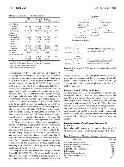
cations (upper 95% CI 4.7%; see Table 2). A total <strong>of</strong> 45<br />
(58%) aspirates were diagnostic for malignancy. Three false<br />
negatives were later discovered by intraoperative findings (n<br />
2) or P-FNA (n 1). One patient with pancreatic adenocarcinoma<br />
(as confirmed by EUS-FNA <strong>of</strong> the pancreas)<br />
had benign cytology <strong>of</strong> a 6-mm left lobe mass that intraoperatively<br />
was confirmed as metastatic adenocarcinoma. A<br />
second patient with pancreatic adenocarcinoma (as confirmed<br />
by EUS-FNA <strong>of</strong> the pancreas) had benign cytology<br />
<strong>of</strong> a 6-mm right lobe mass, which was later confirmed by<br />
percutaneous ultrasound-guided biopsy as metastatic adenocarcinoma.<br />
The third patient with a false negative EUS-FNA<br />
had a 29-mm left lobe mass that was benign by EUS-FNA.<br />
Intraoperative examination, however, confirmed hepatocellular<br />
carcinoma. Overall, 48 <strong>of</strong> 77 (62%) <strong>of</strong> the liver masses<br />
were malignant. Among the remaining 29 subjects with<br />
nonmalignant aspirates, in 22 (76%) the masses were considered<br />
benign by clinical follow-up (n 18; mean 762<br />
days; range: 512–1556 days) or intraoperative evaluation (n<br />
4). Seven (24%) <strong>of</strong> the subjects with nonmalignant<br />
masses with pancreatic adenocarcinoma (as confirmed by<br />
EUS-FNA <strong>of</strong> the pancreas; n 6) or lung cancer (n 1)<br />
died (mean 154 days, range 14–424 days) without follow-up<br />
imaging, biopsy <strong>of</strong> the liver, or autopsy and therefore<br />
cannot be classified as benign or malignant. The 45<br />
diagnostic aspirates for malignancy are considered true positives<br />
(Fig. 1). Assuming that the test results for these seven<br />
patients were all true negatives or all false negatives, the<br />
sensitivity <strong>of</strong> EUS-FNA for the diagnosis <strong>of</strong> malignancy<br />
would range from 82 to 94%.<br />
In comparison between benign and malignant lesions<br />
detected by EUS-FNA (Table 2), no statistically significant<br />
difference was found between the liver lobe aspirated (p <br />
0.57), number <strong>of</strong> passes (p 0.44), lesion size (p 0.70),<br />
Figure 1. Sensitivity <strong>of</strong> EUS-FNA <strong>of</strong> the liver for the diagnosis <strong>of</strong><br />
malignancy.<br />
or echotexture (p 0.36). Malignant masses, however,<br />
were more <strong>of</strong>ten accompanied by the presence <strong>of</strong> multiple<br />
hepatic lesions detected on EUS (38% vs 9%; p 0.03; 2<br />
analysis) and to have regular margins (60% vs 27%; p <br />
0.02; 2 analysis).<br />
Diagnoses from EUS-FNA <strong>of</strong> the Liver<br />
The final diagnoses for the 45 malignant liver aspirates are<br />
recorded in Table 3. Of these, 44 (98%) were metastatic and<br />
one (2%) was a hepatocellular carcinoma. The most common<br />
diagnosis was metastatic adenocarcinoma from the<br />
pancreas, which accounted for 34 <strong>of</strong> 45 (76%). The next<br />
most frequent diagnosis was metastatic neuroendocrine tumor<br />
from the pancreas (n 5; 11%). Of the 32 nonmalignant<br />
aspirates, 25 (33%) were cytologically benign and<br />
seven (9%) were nondiagnostic. One (4%) benign aspirate<br />
demonstrated cytological features consistent with an<br />
abscess.<br />
Previous Imaging <strong>of</strong> Malignancy Diagnosed by<br />
EUS-FNA<br />
Prior radiographic imaging reports were available for 42 <strong>of</strong><br />
45 (93%) malignant hepatic masses diagnosed by EUS-<br />
Table 3. Diagnoses <strong>of</strong> Malignant Aspirates <strong>of</strong> the Liver<br />
Diagnosis No. (%)<br />
Pancreatic adenocarcinoma 34 (76)<br />
Pancreatic neuroendocrine tumor 5 (11)<br />
Renal cell carcinoma 2 (5)<br />
Gallbladder adenocarcinoma 1 (2)<br />
Colon cancer 1 (2)<br />
Hepatocellular carcinoma 1 (2)<br />
Esophageal adenocarcinoma 1 (2)<br />
Total 45