stromatolites and calcareous algae of münder formation
stromatolites and calcareous algae of münder formation
stromatolites and calcareous algae of münder formation

You also want an ePaper? Increase the reach of your titles
YUMPU automatically turns print PDFs into web optimized ePapers that Google loves.
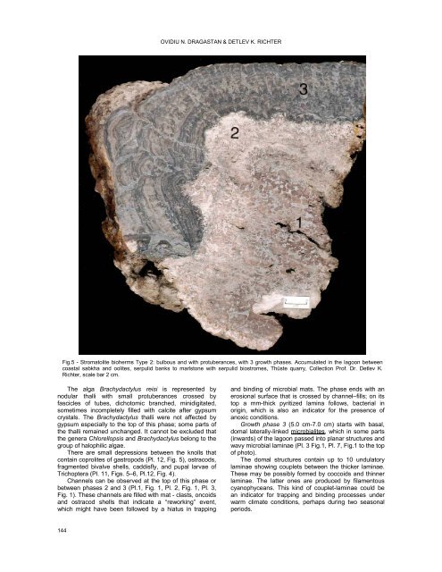
OVIDIU N. DRAGASTAN & DETLEV K. RICHTER<br />
Fig.5 - Stromatolite bioherms Type 2: bulbous <strong>and</strong> with protuberances, with 3 growth phases. Accumulated in the lagoon between<br />
coastal sabkha <strong>and</strong> oolites, serpulid banks to marlstone with serpulid biostromes, Thüste quarry, Collection Pr<strong>of</strong>. Dr. Detlev K.<br />
Richter, scale bar 2 cm.<br />
The alga Brachydactylus reisi is represented by<br />
nodular thalli with small protuberances crossed by<br />
fascicles <strong>of</strong> tubes, dichotomic branched, minidigitated,<br />
sometimes incompletely filled with calcite after gypsum<br />
crystals. The Brachydactylus thalli were not affected by<br />
gypsum especially to the top <strong>of</strong> this phase; some parts <strong>of</strong><br />
the thalli remained unchanged. It cannot be excluded that<br />
the genera Chlorellopsis <strong>and</strong> Brachydactylus belong to the<br />
group <strong>of</strong> halophilic <strong>algae</strong>.<br />
There are small depressions between the knolls that<br />
contain coprolites <strong>of</strong> gastropods (Pl. 12, Fig. 5), ostracods,<br />
fragmented bivalve shells, caddisfly, <strong>and</strong> pupal larvae <strong>of</strong><br />
Trichoptera (Pl. 11, Figs. 5–6, Pl.12, Fig. 4).<br />
Channels can be observed at the top <strong>of</strong> this phase or<br />
between phases 2 <strong>and</strong> 3 (Pl.1, Fig. 1, Pl. 2, Fig. 1, Pl. 3,<br />
Fig. 1). These channels are filled with mat - clasts, oncoids<br />
<strong>and</strong> ostracod shells that indicate a “reworking” event,<br />
which might have been followed by a hiatus in trapping<br />
<strong>and</strong> binding <strong>of</strong> microbial mats. The phase ends with an<br />
erosional surface that is crossed by channel–fills; on its<br />
top a mm-thick pyritized lamina follows, bacterial in<br />
origin, which is also an indicator for the presence <strong>of</strong><br />
anoxic conditions.<br />
Growth phase 3 (5.0 cm-7.0 cm) starts with basal,<br />
domal laterally-linked microbialites, which in some parts<br />
(inwards) <strong>of</strong> the lagoon passed into planar structures <strong>and</strong><br />
wavy microbial laminae (Pl. 3 Fig.1, Pl. 7, Fig.1 to the top<br />
<strong>of</strong> photo).<br />
The domal structures contain up to 10 undulatory<br />
laminae showing couplets between the thicker laminae.<br />
These may be possibly formed by coccoids <strong>and</strong> thinner<br />
laminae. The latter ones are produced by filamentous<br />
cyanophyceans. This kind <strong>of</strong> couplet-laminae could be<br />
an indicator for trapping <strong>and</strong> binding processes under<br />
warm climate conditions, perhaps during two seasonal<br />
periods.<br />
144