stromatolites and calcareous algae of münder formation
stromatolites and calcareous algae of münder formation
stromatolites and calcareous algae of münder formation

Create successful ePaper yourself
Turn your PDF publications into a flip-book with our unique Google optimized e-Paper software.
ACTA PALAEONTOLOGICA ROMANIAE V. 7 (2011), P. 139-168<br />
STROMATOLITES AND CALCAREOUS ALGAE OF MÜNDER FORMATION (TITHONIAN-BERRIASIAN)<br />
FROM NW GERMANY<br />
OVIDIU N. DRAGASTAN 1 & DETLEV K. RICHTER 2<br />
Abstract. The Tithonian <strong>stromatolites</strong> <strong>of</strong> Münder Formation, from Thüste locality (Hils Syncline), NW Germany were<br />
intensively investigated in the last years, as an account <strong>of</strong> their special environment <strong>of</strong> <strong>formation</strong> (Jahnke & Ritzkowski<br />
1980, Dragastan & Richter 2001, Arp et al. 2008).<br />
Descriptions <strong>and</strong> interpretations <strong>of</strong> this paper are focused on the <strong>stromatolites</strong> <strong>of</strong> the lowermost Katzberg Member<br />
(uppermost Tithonian). The <strong>stromatolites</strong> developed in a lagoon with various energy environments, between coastal<br />
sabkha (supra-tidal salt flat) facies <strong>and</strong> intertidal to subtidal facies, including serpulid-reef banks <strong>and</strong> oolitic bars, that<br />
functioned as a "barrier isl<strong>and</strong>" (Dragastan & Richter 2001). Two kinds <strong>of</strong> stromatolite bioherms were described: Type 1<br />
formed in intertidal <strong>and</strong> Type 2 formed in intertidal to supratidal environments. Serpulid reef-banks are more or less the<br />
equivalent <strong>of</strong> oolitic bars, being composed <strong>of</strong> a mass <strong>of</strong> tiny tubes up to 60 % <strong>of</strong> Serpula coacervata (BLUMENBACH 1803)<br />
SCHÖNFELD 1979, during successive growth stages.<br />
In several slabs, the stromatolite bioherms contain diverse morphologies <strong>and</strong> populations characterized by three growth<br />
phases or stages:<br />
- growth phase 1, the basal part composed <strong>of</strong> small (0. 250-0.500 mm in diameter), spheroidal to ellipsoidal microbial<br />
oncolites with serpulid-tubes nuclei <strong>and</strong> few, 2 up to 3 microbial laminae, reduced in thickness. Chlorellopsis coloniata<br />
REIS is rarely present, as bi<strong>of</strong>ilms; in our opinion it is a green alga; however its origin <strong>and</strong> taxonomy are still under<br />
debate.<br />
The initial substratum for the stromatolite bioherms were the serpulid-reef banks <strong>and</strong> biostroms. The tubes <strong>of</strong> serpulids<br />
were selectively transported according to their sizes; in most <strong>of</strong> the cases, they represented nuclei for microbial oncoids.<br />
On the other h<strong>and</strong> it is interesting to specify that very few (pyritized) ooids were found in the stromatolite bioherms, fact<br />
which is an indication that oolities were not in direct "connection" with the <strong>stromatolites</strong> <strong>and</strong> did not serve as substratum.<br />
Biomats occur as mm-sized deposits, while oncolites are found also over large areas on the channels floor.<br />
- growth phase 2 contains microbial macrooncoids (up to 2 cm in diameter) in the lower part, with serpulid-tubes nuclei<br />
fixed by the substratum <strong>and</strong> growing up to form microbial mounds or knolls.<br />
To the upper part, the microbial mounds are consisting <strong>of</strong> up to 10 laminae, thinner in the lower part <strong>and</strong> thicker to the<br />
top <strong>of</strong> this phase. During this phase the green <strong>algae</strong> contributed also to building the edifice <strong>of</strong> <strong>stromatolites</strong>:<br />
Chlorellopsis coloniata REIS (rarely) <strong>and</strong> Brachydactylus reisi DRAGASTAN & RICHTER (frequently). Sometimes, between<br />
the knolls appear vertical "cracking - pockets" filled up with reworked materials (mat debris, intraclasts) <strong>and</strong> small<br />
depressions filled with few coprolites <strong>of</strong> gastropods, ostracods, bivalves fragments <strong>and</strong> caddisfly pupal larvae <strong>of</strong><br />
Trichoptera. This phase ends with a clear erosional surface with partly oxidized framboidal pyrites (bacterial in origin),<br />
which point out to anoxic conditions. Some channels are distributed towards the top <strong>of</strong> growth phase 2 <strong>and</strong> document a<br />
transition zone between the growth phases 2 <strong>and</strong> 3.<br />
- growth phase 3 begins with basal domal laterally linked <strong>stromatolites</strong>, which l<strong>and</strong>wards <strong>of</strong> the lagoon pass into a<br />
tabular stromatolitic structure composed more or less by horizontal, parallel microbial laminae. To the top <strong>of</strong> this final<br />
phase the <strong>stromatolites</strong> present only planar wavy, pustular microbial laminae crossed by upward cracks filled with<br />
coarsely crystalline calcite, rarely ostracods, irregular fenestral fabrics, Brachydactylus sp. (green <strong>algae</strong>),<br />
Pseudorothpletzella" sp. (possible blue-green <strong>algae</strong>) <strong>and</strong> frequently caddisfly cases.<br />
The uppermost Tithonian <strong>stromatolites</strong> <strong>of</strong> Thüste are built mainly by microbial organisms, but also with the contribution<br />
<strong>of</strong> green <strong>algae</strong> such as Chlorellopsis coloniata, Brachydactylus reisi, Brachydactylus.sp. <strong>and</strong> possibly the alga<br />
Pseudorothpletzella sp. in a lagoon with intertidal to supratidal environments under warm climate <strong>and</strong> phases with two<br />
seasonal periods. The growth phases 1 <strong>and</strong> 2 indicate an intertidal depositional facies while growth phase 3<br />
corresponds to supratidal facies. The facies model for Thüste <strong>stromatolites</strong> can be compared with the Recent facies<br />
model <strong>of</strong> the coastal sabkha (supratidal salt flat) from the Trucial Coast (Abu Dhabi), Arabian - Persian Gulf.<br />
The Berriasian deposits <strong>of</strong> Borberg Member, Münder Formation contain marlstone, micritic limestone, bindstone,<br />
oncoids with serpulid-tubes nuclei, <strong>calcareous</strong> <strong>algae</strong>, serpulids <strong>and</strong> ostracods. The <strong>calcareous</strong> <strong>algae</strong> were described<br />
from Deister area, near Springe. Thalli fixed in some cases on serpulid tubes, have hemisphaeroidal, sphaeroidal or<br />
planar shapes <strong>and</strong> belong to cyanophycean <strong>and</strong> chlorophycean <strong>algae</strong>.<br />
The following taxa are re-described <strong>and</strong> described: Springerella bifurcata DRAGASTAN & RICHTER 2001, S. fuchtbaueri<br />
DRAGASTAN & RICHTER 2001, S.westphalica nov. sp., Deisterella germanica nov. gen. nov. sp. (Chlorophyta) <strong>and</strong><br />
Rivularia lissaviensis (BORNEMANN 1887) DRAGASTAN 1985 (Cyanophyta).<br />
The <strong>calcareous</strong> algal assemblage <strong>of</strong> Borberg Member corresponds to lacustrine-brackish marine environments. The<br />
eulittoral freshwater-oligohaline Theriosynoecum ostracods association with <strong>calcareous</strong> <strong>algae</strong> in lith<strong>of</strong>acies 4 included<br />
species <strong>of</strong> genus Springerella <strong>and</strong> Rivularia lissaviensis; sublittoral-miohaline Mantelliana ostracods association<br />
contains only Deisterella germanica nov. gen.nov. sp., in lith<strong>of</strong>acies 5, sensu Arp & Mennerich (2008).<br />
Keywords: Stromatolites, <strong>calcareous</strong> <strong>algae</strong>, new taxa, Tithonian-Berriasian, depositional facies, NW Germany.<br />
INTRODUCTION<br />
The term stromatolite introduced by Kalkowsky (1908)<br />
has now a full recognition as reffering to deposits <strong>of</strong><br />
organo-sedimentary nature <strong>and</strong> including the interpretation<br />
<strong>of</strong> their genesis - i.e. more or less a combination between<br />
biological <strong>and</strong> environmental factors responsible for<br />
morphologies <strong>and</strong> lamination types.<br />
Supratidal <strong>and</strong> intertidal ranges with ephemeral pools<br />
influenced by water supplies induced a special<br />
mineralogy <strong>and</strong> chemistry in various environmental<br />
climates (Horodyski & Van der Haar 1975).<br />
In 1987 Burne & Moore introduced a new category <strong>of</strong><br />
1<br />
University <strong>of</strong> Bucharest, Department <strong>of</strong> Geology <strong>and</strong> Paleontology, Bd. Nicolae Bǎlcescu no.1, 010041 Bucharest, Romania, e-mail:<br />
ovidiud@geo.edu.ro<br />
2<br />
Ruhr - Universität Bochum, Instit. für Geologie, Mineralogie und Geophysik, Lehrstuhl für Sediment und Isotopegeologie, Universitätstr.<br />
no.150, D – 44801 Bochum, Germany, e-mail: Detlev.Richter@rub.de<br />
139
OVIDIU N. DRAGASTAN & DETLEV K. RICHTER<br />
organo-sedimentary deposits, microbialite, related mainly<br />
to microbial carbonate <strong>formation</strong>s. Episodic sedimentation<br />
in relation to periodical microbial growth will produce a<br />
good lamination, characteristic for fine-grained, well<br />
laminated <strong>stromatolites</strong> (Riding 1991). Fine-grained<br />
agglutinated <strong>stromatolites</strong> are dominantly prokaryotic<br />
(bacterial <strong>and</strong> cyanophycean) in origin, being<br />
characteristic for the shallow flat environment composed <strong>of</strong><br />
domal <strong>and</strong> planar-flat, sometimes crinkled laminae.<br />
Microbial mats appear in a variety <strong>of</strong> morphologies as<br />
determined by the dominant species <strong>of</strong> cyanophycean <strong>and</strong><br />
the specific environmental factors. Classical mat types can<br />
be found in the intertidal zone <strong>of</strong> Abu Dhabi, Persian Gulf,<br />
where the tidal conditions provide different environmental<br />
deposits (Golubić 1992). Microbial laminae show irregular<br />
pores or fenestrae <strong>and</strong> radial convex upward cracks filled<br />
with coarsely crystalline calcite or gypsum (?) in case <strong>of</strong><br />
Thüste <strong>stromatolites</strong> (Arp et al. 2008). Sometimes, the<br />
mineral incorporation obliterates the biological structure<br />
(Golubić 1983), but not completely, the initial structures<br />
are still preserved by the evaporative intertidal<br />
environments.<br />
Organisms contributing to the deposition <strong>of</strong> microbial<br />
mats can be assigned to moderate halophiles, e.g.<br />
cyanobacteria, bacteria <strong>and</strong> to extremely halophilic taxa,<br />
e.g. green <strong>algae</strong> <strong>and</strong> halobacteria (Gerdes et al. 2000).<br />
The microbial carbonate <strong>formation</strong>s can be related to the<br />
following main <strong>calcareous</strong> microbialite types:<br />
<strong>stromatolites</strong> <strong>and</strong> oncolites, both found in marine,<br />
freshwater as well as lagoonal brackish environments.<br />
Near the Jurassic–Cretaceous boundary a large<br />
Purbeck inl<strong>and</strong> sea extended from Dorset (Engl<strong>and</strong>),<br />
Netherl<strong>and</strong>s <strong>and</strong> to northwestern Germany, covering the<br />
area <strong>of</strong> the so-called Lower Saxony Basin. The Purbeck<br />
facies contained marlstones, limestone deposits (oolites,<br />
serpulid-reef banks) <strong>and</strong> stromatolite intercalations. Two<br />
locations <strong>and</strong> subunits from the Münder Formation were<br />
re-studied by us: the first one is Katzberg Member,<br />
uppermost Tithonian in age from Thüste, a quarry <strong>of</strong><br />
Schütte Company <strong>and</strong> the second one is Borberg<br />
Member, Berriasian in age, from Deister area, near<br />
Springe (Fig.1 A <strong>and</strong> B).<br />
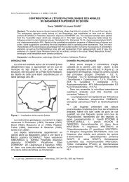
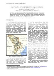
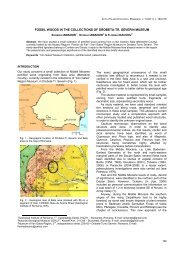
Fig.1 - A. Paleogeographic map showing the distribution <strong>of</strong> Late Jurassic to Early Cretaceous deposits (Purbeck facies) <strong>of</strong> the<br />
Portl<strong>and</strong> Trough, NW Germany ( from Betz et al. 1987).; B. Sketch map with the studied area located southeast <strong>of</strong> Hannover: 1.<br />
Thüste <strong>stromatolites</strong>, 2. Deister <strong>calcareous</strong> <strong>algae</strong>, near Springe.<br />
140
STROMATOLITES AND CALCAREOUS ALGAE OF MÜNDER FORMATION (TITHONIAN-BERRIASIAN) FROM NW GERMANY<br />
Fig.2 - Lithostratigraphy <strong>of</strong> Münder Formation ( Hils Syncline ) with data for (1) after Wolfart (1956), Casey et al. (1975), Waldeck<br />
(1975), Harms (1964), Jordan (1994), Gramann et al. (1997) & Deutsche Stratigraphische Kommission (2002) <strong>and</strong><br />
chronostratigraphy (2) after Remane et al. (2000), Arp et al. (2008),* 1. Thüste <strong>stromatolites</strong>, *2. Deister <strong>calcareous</strong> <strong>algae</strong>.<br />
The goal <strong>of</strong> this study is to contribute with new data on<br />
the role <strong>of</strong> prokaryotic (bacteria <strong>and</strong> cyanophycean) <strong>and</strong><br />
eukaryotic (chlorophycean) organisms to the genesis <strong>of</strong><br />
Thüste <strong>stromatolites</strong> <strong>and</strong> to describe the <strong>calcareous</strong> <strong>algae</strong><br />
from Deister area, including some palaeoenvironmental<br />
considerations.<br />
GEOLOGICAL SETTING<br />
Uppermost Jurassic (Tithonian) to lowermost Cretaceous<br />
(Berriasian) sequences containing serpulids, <strong>algae</strong> <strong>and</strong><br />
sometimes, ooids are typical <strong>of</strong> the final carbonate facies<br />
<strong>of</strong> the E-W striking Portl<strong>and</strong> Trough in northwestern<br />
Germany (Fig. 1). On the southern coast <strong>of</strong> the trough the<br />
two investigated areas Thüste (1) <strong>and</strong> Deister, near<br />
Springe (2) belong to the NW-SE striking Hils Bay. The<br />
configuration <strong>of</strong> this bay was predestinated by a<br />
halotectonic depression formed during the Late Jurassic to<br />
Early Cretaceous (Jordan 1994). The sequences <strong>of</strong> the socalled<br />
Münder Mergel correspond to the environmental<br />
change from the marine Eimbeckhausen Formation to<br />
the nonmarine Bückeburg Formation (Fig. 2). The marl<br />
dominated sequence shows variable thicknesses, between<br />
150 <strong>and</strong> 800 m due to synsedimentary salt tectonics<br />
(lithostratigraphy adapted from Arp et al. 2008).<br />
The first description <strong>of</strong> the serpulids-rich limestones<br />
from the southern coast <strong>of</strong> the Portl<strong>and</strong> Trough was<br />
published by Blumenbach (1803). Later on the<br />
environmental facies including salinity was discussed<br />
several times due to faunal <strong>and</strong> floral assemblages <strong>and</strong><br />
to the distribution <strong>of</strong> salt minerals (e.g. Huckriede 1967,<br />
Gramann et al. 1997). In the last years facies models for<br />
the serpulid-algal outcrops <strong>of</strong> the Hils <strong>and</strong> Deister were<br />
described by Jahnke & Ritzkowski (1980), Ten Hove &<br />
Van den Hurk (1993), Dragastan & Richter (2001) <strong>and</strong><br />
Arp et al. (2008). The localities <strong>of</strong> our study represent<br />
the algal dominated facies <strong>of</strong> the Münder Formation <strong>of</strong><br />
the coastal zone <strong>of</strong> the Portl<strong>and</strong> Trough situated<br />
southwest-south to Hannover (Fig.1).<br />
The Münder Formation (Fig. 2) was divided into<br />
three subunits: in the lower part, the Münder Mergel<br />
Member with marls, oolites <strong>and</strong> serpulids, in the middle<br />
part the Katzberg Member with marlstones <strong>and</strong><br />
<strong>stromatolites</strong> (uppermost Tithonian) followed by the<br />
upper Borberg Member (Berriasian) equivalent <strong>of</strong><br />
Purbeck facies composed <strong>of</strong> marlstones <strong>and</strong> micritic<br />
141
OVIDIU N. DRAGASTAN & DETLEV K. RICHTER<br />
limestones, also with serpulids.<br />
We suggest to change the name <strong>of</strong> Münder Mergel<br />
Member because it has the same name with the whole<br />
Formation, which is in contradiction with IGS rules<br />
becoming invalid.<br />
Locality 1 - Stromatolitic horizon <strong>of</strong> Katzberg Member,<br />
old quarry <strong>of</strong> Schütte Company, ESE <strong>of</strong> Thüste village;<br />
MTB 3923 Salzhemmendorf, H 576565, R 3545100. The<br />
new quarry directly to the west shows the oolitic<br />
limestones with serpulids <strong>of</strong> the upper Münder Mergel<br />
Member (see Fig. 2).<br />
Locality 2 - Purbeck limestones <strong>of</strong> Borberg Member;<br />
three quarries in the Deister Mountains, north <strong>of</strong> Springe;<br />
MTB 3723 Springe; a. H 5789800, R 3538160; b. H<br />
5790210, R 3538480; c. H 5790370, R 3538070<br />
(Dragastan & Richter 2001).<br />
DESCRIPTION OF THÜSTE STROMATOLITES<br />
The <strong>stromatolites</strong> samples from Richter Collection (quarry<br />
<strong>of</strong> Schütte Co.) consist <strong>of</strong> vertical polished slabs <strong>and</strong><br />
oriented thin sections. The dimensions <strong>of</strong> <strong>stromatolites</strong><br />
bioherms are variable between 10 up to 30 cm in width<br />
<strong>and</strong> 10 up to 20 cm in high, as vertical thickness. The<br />
position <strong>of</strong> vertical thin sections is shown in Figure 3.<br />
Two kinds <strong>of</strong> stromatolite bioherms were defined: Type<br />
1, compact-flat, wavy on the surface formed by 4 growth<br />
phases (Fig. 4) <strong>and</strong> Type 2, largeer, bulbous <strong>and</strong> with<br />
protuberances which contain 3 growth phases (Fig. 5).<br />
The different morphologies <strong>of</strong> the stromatolite bioherms<br />
were controlled by the physical environment <strong>and</strong> the<br />
corresponding biological (microbial <strong>and</strong> algal) responses<br />
to that environment.<br />
The first type bioherms with a distinctive inner structure<br />
correspond to a shallow intertidal facies (intertidal algal<br />
flat) accumulated in the lagoon, but disposed seaward on<br />
the serpulid - reef banks <strong>and</strong> oolitic bars, possibly on both<br />
sides <strong>of</strong> the barrier (serpulids <strong>and</strong> oolites). The second<br />
type formed l<strong>and</strong>wards from the lagoon, in shallow<br />
intertidal up to supratidal facies.<br />
The first type stromatolite bioherms are composed <strong>of</strong><br />
a basal growth phase (1) with oncoids <strong>and</strong> serpulid nuclei<br />
followed by phase (2) with columnar, simple or laterally<br />
branched structure; a minor discontinuity to the top <strong>of</strong> this<br />
phase indicates a subaerial exposure with reworked mats.<br />
This discontinuity was followed by growth phase (3), which<br />
contains columnar to domal inner structures; the final<br />
growth phase (4) shows a structure containing only domal<br />
to planar, wavy microbial laminae (Fig. 4).<br />
The second type <strong>of</strong> stromatolite bioherms contains 3<br />
growth phases <strong>and</strong> it is described below, in the subchapter<br />
on internal structure <strong>of</strong> <strong>stromatolites</strong>, because it is more<br />
widespread in the lagoon than the first type disposed<br />
towards the seaward part <strong>of</strong> the lagoon.<br />
The substratum <strong>of</strong> stromatolite bioherms is not so<br />
easy to define, because <strong>of</strong> its transitional facies from the<br />
oolites to serpulid-reef banks <strong>and</strong> from these to marlstone<br />
beds (Fig. 6). Depending on shore line pr<strong>of</strong>iles, the<br />
substratum <strong>of</strong> Thüste <strong>stromatolites</strong> was represented by<br />
either the serpulid-reef banks, or in most <strong>of</strong> the cases by<br />
the marlstone facies. The marlstone facies contains small<br />
lenses <strong>of</strong> serpulid biostroms <strong>and</strong> is positioned on the top <strong>of</strong><br />
the serpulid-reef banks.<br />
The stromatolite bioherms frequently contain in their<br />
basal part tiny tubes <strong>of</strong> Serpula coacervata (BLUMENBACH<br />
1803) SCHÖNFELD 1979. The organisms <strong>of</strong> genus<br />
Serpula are not cemented to its substrate, but they are<br />
not mobile either, partially burying themselves in<br />
carbonatic muds or in s<strong>and</strong>y sediments. The species <strong>of</strong><br />
genus Serpula are suspension deposit feeders.<br />
The serpulid - reef banks contain a mass <strong>of</strong> up to 60<br />
% entire or fragmentary, sometimes, telescopate tubes<br />
<strong>of</strong> Serpula coacervata (after Ten Hove & Hurk 1993);<br />
they are not fully equivalent to the oolitic bar. The<br />
serpulid-reef banks with variable thickness from 1.0 up to<br />
6.0 m (Schönfeld 1979) were formed exclusively by<br />
tubes <strong>of</strong> Serpula disposed more or less in a micritic<br />
matrix, parallel to the bedding plane. Ooids occur<br />
extremely rarely in the stromatolite bioherms, suggesting<br />
that there was no direct relation between oolites <strong>and</strong> the<br />
substratum <strong>of</strong> <strong>stromatolites</strong>. In exchange, mat debris,<br />
(marly-limestone) intraclasts, serpulid tubes, rarely<br />
ostracod shells, were all stored to the basal part <strong>of</strong> the<br />
<strong>stromatolites</strong> <strong>and</strong> (partly) contributed to the initial growth<br />
phase 1.<br />
Internal structure <strong>of</strong> <strong>stromatolites</strong>.<br />
Studied in several slabs <strong>and</strong> in oriented thin sections,<br />
the stromatolitic bioherms showed diverse morphologies<br />
<strong>and</strong> populations, characteristic for three or four growth<br />
phases or stages, in relationship with the specific<br />
depositional environments. Here we describe in detail<br />
type 2 <strong>of</strong> stromatolite bioherms (Fig. 5 <strong>and</strong> Plates 1-7),<br />
which is relatively more extended in the lagoon,<br />
l<strong>and</strong>wards:<br />
Growth phase 1 (3.0-5.0 cm) in the basal part,<br />
consists <strong>of</strong> different particles (mat debris, marlylimestone<br />
intraclasts with diameters between 1. 0 mm up<br />
to 5.0 mm, <strong>and</strong> rarely ostracod shells <strong>and</strong> serpulid<br />
tubes).<br />
The main components <strong>of</strong> this growth phase are<br />
spheroidal to ellipsoidal microbial oncolites (with<br />
diameters from 0.25-0.50 up to 3.0 mm) with only<br />
serpulid tubes nuclei <strong>and</strong> 2 up to 5 microbial laminae,<br />
variable in thickness from 0.090 mm up to 0.15 mm. The<br />
spaces between oncoids are filled up with mat-debris,<br />
small intraclasts, rare ostracod shells <strong>and</strong> pyritized<br />
minicrusts. To the upper part <strong>of</strong> this phase fine, vertical<br />
or irregular cracks were formed during the lithification<br />
processes. The oncolites, radial calcitic ooids <strong>and</strong> shells<br />
represent, in this case, mobile built-ups.<br />
Rarely, bi<strong>of</strong>ilm-like Chlorellopsis coloniata REIS is<br />
present; this form, considered a green alga after Freytet<br />
(2000) <strong>and</strong> confirmed also in our opinion, is still under<br />
debate as far as origin <strong>and</strong> taxonomy are concerned.<br />
This species is not very abundant at this level, which<br />
contains small spheres <strong>of</strong> sparry calcite, 0.10-0.15 mm<br />
in diameter (Pl. 8, Figs. 1- 4).<br />
The boundary between growth phases 1 <strong>and</strong> 2<br />
corresponds to an easily-noticeable irregular plane; the<br />
oncoids <strong>of</strong> the two stages are also different in sizes.<br />
Growth phase 2 (4.0-5.0 cm) contains in the lower<br />
part microbial macrooncoids sensu Richter (1983a,<br />
1983b) with diameters up to 3.0 - 4.0 cm. The<br />
macrooncoids have spheroidal or ellipsoidal shape,<br />
sometimes more elongated on the vertical plane, when<br />
they incorporate 2 or 3 serpulid-nuclei. The<br />
macrooncoids present concentrical microbial laminae.<br />
The variable number <strong>of</strong> microbial laminae have different<br />
thicknesses, from 0. 050 to 0. 20 mm.<br />
142
STROMATOLITES AND CALCAREOUS ALGAE OF MÜNDER FORMATION (TITHONIAN-BERRIASIAN) FROM NW GERMANY<br />
Fig.3 - Location <strong>of</strong> the oriented thin sections within the studied stromatolite bioherms Type 2 from Thüste (not to scale).<br />
Fig.4 - Stromatolite bioherms Type 1: compact, flat, wavy at the surface, with 4 growth phases. Accumulated on the intertidal algal flat<br />
(intertidal facies) disposed seawards from the lagoon on serpulid-reef banks <strong>and</strong> oolitic bars, Thüste quarry, Collection Pr<strong>of</strong>. Dr.<br />
Detlev K. Richter, scale bar 2 cm.<br />
Given their large sizes <strong>and</strong> the moderately high level <strong>of</strong><br />
hydrodynamism, the macrooncoids were fixed to the<br />
substratum during the growth phase 1. After fastening on<br />
the substratum, the macrooncoids were covered by<br />
microbial laminae which continue to grow <strong>and</strong> to built<br />
mounds or knolls (Fig. 6). To the upper part <strong>of</strong> this phase,<br />
the microbial mounds (= knolls) contain up to 10 microbial<br />
laminae, parallel <strong>and</strong> wavy, thinner in the lower part (7<br />
laminae) <strong>and</strong> thicker to the top (3 or 4 laminae) - (Pl. 2<br />
Fig. 1, Pl. 3, Fig. 1, Pl. 5, Fig. 1).<br />
Additional to this phase contributed the green <strong>algae</strong><br />
Chlorellopsis coloniata REIS, rarely (Pl. 8 Fig.1 - 4) <strong>and</strong><br />
Brachydactylus reisi DRAGASTAN & RICHTER, frequently<br />
(Pl. 8, Figs. 5 -7), being disposed on the top <strong>of</strong> the layer,<br />
between the knolls.<br />
143
OVIDIU N. DRAGASTAN & DETLEV K. RICHTER<br />
Fig.5 - Stromatolite bioherms Type 2: bulbous <strong>and</strong> with protuberances, with 3 growth phases. Accumulated in the lagoon between<br />
coastal sabkha <strong>and</strong> oolites, serpulid banks to marlstone with serpulid biostromes, Thüste quarry, Collection Pr<strong>of</strong>. Dr. Detlev K.<br />
Richter, scale bar 2 cm.<br />
The alga Brachydactylus reisi is represented by<br />
nodular thalli with small protuberances crossed by<br />
fascicles <strong>of</strong> tubes, dichotomic branched, minidigitated,<br />
sometimes incompletely filled with calcite after gypsum<br />
crystals. The Brachydactylus thalli were not affected by<br />
gypsum especially to the top <strong>of</strong> this phase; some parts <strong>of</strong><br />
the thalli remained unchanged. It cannot be excluded that<br />
the genera Chlorellopsis <strong>and</strong> Brachydactylus belong to the<br />
group <strong>of</strong> halophilic <strong>algae</strong>.<br />
There are small depressions between the knolls that<br />
contain coprolites <strong>of</strong> gastropods (Pl. 12, Fig. 5), ostracods,<br />
fragmented bivalve shells, caddisfly, <strong>and</strong> pupal larvae <strong>of</strong><br />
Trichoptera (Pl. 11, Figs. 5–6, Pl.12, Fig. 4).<br />
Channels can be observed at the top <strong>of</strong> this phase or<br />
between phases 2 <strong>and</strong> 3 (Pl.1, Fig. 1, Pl. 2, Fig. 1, Pl. 3,<br />
Fig. 1). These channels are filled with mat - clasts, oncoids<br />
<strong>and</strong> ostracod shells that indicate a “reworking” event,<br />
which might have been followed by a hiatus in trapping<br />
<strong>and</strong> binding <strong>of</strong> microbial mats. The phase ends with an<br />
erosional surface that is crossed by channel–fills; on its<br />
top a mm-thick pyritized lamina follows, bacterial in<br />
origin, which is also an indicator for the presence <strong>of</strong><br />
anoxic conditions.<br />
Growth phase 3 (5.0 cm-7.0 cm) starts with basal,<br />
domal laterally-linked microbialites, which in some parts<br />
(inwards) <strong>of</strong> the lagoon passed into planar structures <strong>and</strong><br />
wavy microbial laminae (Pl. 3 Fig.1, Pl. 7, Fig.1 to the top<br />
<strong>of</strong> photo).<br />
The domal structures contain up to 10 undulatory<br />
laminae showing couplets between the thicker laminae.<br />
These may be possibly formed by coccoids <strong>and</strong> thinner<br />
laminae. The latter ones are produced by filamentous<br />
cyanophyceans. This kind <strong>of</strong> couplet-laminae could be<br />
an indicator for trapping <strong>and</strong> binding processes under<br />
warm climate conditions, perhaps during two seasonal<br />
periods.<br />
144
STROMATOLITES AND CALCAREOUS ALGAE OF MÜNDER FORMATION (TITHONIAN-BERRIASIAN) FROM NW GERMANY<br />
Fig.6 - Substratum, biogenic <strong>and</strong> sedimentary structures <strong>of</strong> stromatolite bioherms Type 2 <strong>and</strong> the evolution <strong>of</strong> growth phases (1 - 3),<br />
based on vertical slabs, Thüste quarry <strong>of</strong> Schütte Co. (not to scale).<br />
On the top <strong>of</strong> the domal microbialites a possible<br />
cyanophycean thin algal crust occurs, similar as inner<br />
morphology is concerned with the thallus-crust <strong>of</strong> the<br />
genus Pseudorothpletzella SCHLAGINTWEIT & GAWLICK 2007<br />
described here under the name <strong>of</strong> Pseudorothpletzella sp.<br />
(Pl. 11, Figs. 3, 5, Pl. 12, Fig. 3). Between the domal<br />
structures, in small depression also the alga<br />
Brachydactylus sp. (Pl. 11, Figs. 3 - 4) was noticed, with<br />
small thalli <strong>and</strong> only one or two fan-haped fascicles<br />
crossed by dichotomously branched tubes that differ<br />
from those <strong>of</strong> species B. reisi DRAGASTAN & RICHTER.<br />
Also, in the domal linked depressions ostracods shells,<br />
caddisfly pupal larvae (Pl. 11, Figs. 5 - 6), mat clasts <strong>and</strong><br />
microbial peloids are present. Towards the margin <strong>of</strong> this<br />
145
OVIDIU N. DRAGASTAN & DETLEV K. RICHTER<br />
stromatolitic bioherm, between the growth phase 2 <strong>and</strong> the<br />
basal part <strong>of</strong> growth phase 3 vertical, irregular channels<br />
can be observed (Pl. 1, Fig. 1, Pl. 2, Fig. 2). The channels<br />
are filled with microbial mat-clasts, intraclasts with algal<br />
fragments, oncoids <strong>and</strong> rarely radial calcitic ooid-clasts. To<br />
the top <strong>of</strong> this final phase, the <strong>stromatolites</strong> present only<br />
planar, wavy, pustular microbial laminae crossed by<br />
upward cracks, irregular fenestral fabric <strong>and</strong> rare ostracod<br />
shells (Pl. 2, Fig. 1, Pl. 7, Fig. 1).<br />
This top planar layer consists <strong>of</strong> microbial fine-grained<br />
agglutinated sediments <strong>and</strong> it is well laminated.<br />
The phase contains a variable number (from 14 up to<br />
20) <strong>of</strong> laminae. Here, two types <strong>of</strong> laminae were noticed:<br />
thicker – with thicknesses <strong>of</strong> 0.15 up to 0.60 mm, <strong>and</strong><br />
thinner – with thicknesses between 0.025 mm up to 0.10<br />
mm.<br />
As conclusion to this general presentation we can state<br />
the following:<br />
1. The uppermost Tithonian <strong>stromatolites</strong> <strong>of</strong> Thüste,<br />
Katzberg Member <strong>of</strong> Münder Formation were mainly built<br />
by the microbial organisms, with the contribution <strong>of</strong> green<br />
<strong>algae</strong> Chlorellopsis coloniata REIS, rarely found in growth<br />
phases 1 <strong>and</strong> 2, Brachydactylus reisi DRAGASTAN &<br />
RICHTER, frequently found in growth phase 2 <strong>and</strong><br />
Brachydactylus sp., rarely found in growth phase 3. The<br />
cyanophycean (?) thalli crusts <strong>of</strong> Pseudorhotpletzella sp.<br />
were rarely encountered in growth phases 2 <strong>and</strong> 3.(Pl. 11,<br />
Figs. 3, 5, Pl. 12, Fig. 3). In-between the domal link<br />
depressions coprolites <strong>of</strong> gastropods (Pl.12, Fig.5),<br />
caddishfly pupal larvae (Pl.11, Figs. 3, 5 - 6, Pl.12, Fig. 4),<br />
ostracods <strong>and</strong> bivalves shells, microbial mats occur. All<br />
the components contributed in small amounts to building<br />
the edifice <strong>of</strong> the stromatolitic bioherms.<br />
2. Two types <strong>of</strong> stromatolitic bioherms are described:<br />
first type (1) massive, compact with a flat, wavy surface<br />
composed <strong>of</strong> 4 growth phases with microbial columnar <strong>and</strong><br />
domal inner structure formed in the intertidal environments<br />
on the serpulid-reef banks <strong>and</strong> oolites. They are disposed<br />
towards the seaward edges <strong>of</strong> the lagoon or possibly to<br />
both edges <strong>of</strong> serpulid <strong>and</strong> oolites deposits which<br />
functioned as an isl<strong>and</strong> barrier; second type (2), larger,<br />
bulbous with protuberances containing three growth<br />
phases, with microbial oncoids (1), microbial<br />
macrooncoids <strong>and</strong> green <strong>algae</strong> (2), <strong>and</strong> domal to planar,<br />
wavy, pustular (3), inner structure.<br />
This type <strong>of</strong> bioherms, disposed inwards or l<strong>and</strong>wards<br />
<strong>of</strong> the lagoon, has developed in intertidal to supratidal<br />
environments.<br />
3. The stromatolitic bioherms present internally a<br />
variety <strong>of</strong> morphologies controlled by the physical<br />
environment (water movements <strong>and</strong> current actions)<br />
including a characteristic biological content.<br />
4. The internal morphologies <strong>of</strong> the stromatolitic<br />
bioherms showed that the organo-sedimentary<br />
associations from growth phases 1, 2, 3 <strong>and</strong> 4 contain<br />
microbial, green <strong>and</strong> blue-green <strong>algae</strong>, the latter ones as<br />
halophilic biota. The growth phases 1, 2, 3 <strong>and</strong> 4 from the<br />
first type (1) bioherm developed in a moderately highenergy<br />
environment, only on the intertidal algal-flat<br />
including both sides <strong>of</strong> the „isl<strong>and</strong> barrier” with columnar,<br />
simple or branched structures; to the top, this continued<br />
exclusively with the domal stuctures.<br />
The growth phases 1, 2, 3 from the second type (2)<br />
bioherm developed in a moderately low-energy<br />
environment, inward or l<strong>and</strong>ward <strong>of</strong> the lagoon is<br />
represented by domal or knolls structure, which<br />
correspond to the intertidal environment <strong>and</strong> planar,<br />
wavy, pustular structure to the supratidal environment.<br />
5. The growth phase 2 <strong>and</strong> the basal part <strong>of</strong> phase 3<br />
were crossed by channel systems filled with reworked<br />
materials from the <strong>of</strong>fshore-sea bottom <strong>and</strong> from the<br />
seaward edges <strong>of</strong> the lagoon.<br />
6. Between the growth phases 2 <strong>and</strong> 3, an erosional<br />
surface was outlines, as discontinuity or hiatus<br />
corresponding to the lack <strong>of</strong> accretionary processes.<br />
This surface, which was impregnated with framboidal<br />
pyrites <strong>of</strong> bacterial origin during an anoxic event, is<br />
probably a marker for a transitional phase from intertidal<br />
to supratidal environments.<br />
7. The marlstone with serpulid biostroms, the<br />
serpulid-reef banks <strong>and</strong> oolites were accumulated in<br />
intertidal to subtidal environments.<br />
8. The presence <strong>of</strong> two types <strong>of</strong> microbial laminae in<br />
the stromatolitic bioherms – some thicker (possibly<br />
coccoidal in origin) <strong>and</strong> others thinner (possible<br />
cyan<strong>of</strong>ilamentous in origin), could be an indicator for the<br />
fact that the microbialites <strong>and</strong> the associated populations<br />
evolved in a lagoon environment (<strong>of</strong> the tidal flat), under<br />
warm climate <strong>and</strong> perhaps during two-seasonal periods.<br />
9. The facies model <strong>of</strong> Thüste <strong>stromatolites</strong> including<br />
the serpulid-reef banks <strong>and</strong> oolites which functioned like<br />
an isl<strong>and</strong> barrier <strong>and</strong> associated deposits can be<br />
compared with the Recent facies model <strong>of</strong> the Trucial<br />
Coast (Abu Dhabi) from the Arabian/Persian Gulf (Fig.<br />
7).<br />
In the case <strong>of</strong> Thüste <strong>stromatolites</strong>, the coastal<br />
sabkha corresponds to supratidal salt-flat, including<br />
gypsum <strong>and</strong> anhydrite deposits (early diagenetic<br />
minerals, like widespread pyrites <strong>and</strong> dolomites) <strong>and</strong> to<br />
the lagoon with <strong>stromatolites</strong> disposed between the<br />
sabkha flat <strong>and</strong> the oolites-serpulid-reef banks up to<br />
marlstones with serpulid biostroms comparable with a<br />
barrier isl<strong>and</strong>, the last deposits being accumulated in<br />
intertidal to subtidal environments. The Thüste<br />
stromatolite bioherms were formed in the lagoon, in<br />
intertidal <strong>and</strong> supratidal enviroments.<br />
10. The serpulid-reef banks, the oolites <strong>and</strong><br />
marlstones with serpulid biostroms functioned, more or<br />
less, like a barrier isl<strong>and</strong>; associate deposits were<br />
accumulated, corresponding to intertidal <strong>and</strong> subtidal<br />
environments.<br />
11. In the case <strong>of</strong> environmental interpretations <strong>of</strong><br />
the Thüste <strong>stromatolites</strong>, the <strong>algae</strong> <strong>and</strong> bacteria are<br />
more sensitive than the assemblages <strong>of</strong> minerals,<br />
because <strong>of</strong> the direct contact <strong>of</strong> the organisms with the<br />
waters <strong>of</strong> the Portl<strong>and</strong> Basin, whereas the minerals, e.g.<br />
sulphates, pyrite, or dolomite have crystallized under<br />
diagenetic conditions – after sedimentation <strong>and</strong><br />
lithification.<br />
CALCAREOUS ALGAE OF DEISTER AREA, NEAR<br />
SPRINGE<br />
The Berriasian deposits <strong>of</strong> the Borberg Member contain<br />
marlstones, micritic limestones, bindstones, oncoids with<br />
serpulid-tubes nuclei, <strong>calcareous</strong> <strong>algae</strong>, serpulids <strong>and</strong><br />
ostracods. The deposits are also known as Purbeck<br />
limestones <strong>and</strong> marls; their thicknesses vary between 50<br />
m <strong>and</strong> 185 m (Fig. 2). The Borberg Member defined by<br />
Wolfart in Waldeck (1975) contains marls intercalated<br />
with micritic limestones.<br />
146
STROMATOLITES AND CALCAREOUS ALGAE OF MÜNDER FORMATION (TITHONIAN-BERRIASIAN) FROM NW GERMANY<br />
Fig.7 - Adapted Recent facies model <strong>of</strong> coastal sabkha (supratidal salt flat), lagoon <strong>and</strong> barrier isl<strong>and</strong> (intertidal to subtidal facies)<br />
from the Trucial Coast, Abu Dhabi (after Purser 1985, in Einsele 2000) reconsidered in the case <strong>of</strong> Thüste <strong>stromatolites</strong>.<br />
Arp & Mennerich (2008) investigated a typical section<br />
for this subunit, which contains six characteristic lith<strong>of</strong>acies<br />
types (Lithos 1-6), as follows: lith<strong>of</strong>acies 1 includes<br />
marlstones <strong>and</strong> limestones with grey nodules, laminated<br />
fabric <strong>and</strong> ostracods, considered as massive limestones;<br />
lith<strong>of</strong>acies 2 with bedded marlstones, mud cracks, stem<br />
<strong>and</strong> oogonia <strong>of</strong> charophycean <strong>and</strong> ostracods; lith<strong>of</strong>acies<br />
3 with dark-grey marlstones, ostracods, charophyceans<br />
<strong>and</strong> gastropods; lith<strong>of</strong>acies 4, also consisting <strong>of</strong> massive<br />
limestones, which contain <strong>calcareous</strong> <strong>algae</strong><br />
(cyanophyceans <strong>and</strong> chlorophyceans, based on our data),<br />
ostracods, bivalves, gastropods <strong>and</strong> rare oncoids;<br />
lith<strong>of</strong>acies 5 composed by grey, micritic limestones with<br />
ostracods, gastropods <strong>and</strong> <strong>calcareous</strong> <strong>algae</strong><br />
(chlorophyceans) <strong>and</strong> lith<strong>of</strong>acies 6 with grey, clayely<br />
marlstones with ostracods, miliolids <strong>and</strong> authigenic<br />
aggregates <strong>of</strong> pyrite.<br />
The <strong>calcareous</strong> <strong>algae</strong> <strong>of</strong> the Deister area include the<br />
following cyanophyceans: Rivularia lissaviensis<br />
(BORNEMANN 1887) DRAGASTAN 1985, Rivularia sp. <strong>and</strong><br />
chlorophyceans: Springerella bifurcata DRAGASTAN &<br />
RICHTER 2001, S. fuchtbaueri DRAGASTAN & RICHTER 2001,<br />
S. westphalica nov. sp. <strong>and</strong> Deisterella germanica<br />
nov.gen. nov.sp.<br />
The eulittoral freshwater-oligohaline association<br />
(Theriosynoecum ostracod association) from lith<strong>of</strong>acies 4<br />
contains the following <strong>algae</strong>: species <strong>of</strong> genus<br />
Springerella, Rivularia lissaviensis, Rivularia sp. In the<br />
sublittoral-miohaline association (Mantelliana ostracod<br />
association) from lith<strong>of</strong>acies 5 only the alga Deisterella<br />
germanica nov. gen. nov. sp. was found.<br />
Dragastan & Richter (2001) described some<br />
<strong>calcareous</strong> green <strong>algae</strong> (Chlorophyta) from Deister area,<br />
near Springe: genus Springerella with two new species,<br />
S. bifurcata <strong>and</strong> S. fuchtbaueri.<br />
The thalli <strong>of</strong> the <strong>algae</strong>, in most <strong>of</strong> the cases, were<br />
fixed on the serpulid tubes; they show various<br />
hemispherical or spheroidal shapes, when they<br />
completely surround one serpulid tube nucleus, or<br />
ellipsoidal shapes, when they include two serpulid-tubes<br />
nuclei (Pl. 10, Fig. 2), in case <strong>of</strong> Springerella species,<br />
<strong>and</strong> flat-planar crustose thalli, when they are fixed only<br />
on one, "upper" surface <strong>of</strong> the serpulid tubes, in case <strong>of</strong><br />
Rivularia lissaviensis (Pl. 9, Fig. 5, Pl. 10, Fig. 1).<br />
In this paper, the following taxa are redescribed or<br />
described: Springerella bifurcata DRAGASTAN & RICHTER<br />
2001, S. fuchtbaueri DRAGASTAN & RICHTER 2001, S.<br />
westphalica nov. sp., Deisterella germanica nov. gen.<br />
nov.sp. (Chlorophyta), Rivularia lissaviensis (BORNEMANN<br />
1887) DRAGASTAN 1985 <strong>and</strong> Rivularia sp. (Cyanophyta).<br />
The <strong>calcareous</strong> algal assemblage from Deister area<br />
corresponds to lacustrine-freshwater-brackish<br />
environments.<br />
PALEOALGOLOGICAL DESCRIPTION OF THÜSTE<br />
STROMATOLITES<br />
Phylum CHLOROPHYTA<br />
Genus Chlorellopsis REIS 1923<br />
Chlorellopsis coloniata REIS 1923<br />
Pl.8, Figs. 1-4<br />
1923 Chlorellopsis coloniata n.gen.n.sp. Reis, p.107,<br />
Pl. III, Figs. 1-2, 9, Pl. IV,<br />
Figs. 3, 6,Pl. V, Figs. 2 - 6 <strong>and</strong> Text - Fig 1, p. 105.<br />
1995 Problematicum Chlorellopsis, Arp, p. 83, Pl.17/ 6.<br />
1997 Ch. coloniata, Freytet, p.13, Pl.1, Figs. a - c.<br />
147
OVIDIU N. DRAGASTAN & DETLEV K. RICHTER<br />
1999 Ch. coloniata, Freytet et al., p.121, p.126, Figs. 8<br />
e - f.<br />
2000 Ch. coloniata, Freytet, p.11, Pl. I, Fig. c.<br />
2001 Ch. coloniata, Freytet et al., p.161, Pl. III, Figs. a<br />
- b.<br />
2001 Ch.coloniata, Dragastan & Richter, p.314, Figs<br />
12/ 1-2.<br />
2007 Ch. coloniata, Leggitt et al., p. 673 (mentioned<br />
without description).<br />
2008 Ch.coloniata, Arp et al., p. 1224 (mentioned<br />
without description).<br />
Paratypes: Pl. 8, Figs. 1-4, Collection MMPP<br />
(Micr<strong>of</strong>acies, Micropaleobotany, Paleobotany &<br />
Palynology), Laboratory <strong>of</strong> Paleontology, University <strong>of</strong><br />
Bucharest, No. 1187, 1188, 1189, Uppermost Tithonian,<br />
Thüste <strong>stromatolites</strong>, Katzberg Member, Münder<br />
Formation.<br />
Description: Thalli irregular, sometimes included in<br />
nodular microbial mats or in ellipsoidal oncoids with<br />
superficial laminae formed by microbial peloids (Pl. 8, Fig.<br />
4). Thalli contain spherical cells, variable in diameter<br />
between 0.10 mm <strong>and</strong> 0.15 mm, preserved in coarsely<br />
crystalline calcite.<br />
The spherical cells are surrounded by a micritic layer<br />
which is, in its turn surrounded by sparry polygonal or<br />
hexagonal networks, muff-like. The polygonal or<br />
hexagonal sparry network is an important character, which<br />
confers to this taxon an algal origin. The Recent genus<br />
Chlorella also presents a hexagonal network disposition<br />
around the spherical-ball cells; thus this feature <strong>of</strong> the<br />
fossil genus is very similar with that <strong>of</strong> this Recent taxon.<br />
This species was not so frequently found in growth<br />
phases 1 <strong>and</strong> 2 <strong>of</strong> the Thüste <strong>stromatolites</strong>.<br />
Dimensions in mm: size <strong>of</strong> thallus: 1.0-4.0; diameter<br />
<strong>of</strong> spherical-ball cells: 0.10-0.150.<br />
Remarks: in spite <strong>of</strong> its disputed origin, Chlorellopsis<br />
coloniata REIS is a frequent algal component <strong>of</strong> brackish<br />
<strong>and</strong> freshwater environments, mainly associated with<br />
stromatolite built-ups. A discussion <strong>and</strong> interpretation <strong>of</strong><br />
Ch. coloniata was presented in detail at page 11 by Freytet<br />
(2000) showing that: Chlorellopsis are never free in the<br />
sediments, they are always associated with stromatolitic<br />
built-ups.<br />
An argument in favour <strong>of</strong> the algal origin remains the<br />
hexagonal or polygonal outline (Pl. 8, Figs. 1-3)<br />
surrounded the spheric cells, like in the Recent genus<br />
Chlorella.<br />
The systematic position <strong>of</strong> this taxon is <strong>and</strong> remains<br />
controversial:<br />
‐ Reis (1923) concluded that this genus belongs to<br />
Chlorophyta, Order Protococcales, being compared with<br />
the marine genus Halosphaera <strong>and</strong> with freshwater genera<br />
Eremosphaera <strong>and</strong> Chlorella;<br />
‐ Nathan (1925) concluded that Chlorellopsis is an<br />
alga;<br />
‐ Bradley (1929) considered this taxon as a potential<br />
index for algal remains;<br />
‐ Bolten (1977) considered also that this taxon is an<br />
alga;<br />
‐ Stapf (1988) proposed for Chlorellopsis an algal<br />
origin;<br />
‐ Bertr<strong>and</strong>-Sarfati et al. (1994) <strong>and</strong> Arp (1995)<br />
assigned the genus Chlorellopsis to insect eggs or<br />
arthropods, but without arguments;<br />
‐ Lundquist (1994) considers that these thalli with<br />
spheres represent endogonaceous fungal spores;<br />
‐ Freytet (1997) shows that a precise attribution <strong>of</strong><br />
Chlorellopsis remains open; in 2000, the same author<br />
preferred the hypothesis <strong>of</strong> an algal origin;<br />
‐ Dragastan & Richter (2001) also assigned genus<br />
Chlorellopsis to the green <strong>algae</strong>.<br />
Stratigraphic range: Permian (?), Lower Triassic,<br />
Upper Jurassic (uppermost Tithonian <strong>of</strong> Thüste<br />
<strong>stromatolites</strong>), Eocene <strong>and</strong> Oligo-Miocene.<br />
Genus Brachydactylus REIS 1923<br />
Brachydactylus reisi DRAGASTAN & RICHTER 2001<br />
Pl. 8, Figs. 5 – 7<br />
2001 Brachydactylus reisi n.sp. Dragastan & Richter,<br />
p.314, Fig.12.1 <strong>and</strong> Fig.13.1-6.<br />
Holotype in 2001, Pl. 8, Fig. 5, Collection MMPP –<br />
Bucharest, No. 1138.<br />
Paratypes: Pl.8, Figs. 6 - 7, Collection MMPP –<br />
Bucharest, No. 1190 - 1191, uppermost Tithonian,<br />
Thüste <strong>stromatolites</strong>, Katzberg Member, Münster<br />
Formation.<br />
Description: Nodular thalli with small protuberances<br />
(see Holotype) sometimes covered by microbial laminae.<br />
Thalli composed <strong>of</strong> small fan-shaped bundles crossed by<br />
dichotomously branched tubes. To the distal ends, the<br />
tubes appear short <strong>and</strong> minidigitate as structure. In the<br />
transverse section, the fan-shaped bundles contain up to<br />
four fascicles, all together round in shape <strong>and</strong> looking<br />
like a cauliflower (Pl. 8, Figs. 6- 7). Some bundles<br />
preserved completely the inner structure, showing the<br />
radially disposed minidigitate tubes, the dichotomously<br />
branched remaining unaffected by the <strong>formation</strong> <strong>of</strong> postdiagenetic<br />
gypsum crystals (Pl.8, Fig. 7).<br />
In vertical section (Pl. 8, Fig. 6) the fan-shaped<br />
bundles are disposed bilaterally <strong>and</strong> present<br />
dichotomously branched tubes, which can be seen only<br />
to the distal ends <strong>of</strong> the fascicles. However, in some<br />
parts <strong>of</strong> the thalli-bundles, the terminal traces <strong>of</strong> the<br />
tubes can be observed (Pl. 8, Fig.7).<br />
Dimensions in mm: diameter <strong>of</strong> nodular thallus: 2.0<br />
- 3.0, diameter <strong>of</strong> the isolate protuberance: 1.0 -1.5,<br />
width <strong>of</strong> fan-shaped bundles: 0.50 - 0.65, heigth <strong>of</strong> the<br />
fan-shaped bundles: 0.35 - 0.50, proximal diameter <strong>of</strong><br />
dichotomously branched tubes: 0.040 - 0.060, distal<br />
diameter <strong>of</strong> dichotomously branched tubes: 0.010 -<br />
0.030.<br />
Remarks: Brachydactylus reisi DRAGASTAN &<br />
RICHTER 2001 was compared with, but differs from the<br />
Miocene Brachydactylus radialis REIS 1923, by the<br />
shape <strong>of</strong> the thallus – nodular with protuberances, by the<br />
disposition <strong>of</strong> the dichotomously tubes into fan-shaped<br />
bundles with fascicles <strong>of</strong> tubes <strong>and</strong> also by a lesser<br />
number <strong>of</strong> bundles in comparison with the Reis species,<br />
which presents a more compact inner thallus structure<br />
<strong>and</strong> has a different shape <strong>of</strong> the bundles.The<br />
Brachydactylus reisi was frequently identified in growth<br />
phase 2 <strong>of</strong> the Thüste <strong>stromatolites</strong>.<br />
Stratigraphic range: uppermost Tithonian, Katzberg<br />
Member, Münder Formation.<br />
Brachydactylus sp.<br />
Pl.11, Figs. 3-4<br />
Material: Two specimens, Collection MMPP-<br />
Bucharest, No. 1205-1206, uppermost Tithonian, Thüste<br />
148
STROMATOLITES AND CALCAREOUS ALGAE OF MÜNDER FORMATION (TITHONIAN-BERRIASIAN) FROM NW GERMANY<br />
<strong>stromatolites</strong>, Katzberg Member, Münder Formation.<br />
Description: Thallus very small, fan-shaped,<br />
corresponding to one bundle crossed by dichotomouslybranched<br />
tubes. The tubes are short <strong>and</strong> have a larger<br />
diameter in the base, before branching <strong>and</strong> smaller in the<br />
distal parts <strong>of</strong> the tubes.<br />
Dimensions in mm: heigth <strong>of</strong> thallus: 0.30-0.40, width<br />
<strong>of</strong> thallus: 0.40-0.50, proximal diameter <strong>of</strong> tubes before<br />
branching: 0.030-0.040 <strong>and</strong> distal diameter <strong>of</strong><br />
dichotomously branched tubes: 0.015-0.025.<br />
Remarks: This alga occurs rarely, only in the growth<br />
phase 3 <strong>of</strong> Thüste <strong>stromatolites</strong> bioherms Type 2. When<br />
comparing with species Brachydactylus radialis REIS <strong>and</strong><br />
B. reisi DRAGASTAN & RICHTER, this alga shows a very<br />
small incipient thallus with only one or two bundles<br />
crossed by dichotomously branched tubes. The alga grew<br />
on the top <strong>of</strong> microbial domal structures, between the<br />
laminae (Pl. 11, Fig. 3), but also in the middle <strong>of</strong> the<br />
”depressions” formed between the domal structures (Pl.11,<br />
Fig. 4).<br />
Stratigraphic range: uppermost Tithonian, Thüste<br />
<strong>stromatolites</strong>, Katzberg Member, Münder Formation.<br />
PALEOALGOLOGICAL DESCRIPTION OF DEISTER<br />
AREA<br />
Genus Springerella DRAGASTAN & RICHTER 2001<br />
Springerella bifurcata DRAGASTAN & RICHTER 2001<br />
Pl. 9, Figs. 1 - 4, Pl.10, Figs. 2 – 3<br />
2001 Springerella bifurcata nov. gen.nov. sp.<br />
Dragastan & Richter, p.313, Fig.10.1-3, Fig.11.1-4.<br />
Paratypes: Pl. 9, Figs.1 – 4, Pl.10, Fig. 2–3, Collection<br />
MMPP- Bucharest, No. 1192, 1193, 1194, 1194 a,<br />
Berriasian, Borberg Member, Münder Formation.<br />
Description: Nodular, spheroidal, ellipsoidal to<br />
hemispherical thallus. The morphology <strong>of</strong> the thalli is<br />
influenced by the shape <strong>of</strong> the nuclei <strong>and</strong> by the<br />
substratum. If the alga is attached on a single serpulidtube<br />
nucleus, the shape is spheroidal (Pl. 9, Fig. 2) while<br />
when it is attached on two serpulid-tubes nuclei the thallus<br />
become ellipsoidal (Pl. 10, Fig. 2). Sometimes the thalli<br />
present also hemispherical shapes (Pl. 9, Fig. 1).<br />
In vertical-longitudinal sections, the thalli are<br />
composed <strong>of</strong> long Y- shaped, open, dichotomously<br />
branched tubes having a strongly calcified swelling in the<br />
area <strong>of</strong> branching, <strong>and</strong> occasionally a swelling along the<br />
tubes between the branched areas (Pl. 9, Figs. 1-2). The<br />
swellings are ovoidal to ellipsoidal in shape, between the<br />
dichotomies <strong>of</strong> the tubes. The angle <strong>of</strong> divergence<br />
between the Y- branched tubes is variable, from 30° to 40°<br />
(Pl. 9, Figs. 3-4, Pl. 10, Fig. 3).<br />
In transverse sections, the tubes are disposed in a<br />
regular quadrangular or polygonal network, grouped into 6<br />
up to 8 tubes (Pl. 9, Figs. 1, 3 see in the lower part <strong>of</strong> the<br />
photo) <strong>and</strong> 4, in a broken thallus (Pl. 10, Fig. 2). The tubes<br />
show a petaloid, more or less regular disposition in<br />
transverse section <strong>of</strong> the broken thallus fragment (Pl. 9,<br />
Fig. 4 in the upper part <strong>of</strong> the photo). Also, in transverse<br />
section the swellings have a circular shape with large<br />
diameter; their appearance is similar to white sparitic spots<br />
(Pl. 9, Fig. 3 arrows).<br />
Dimensions in mm: maximum diameter <strong>of</strong> thallus:<br />
3.5-4.0, normal diameter <strong>of</strong> thallus: 2.20-3.0, diameter <strong>of</strong><br />
tubes in the branching area: 0.075-0.080, diameter <strong>of</strong> tube<br />
after the dichotomously branching area: 0.030-0.045,<br />
diameter <strong>of</strong> occasional swellings along the tubes: 0.040-<br />
0.050.<br />
Remarks: This species can be compared with<br />
Sarfatigirella fallacia FREYTET & VERRECHIA, 1998 from<br />
the Campanian (Late Cretaceous). The difference<br />
consists <strong>of</strong> the smaller diameter <strong>of</strong> its erect ”filaments” (=<br />
tubes) that are not undulose. The common features<br />
consist in the presence <strong>of</strong> the swellings, but they are not<br />
distributed in the branching area. The marine species<br />
Mitcheldeania americana (Johnson 1961) Dragastan<br />
1985 from the Late Jurassic <strong>of</strong> Family Avrainvilleaceae<br />
DRAGASTAN et al. 1999 non 1997, differs from<br />
Springerella bifurcata by the presence <strong>of</strong> siphons,<br />
dichotomously branched after an angle <strong>of</strong> divergence <strong>of</strong><br />
less than 10° <strong>and</strong> by the presence <strong>of</strong> many swellings<br />
along the siphons The marine species <strong>of</strong> genus<br />
Pseudomitcheldeania SCHLAGINTWEIT 1990, P.<br />
dragastani SCHLAGINTWEIT 1990 from the Upper Aptian ,<br />
P. akrokorinthiaca DRAGASTAN & RICHTER 1999 from the<br />
Tithonian <strong>of</strong> Acrocorinth (Greece) <strong>and</strong> P.sp. from the<br />
Valanginian <strong>of</strong> Ghilcoş Massif, Transylvanian Carbonate<br />
Platform (Dragastan et al. 1997) differ from S. bifurcata<br />
by the presence <strong>of</strong> many swellings along the siphons,<br />
variable in shape. All these marine species belong to the<br />
Family Avrainvillaceae DRAGASTAN et al. 1999, Class<br />
Bryopsidophyceae, Chlorophyta.<br />
Stratigraphic range: Berriasian <strong>of</strong> Dreister area,<br />
lith<strong>of</strong>acies 4, eulittoral, freshwater-oligohaline,<br />
Theriosynoecum ostracods association.<br />
Springerella fuchtbaueri DRAGASTAN & RICHTER 2001<br />
Pl.10, Figs. 4-5<br />
2001 Springerella bifurcata nov.sp. Dragastan &<br />
Richter, p. 313, Fig. 11. 5 - 7.<br />
2008 undescribed porostromate alga attached to a<br />
charophyte stem fragment, Arp & Mennerich, p. 23, Fig.<br />
4 C, lith<strong>of</strong>acies 4.<br />
Paratypes: Pl.10, Figs. 4 - 5, Collection MMPP-<br />
Bucharest, No. 1199, 1200, Berriasian, Borberg<br />
Member, Deister area, near Springe.<br />
Description: Thallus small, hemispheroidal to<br />
spheroidal crossed by claviform, dichotomously<br />
branched <strong>and</strong> strongly calcified tubes, with larger<br />
diameter to the distal part (Pl.10, Figs. 4-5). Thalli<br />
attached in many cases on the serpulid-tubes;<br />
sometimes, together also with thalli <strong>of</strong> Springerella<br />
bifurcata.<br />
Dimensions in mm: heigth <strong>of</strong> thallus: 0.30 - 0.70,<br />
width <strong>of</strong> thallus: 0.40-0.90, diameter <strong>of</strong> tubes in the<br />
proximal parts: 0.020-0.040, diameter <strong>of</strong> tubes in the<br />
distal parts: 0.050-0.090.<br />
Remarks: The new material <strong>of</strong> Springerella<br />
fuchtbaueri shows the claviform shape <strong>of</strong> tubes, but<br />
without swellings. The claviform shape <strong>of</strong> the tubes<br />
remains a characteristic feature <strong>of</strong> this species.<br />
Stratigraphic range: Berriasian <strong>of</strong> Deister area,<br />
lith<strong>of</strong>acies 4, eulittoral, freshwater -oligohaline,<br />
Theriosynoecum ostracods association.<br />
Springerella westphalica nov.sp.<br />
Pl. 10, Figs. 6 – 8<br />
Derivatio nominis: ”westphalica” from Westphalia,<br />
the German historical part (l<strong>and</strong>) <strong>of</strong> NW Germany.<br />
Holotype: Pl.10, Fig. 6, Collection MMPP-<br />
Bucharest, No. 1201, Berriasian <strong>of</strong> Borberg Member,<br />
149
OVIDIU N. DRAGASTAN & DETLEV K. RICHTER<br />
Deister area, near Springe.<br />
Isotypes: Pl. 10, Figs. 7 - 8, Collection MMPP -<br />
Bucharest, No. 1202, 1203, Berriasian <strong>of</strong> Borberg<br />
Member, Deister area, near Springe.<br />
Dimensions in mm: height <strong>of</strong> thallus: 1.50 - 2.0, width<br />
<strong>of</strong> thallus: 2.0-3.0 when they have lobes (diameter <strong>of</strong> lobe<br />
1.0); proximal diameter <strong>of</strong> tubes before branching: 0.030-<br />
0.040, distal diameter <strong>of</strong> tubes: 0.035-0.047, diameter <strong>of</strong><br />
tubes in transverse section: 0.040-0.050, diameter <strong>of</strong><br />
swellings: 0.060-0.090.<br />
Description: Thalli hemispheroidal or spheroidal in<br />
shape, sometimes with lobes, not very compact in the<br />
inner structure, crossed by very laxly disposed<br />
dichotomously branched tubes. The tubes have a large<br />
angle <strong>of</strong> divergence; their opening varies from 30 up to<br />
40°.<br />
In vertical-longitudinal section (Pl. 10, Fig. 6), the<br />
thallus is composed <strong>of</strong> dichotomously branched tubes with<br />
a very open angle <strong>of</strong> divergence. The tubes present<br />
swellings, which appear like large sparry calcite spots, oval<br />
to ellipsoidal in shape. In the oblique-longitudinal section<br />
(Pl. 10, Fig. 7) the thallus is also crossed by dichotomously<br />
branched tubes.<br />
The tubes present obvious sparitic oval to ellipsoidal<br />
swellings, located very close to each other. In transverse<br />
section (Pl.10, Fig. 8), the thallus has a round shape with<br />
some lobes; it presents the lax disposition <strong>of</strong> the tubes,<br />
with large spaces in-between. Because <strong>of</strong> the lax<br />
disposition, there are only four tubes arranged into a more<br />
or less quadrangular, or even irregular ”network” (Pl. 10,<br />
Fig. 8).<br />
Remarks: The new species is comparable with the<br />
stock <strong>of</strong> Springerella species. The differences from the<br />
species <strong>of</strong> genus Springerella already described are<br />
represented by the lax inner structure <strong>of</strong> the thalli with<br />
lesser dichotomously branched tubes, <strong>and</strong> the large angle<br />
<strong>of</strong> divergence <strong>and</strong> the quadrangular up to irregular<br />
disposition <strong>of</strong> tubes – feature visible in transverse section.<br />
The presence <strong>of</strong> swellings along the tubes is a feature<br />
similar to that in Springerella bifurcata, but the distribution<br />
<strong>of</strong> the tubes with large spaces in-between <strong>and</strong> the<br />
quadrangular disposition represent the differences<br />
between these two species.<br />
S. fuchtbaueri differs from both species by the<br />
claviform shape <strong>of</strong> the tubes with large diameters in the<br />
distal parts <strong>and</strong> by the compact disposition <strong>of</strong> the tubes<br />
along the thallus. The new species is frequently attached<br />
on serpulid-tubes.<br />
Stratigraphic range: Berriasian <strong>of</strong> Deister area,<br />
lith<strong>of</strong>acies 4, eulittoral, freshwater-oligohaline,<br />
Theriosynoecum ostracods association.<br />
Deisterella nov.gen. DRAGASTAN & RICHTER<br />
Derivation nominis: from Deister Mountains, NW<br />
Germany.<br />
Type species: Deisterella germanica nov.gen.nov.sp.<br />
Diagnosis: Thallus hemispherical crossed by Y-<br />
shaped <strong>and</strong> V-shaped dichotomously branched tubes,<br />
which show a sparitic, elongate or passing to conical in<br />
shape swelling in area <strong>of</strong> branching,. In the lower part <strong>of</strong><br />
the thallus spheroidal ”calcitic bodies” - considered to<br />
represent possible reproductive organs - are disposed.<br />
Remarks: The new taxon is comparable with the<br />
marine genera Niteckiella Dragastan 1988 (Tithonian <strong>of</strong><br />
Bihor <strong>and</strong> Getic Carbonate Platforms) <strong>and</strong> Hansiella<br />
Dragastan 1990 (Late Oxfordian-Kimmeridgian <strong>of</strong> Getic<br />
Carbonate Platform) from Family Pseudoudoteaceae<br />
DRAGASTAN et al. 1997, Class Bryopsidophyceae,<br />
Chlorophyta revised by Dragastan (2002).<br />
Although these genera have different branched - V-<br />
<strong>and</strong> Y-type shaped tubes, they can be compared with<br />
the new taxon that presents similar but not identical<br />
branched tubes. It misses the fine, long, simple or<br />
bifurcate tubes present in the case <strong>of</strong> genus Hansiella<br />
<strong>and</strong> the more diversified inner structure with different<br />
branched tubes present in the case <strong>of</strong> genus Niteckiella.<br />
The new genus is morphologically similar with genus<br />
Hansiella, which has a thallus crossed by Y-shaped<br />
tubes, but also by V-shaped <strong>and</strong> long, fine, simple or<br />
bifurcate tubes.<br />
The Y- <strong>and</strong> V-shaped branched tubes <strong>and</strong> the<br />
spherical calcitic bodies – as possible reproductive<br />
organs –, provided a striking resemblance <strong>of</strong> the two<br />
genera.<br />
The new taxon can be compared also with freshwater<br />
genera Cladophorites REIS 1921 (Miocene ), Ries-<br />
Impact-Crater from southern Germany <strong>and</strong> Purserella<br />
FREYTET 1997 (Oligo-Miocene lake) from Limagne <strong>of</strong><br />
Allier, France, based on the presence <strong>of</strong> V- <strong>and</strong> Y-<br />
shaped dichotomously branched tubes; however it differs<br />
from these genera by the absence <strong>of</strong> spheroidal calcitic<br />
bodies in the latter. Both genera are considered green<br />
<strong>algae</strong> (Chlorophyta).<br />
Deisterella germanica nov.sp.<br />
(Pl. 11, Figs. 1 - 2)<br />
Derivatio nominis: ”germanica” from the Germanic<br />
ancient populations that inhabited the territory <strong>of</strong> present<br />
Germany.<br />
Holotype: Pl. 11, Fig.1, Collection MMPP-<br />
Bucharest, No. 1204, Berriasian <strong>of</strong> Borberg Member,<br />
Deister area, near Springe.<br />
Description: Thallus hemispherical attached on<br />
serpulid tubes <strong>and</strong> covered by thin microbial mats. The<br />
thallus is composed <strong>of</strong> Y- shaped dichotomously<br />
branched tubes with an angle <strong>of</strong> divergence between 30°<br />
up to 40° <strong>and</strong> <strong>of</strong> V - shaped dichotomously branched<br />
tubes with an angle <strong>of</strong> divergence between 10° up to<br />
20°; the last ones were more frequently identified in the<br />
composition <strong>of</strong> the thallus. At the branching point, a<br />
small conical or elongate swelling is visible, similar to a<br />
white spot. In the basal part <strong>of</strong> the thallus, between the<br />
Y- shaped dichotomous tubes the spheroidal ”calcitic<br />
bodies” considered as possible reproductive organs<br />
occur (Pl. 11, Fig. 2).<br />
Also in the basal part <strong>of</strong> the thallus, similar to the<br />
features <strong>of</strong> the transverse section, the disposition <strong>of</strong> the<br />
tubes in pentagonal or hexagonal frame can be noticed;<br />
each tube is set up to the angle <strong>of</strong> geometric disposition<br />
(Pl. 11, Fig.1, arrows).<br />
Dimensions in mm: height <strong>of</strong> thallus: 3.0-3.50,<br />
width <strong>of</strong> thallus: 2.0-2.50, diameter <strong>of</strong> tubes in the<br />
branching area: 0.045-0.070, diameter <strong>of</strong> dichotomic<br />
tubes: in the proximal part, 0.025-0.030 while in the<br />
distal part, 0.035-0.050-(0.090), diameter <strong>of</strong> calcitic<br />
bodies: 0.10-0.12.<br />
Remarks: Deisterella germanica nov.sp. differs from<br />
the marine species Niteckiella flabelliformis DRAGASTAN<br />
1988, by the presence <strong>of</strong> only two types <strong>of</strong> tubes<br />
dichotomously Y- <strong>and</strong> V - shaped branched, <strong>and</strong> by the<br />
150
STROMATOLITES AND CALCAREOUS ALGAE OF MÜNDER FORMATION (TITHONIAN-BERRIASIAN) FROM NW GERMANY<br />
presence <strong>of</strong> spheroidal calcitic bodies. N. flabelliformis<br />
shows several types <strong>of</strong> tubes <strong>and</strong> does not display<br />
spheroidal calcitic bodies. As compared with the marine<br />
species Hansiella fibrata DRAGASTAN 1990, which also has<br />
Y- <strong>and</strong> V-shaped dichotomously branched tubes, it also<br />
shows fine, long, simple or bifurcate, parallel tubes<br />
disposed in bundles <strong>and</strong> spherical calcitic bodies as<br />
reproductive organs. The only difference in the new<br />
species is the absence <strong>of</strong> fine, long, simple or bifurcate<br />
tubes disposed in bundles.<br />
Both marine species were found in the Tithonian<br />
(Niteckiella flabelliformis) <strong>of</strong> the Transylvanian, Getic <strong>and</strong><br />
Bihor Carbonate Platforms <strong>and</strong> in the Late Oxfordian-<br />
Kimmeridgian (Hansiella fibrata) <strong>of</strong> the Getic Carbonate<br />
Platform.<br />
The freshwater species Cladophorites incrustans<br />
(LUDWIG 1858) REIS 1921 showing hemispherical thallus<br />
crossed by V– shaped <strong>and</strong> rarely Y - shaped<br />
dichotomously branched tubes differs from the new taxon<br />
by a different disposition <strong>of</strong> the tubes in transverse section<br />
<strong>and</strong> by the absence <strong>of</strong> spheroidal calcitic bodies. In the<br />
Oligo-Miocene species Purserella gracilis FREYTET 1997,<br />
the differences consist <strong>of</strong> the disposition <strong>of</strong> dichotomic<br />
tubes into fascicles <strong>and</strong> the absence <strong>of</strong> calcitic bodies.<br />
Stratigraphic range: Berriasian <strong>of</strong> Deister area,<br />
lith<strong>of</strong>acies 5, sublittoral-miohaline, Mantelliana ostracod<br />
association.<br />
Genus Rivularia (ROTH 1802) AGARDH 1824<br />
Rivularia lissaviensis (BORNEMANN 1887) DRAGASTAN<br />
1985<br />
Pl. 9, Fig. 5, Pl.10, Fig. 1, Pl.12, Figs.1-2<br />
1887 Zonotrichites lissaviensis n. gen. n.sp.,<br />
Bornemann, p.126, Pl. V, Figs.1-2, Pl. VI, Figs.1-2.<br />
1985 Rivularia lissaviensis (Bornemann 1887)<br />
Dragastan 1985, p. 106-109, Text-Figs. 1-2, Pl. I, Figs. 1-<br />
3, Pl. III, Figs. 1-2, Pl. IV, Figs. 1-6, Pl. V, Figs.1-5, Pl. VI,<br />
Figs. 1-2, Pl. VII, Figs. 1-4, Pl. VIII, Figs. 1-5, see a<br />
complete list with synonymies in Dragastan 1985.<br />
1988 Rivularia lissaviensis - Dragastan, p.253, Fig. 1<br />
(comparative analyses between different species <strong>of</strong> genus<br />
Rivularia).<br />
1992 Rivularia lissaviensis - Dragastan, p.98, Fig. 4,<br />
Pl. I, Figs. 1-5, Pl. II, Figs.1-3.<br />
2008 Charophyte stem with the cyanobacterial filament<br />
- Arp & Mennerich, p. 23, Fig. 4 A.<br />
Paratypes: Pl. 9, Fig. 5, Pl. 10, Fig.1, Pl. 12, Figs. 1-2,<br />
Collection MMPP - Bucharest No. 1195, 1196, 1197, 1198,<br />
Berriasian <strong>of</strong> Borberg Member, Deister area, near<br />
Springe.<br />
Description: Thalli hemispherical or as planar crusts<br />
(Pl. 9, Fig. 5, Pl. 10, Fig.1, Pl. 12, Figs. 1-2) attached or<br />
not on serpulid tubes. The thalli crossed by V -shaped<br />
dichotomously branched tubes show compact inner<br />
structure. Sometimes the disposition <strong>of</strong> the branched<br />
tubes corresponds to the growth stages <strong>of</strong> the thalli,<br />
imprinting microstrata-like aspect (Pl. 10, Fig. 1). The<br />
angle <strong>of</strong> divergence varies between 6° <strong>and</strong> 10° (Dragastan<br />
1985, 1988).<br />
Dimensions vary in large limits, given the influence <strong>of</strong><br />
diverse facies types (marine, brackish or freshwater) <strong>and</strong><br />
<strong>of</strong> different types <strong>of</strong> substratum.<br />
Remarks: Bornemann (1887) at Pl. V, Figs.1-2 figured<br />
a macrooncoid with a bivalve shells nucleus surrounded by<br />
Rivularia lissaviensis thallus-crusts, which finally is<br />
covered by microbial mats. The thallus which covered<br />
the charophyte stem in an oncoid structure figured by<br />
Arp & Mennerich (2008) also belongs to Rivularia<br />
lissaviensis ( BORNEMANN ) DRAGASTAN 1985.<br />
Stratigraphic range: Berriasian <strong>of</strong> Deister area,<br />
lith<strong>of</strong>acies 4, eulittoral freshwater-oligohaline,<br />
Theriosynoecum ostracods association.<br />
MICROPROBLEMATICAE<br />
Genus Pseudorothpletzella SCHLAGINTWEIT &<br />
GAWLICK 2007<br />
Pseudorothpletzella sp.<br />
Pl. 11, Figs. 3, 5 <strong>and</strong> Pl. 12, Fig. 3<br />
Material: Two thin sections with three specimens,<br />
Collection MMPP - Bucharest, No. 1207, 1208, 1208 a,<br />
Thüste <strong>stromatolites</strong> bioherms, uppermost Tithonian,<br />
Katzberg Member, Münder Formation.<br />
Description: Thalli flat, undulatory, attached on<br />
microbial domal structures (Pl. 11, Figs. 3, 5). It<br />
resembles a thin encrusting ”sheet” consisting <strong>of</strong><br />
juxtaposed tubes or cells disposed horizontally, in one<br />
plane; possibly dichotomously branched ?.<br />
Dimensions in mm: thickness <strong>of</strong> thallus crust: 0.20 -<br />
0.30, thickness <strong>of</strong> the juxtaposed undulatory ”sheet”:<br />
0.075 - 0.10, diameter <strong>of</strong> cells–tubes: 0.005-0. 007,<br />
length <strong>of</strong> cells: 0.010-0.020.<br />
Remarks: The encrusting alga with a reduced<br />
thickness <strong>of</strong> the thalli <strong>and</strong> horizontal disposition <strong>of</strong><br />
dichotomously branched tube-cells occurs preferentially<br />
on the top <strong>of</strong> the microbial domal structures (Pl. 11, Figs.<br />
3, 5, Pl.12, Fig. 3). This taxon was found in the growth<br />
phase 3 <strong>of</strong> the Thüste stromatolite bioherms Type 2.<br />
Wood (1948) considered Rothpletzella gotl<strong>and</strong>ica<br />
(WOOD 1948) as a potential alga without septation<br />
belonging to Myxophyceae (= Cyanophyceae), or<br />
possibly to the group <strong>of</strong> Siphonales from Chlorophyceae.<br />
The walls <strong>of</strong> the tubes-cells consist <strong>of</strong> microcrystalline<br />
calcite. Riding (1991) showed that these arguments are<br />
not conclusive, genus Rothpletzella being more probably<br />
regarded as a cyanophycean (= cyanobacterium). It is<br />
common as reefal crusts in <strong>stromatolites</strong> or oncoids, <strong>of</strong><br />
the middle Paleozoic, <strong>and</strong> in the fore-reef <strong>stromatolites</strong>.<br />
The new genus Pseudorothpletzella with new species<br />
P. schmidi introduced recently by Schlagintweit &<br />
Gawlick (2007) consisting <strong>of</strong> dome-shaped crusts<br />
composed <strong>of</strong> subparallel tubes or cells disposed in<br />
juxtaposed sheets is close, but not similar with the inner<br />
structure is concerned, with P. sp.<br />
The taxon described here is treated as<br />
microproblematicum or incertae sedis with a possible<br />
microbial origin. The specimens found in growth phase 3<br />
on microbial domal structures <strong>of</strong> Thüste <strong>stromatolites</strong><br />
differ in the oval to ellipsoidal shape <strong>of</strong> the horizontal<br />
cells, <strong>and</strong> in the thin juxtaposed „sheets”. In Thüste<br />
<strong>stromatolites</strong>, this organism contributed to the binding <strong>of</strong><br />
the microbial domal structures <strong>and</strong> is disposed on the<br />
top <strong>of</strong> these structures.<br />
Stratigraphic range: uppermost Tithonian, Thüste<br />
<strong>stromatolites</strong>, Katzberg Member, Münder Formation.<br />
Coprolite<br />
Pl. 12, Fig. 5<br />
Material: One thin section, Collection MMPP -<br />
Bucharest, No. 1212, uppermost Tithonian, Thüste<br />
151
OVIDIU N. DRAGASTAN & DETLEV K. RICHTER<br />
<strong>stromatolites</strong>, Katzberg Member, Münder Formation.<br />
Description: Rectangular body currently without<br />
internal channels or furrows. The furrows in this case were<br />
(probably) disposed in the external part <strong>of</strong> the body; they<br />
were removed as a result <strong>of</strong> erosion during sedimentation.<br />
It is assigned as a product <strong>of</strong> gastropods’ activity.<br />
Dimensions in mm: length <strong>of</strong> the body: 0.30-0.45,<br />
diameter <strong>of</strong> the body: 0.10-0.150.<br />
Remarks: It was found only in growth phase 3 in<br />
Thüste stromatolite bioherms Type 2.<br />
Stratigraphic range: uppermost Tithonian, Thüste<br />
<strong>stromatolites</strong>, Katzberg Member, Münder Formation.<br />
Caddisfly pupal larvae<br />
Pl.11, Figs. 3, 5-6, Pl. 12, Fig. 4<br />
Material: Three thin sections, Collection MMPP -<br />
Bucharest, No. 1209, 1210, 1211, uppermost Tithonian,<br />
Thüste <strong>stromatolites</strong>, Katzberg Member, Münder<br />
Formation.<br />
Description: The cylindrical straight, slightly tapering<br />
caddisfly cases from Thüste <strong>stromatolites</strong> appear as tubes<br />
having the walls consisting <strong>of</strong> grains <strong>of</strong> microbial peloids.<br />
The grains are disposed more or less parallel;<br />
sometimes they show an irregular arrangement, visible in<br />
longitudinal <strong>and</strong> oblique-longitudinal sections (Pl. 11, Figs.<br />
5-6).The peloid grains were cemented by the micritic<br />
matrix.<br />
In transverse sections (Pl. 11, Fig. 3, Pl. 12, Fig. 4),<br />
the cases show a round disposition <strong>of</strong> the peloid grains,<br />
which is not that regular in the case <strong>of</strong> all caddisfly larvae.<br />
The cases are cut at various angles, some appear circular,<br />
others elliptical or with irregular outline in transverse<br />
section – the latter due to de<strong>formation</strong> processes during<br />
lithification in the frame <strong>of</strong> microbial domal structures.<br />
The caddisfly larval cases are arranged in microlayers,<br />
thicker at centre <strong>and</strong> a little taper towards the edges. The<br />
caddisfly microlayers are separated from each other by a<br />
very thin carbonate "laminae" repeated several times<br />
during the growth phases.<br />
The caddisfly was scarcely identified in growth phase 2<br />
<strong>and</strong> more frequently in growth phase 3 in the frame <strong>of</strong> the<br />
stromatolite bioherms Type 2. The caddisfly accumulated<br />
preferentially at the top <strong>of</strong> microbial domal structures (Pl.<br />
11, Figs. 5-6, Pl. 12, Fig. 4) <strong>and</strong> also in the small<br />
"depressions" formed between the domal structures (Pl.<br />
11, Fig. 3).<br />
Dimensions in mm: length <strong>of</strong> caddisfly cases: 1.0-2.0,<br />
diameter <strong>of</strong> caddisfly tubes or cases: 0.60, 0.80, 1.0 up to<br />
1.80, length <strong>of</strong> peloidal grains: 0.30-0.50, diameter <strong>of</strong><br />
peloidal grains: 0.10-0.15 (0.20).<br />
Remarks: Until now, the oldest record <strong>of</strong> microbial<br />
caddisfly bioherms was reported from the Early<br />
Cretaceous Jinju Formation, Korea by Paik (2005). Also,<br />
caddisfly were described from Turonian, Upper<br />
Cretaceous amber <strong>of</strong> New Jersey by Botosaneanu (1995),<br />
by Bradley (1924,1926, 1929) <strong>and</strong> by Leggitt et al. (2007)<br />
from the Eocene Green River Formation. In the Eocene<br />
Green River Formation caddisfly dominated the microbialcarbonate<br />
mounds, 30 cm thick <strong>and</strong> 70 cm in diameter.<br />
The coated grains from the walls <strong>of</strong> caddisfly casts consist<br />
<strong>of</strong> different types <strong>of</strong> grains such as ostracods shells, ooids,<br />
peloids, quartz <strong>and</strong> rock fragments.<br />
A study on Recent larvae from the North American<br />
caddisfly genera (Trichoptera) was performed by Wiggins<br />
(1998). This author distinguished 7 families producing<br />
caddisfly, <strong>of</strong> which only 4 families build cases made <strong>of</strong><br />
rock fragments <strong>and</strong> peloidal grains with particular sizes,<br />
similar in shape with the caddisfly cases described from<br />
the Thüste <strong>stromatolites</strong> bioherms Type 2.<br />
The presence <strong>of</strong> caddisfly in Thüste <strong>stromatolites</strong><br />
bioherms Type 2, mostly in growth phase 3 is an<br />
indicator that the nearshore or l<strong>and</strong>ward lagoon was<br />
represented by a freshwater, lacustrine environment<br />
with a lentic character, or by a slow-moving aquatic<br />
habitat disposed near the coastline.<br />
Until now, the discovery <strong>of</strong> caddisfly pupal larvae in<br />
the uppermost Tithonian <strong>of</strong> Thüste <strong>stromatolites</strong><br />
represents the oldest record <strong>of</strong> this type <strong>of</strong> organic<br />
product.<br />
Stratigraphic range: uppermost Tithonian, Thüste,<br />
stromatolite bioherms Type 2, Katzberg Member,<br />
Münder Formation.<br />
Algal crust<br />
Pl. 12, Fig. 6<br />
Material: One thin section, Thüste stromatolite<br />
bioherms Type 2, Katzberg Member, Münder Formation.<br />
Description: Thallus as a thin crust, attached <strong>and</strong><br />
growing on the microbial planar-wavy <strong>stromatolites</strong> from<br />
the top <strong>of</strong> growth phase 3. The crust crossed by primary<br />
tubes with large diameter before reaching the branching<br />
area (Pl. 12, Fig. 6, see arrows). After branching, the<br />
secondary tubes continue into two or three ? divergently<br />
disposed, short tubes with small diameter.<br />
Remarks: This alga can be assigned to<br />
cyanophycean <strong>algae</strong>, if it shows only dichotomously<br />
branched tubes or to chlorophycean ones if it displays<br />
trichotomic ? branched tubes. The final assignment<br />
remains open.<br />
Stratigraphic range: uppermost Tithonian, Thüste<br />
stromatolite bioherms Type 2, Katzberg Member,<br />
Münder Formation.<br />
Acknowledgments<br />
The authors thank Pr<strong>of</strong>. Dr. Adrian Immenhauser,<br />
Chair <strong>of</strong> Sediment <strong>and</strong> Isotopegeology, Ruhr University<br />
Bochum for his logistic <strong>and</strong> scientific support. One <strong>of</strong> the<br />
authors (O.N.D) acknowledges the Alex<strong>and</strong>er von<br />
Humboldt-Foundation for financial support <strong>and</strong> is<br />
thankful to the Staff <strong>of</strong> the foundation for giving the<br />
opportunity to continue the microbial research project<br />
concerning the Jurassic-Cretaceous transition zone in<br />
NW Germany, in collaboration with Pr<strong>of</strong>. Dr. Detlev K.<br />
Richter. We would like to thank Dr. Thomas Götte, Dr.<br />
Niels Rameil from the same chair for microscope<br />
scanning photos <strong>and</strong> Ann Schäffer for technical design.<br />
REFERENCES<br />
Arp, G., 1995. Lacustrine Bioherms, Spring Mounds, <strong>and</strong><br />
Marginal Carbonates <strong>of</strong> the Ries-Impact-Crater ( Miocene,<br />
Southern Germany). Facies, 33: 35 -90.<br />
Arp, G. & Mennerich, C., 2008. Ostracod assemblages,<br />
palaeoenvironment <strong>and</strong> cyclicity <strong>of</strong> Purbeck-type sediments<br />
<strong>of</strong> the Münder Formation (Lower Cretaceous, Hils Syncline,<br />
N - Germany). Palaeogeography, Palaeoclimatology,<br />
Palaeoecology, 264: 230 -249.<br />
Arp, G., Henning–Ostertag, Ch., Yücekent, S., Reitner, J. &<br />
Thiel, V., 2008. Methane-related microbial gypsum<br />
calcitization in <strong>stromatolites</strong> <strong>of</strong> marine evaporative setting<br />
(Münder Formation, Upper Jurassic, Hils Syncline, north<br />
152
STROMATOLITES AND CALCAREOUS ALGAE OF MÜNDER FORMATION (TITHONIAN-BERRIASIAN) FROM NW GERMANY<br />
Germany). Sedimentology, 55: 1227-1251.<br />
Bertr<strong>and</strong>–Sarfati, J., Freytet, P. & Plaziat, J. C., 1994.<br />
Microstructures in Tertiary nonmarine <strong>stromatolites</strong> (France).<br />
Comparison with Proterozoic. In: Phanerozoic <strong>stromatolites</strong> II<br />
(Eds. Bertr<strong>and</strong>–Sarfati, J. & Monty, C.). Kluwer Acad. Publ.<br />
Dordrecht, The Netherl<strong>and</strong>s: 155 -191,<br />
Betz, D., Fürer, F., Greiner, G. & Plein, E., 1987. Evolution <strong>of</strong><br />
Lower Saxony Basin. Tectonophysics, 137: 127-170.<br />
Blumenbach, J. F., 1803. Specimen Archaeologiae telluris<br />
terrarum - que imprimis Hannoveranarum, H. Dietrich,<br />
Göttingen: 1-28.<br />
Bolten, R. H., 1977. Die Karbonatischen Ablagerungen des<br />
obermiozänen Krater-see Nördlinger Ries, Ludwig -<br />
Maximilians - Universität, München, Ph. D. Thesis, 228p.<br />
Botosaneanu, L. 1995,. Caddisflies (Trichoptera) from Turonian<br />
(Upper Cretaceous amber <strong>of</strong> New Jersey). Am. Mus.<br />
Novitates, 3140: 1-7.<br />
Bradley, W. H., 1924. Fossil caddis fly cases from Green River<br />
Formation <strong>of</strong> Wyoming. Am. J. Sci., Fifth Ser.7: 310-313.<br />
Bradley, W. H., 1926. Shore phases <strong>of</strong> the Green River Formation<br />
in northern Sweetwater, Wyoming. U.S. Geol.Surv. Pr<strong>of</strong>.<br />
Paper, 140:121-131.<br />
Bradley, W. H., 1929. The varves <strong>and</strong> climates <strong>of</strong> the Green River<br />
Epoch. U.S. Geol. Surv. Pr<strong>of</strong>. Paper, 158 E: 87-110.<br />
Burne, R. V. & Moore, L. S. 1987. Microbialites organosedimentary<br />
deposits <strong>of</strong> benthic microbial communities.<br />
Palaios, 2: 241 - 254.<br />
Casey, R., Allen, P., Dörhöfer, G., Gramann, G., Hughes, N. F.,<br />
Kemper, E., Rawson, P. F. & Surlyk, F.,1975. Stratigraphical<br />
subdivision <strong>of</strong> the Jurassic-Cretaceous boundary beds in NW<br />
Germany. Newsl. Stratigr., 4: 4– 5..<br />
Deutsche Stratigraphische Kommission, 2002. Stratigraphische<br />
Tabelle von Deurschl<strong>and</strong>.<br />
Dragastan, O., 1985. Review <strong>of</strong> Tethyan Mesozoic Algae <strong>of</strong><br />
Romania. In: Palaeoalgology (Eds Toomey, D. F & Nitecki, N.<br />
H), Springer Verlag Berlin, Heidelberg, N.Y: 101 -161.<br />
Dragastan, O., 1988. Some „Porostromata” <strong>algae</strong>, an attempt<br />
toward their classification. Revista Espanola de<br />
Micropaleontologia, XX /2: 251- 272.<br />
Dragastan, O., 1988 a. New „ Porostromata” <strong>algae</strong> <strong>of</strong> the<br />
Mesozoic (I). Revista Espanola de Micropaleontologia, XX /3:<br />
355-388.<br />
Dragastan, O., 1990. New „Porostromata” <strong>algae</strong> <strong>of</strong> the Mesozoic<br />
(III). Revista Espanola de Micropaleontologia, XXII /1: 5-32.<br />
Dragastan, O., 1992. Calcitic bodies - possible heterocysts,<br />
akinetes <strong>and</strong> hormogonia at some fossil Cyanophyta taxa.<br />
Revista Espanola de Micropaleontologia, XXIV /3: 83-110.<br />
Dragastan, O., 2002. New Jurassic <strong>calcareous</strong> <strong>algae</strong> from<br />
Carpathian Carbonate Platforms <strong>and</strong> new taxonomical<br />
subdivisions <strong>of</strong> Class Bryopsidophyceae. Acta<br />
Palaeontologica Romaniae , 3: 111-134<br />
Dragastan, O., 2005. Mesozoic <strong>algae</strong> <strong>of</strong> Family<br />
Protohalimedaceae Dragastan, Littler & Littler 2002 (<br />
Chlorophycota ): a crtitical review. Acta Palaeontologica<br />
Romaniae, 5: 107-140.<br />
Dragastan, O., Richter, K. D., Kube, B., Popa, M., Sârbu, A. &<br />
Ciugulea, I., 1997. A new family <strong>of</strong> Paleo - Mesozoic<br />
<strong>calcareous</strong> green siphons-<strong>algae</strong> (Order Bryopsidales, Class<br />
Bryopsidophyceae, Phylum Siphonophyta). Revista Espanola<br />
de Micropaleontologia, XIX: 69 -135.<br />
Dragastan, O. & Richter, K. D., 1999. Late Jurassic oolites from<br />
the Acrocorinth (NE- Peloponnesus): Calcareous micro<strong>algae</strong><br />
as an exceptional paleoecologic indicator.In. Beiträge zur<br />
Sedimentologie von Griechenl<strong>and</strong> I( Eds Richter, K. D. &<br />
Wurth, G.). Bochumer Geol. und Geotechn. Arbeiten, 53: 149-<br />
172.<br />
Dragastan, O. & Richter, K. D., 2001. Non-marine <strong>calcareous</strong><br />
<strong>algae</strong> <strong>of</strong> Upper Jurassic to Lower Cretaceous sequences from<br />
the Weserbergl<strong>and</strong> (northwest Germany). Geologica<br />
Carpathica, 52/5: 301-318.<br />
Einsele, G., 2000. Sedimentary Basins. Evolution, Facies <strong>and</strong><br />
Sediment Budget. Springer Verlag, , 792 p.<br />
Flügel, E., 2004. Micr<strong>of</strong>acies <strong>of</strong> Carbonate Rocks. Analyses<br />
Interpretation, Applications, Springer, Berlin, Heidelberg,<br />
N.Y., 997 p.<br />
Folk, R. L., 1959. Practical classification <strong>of</strong> limestones. AAPG<br />
Bull. 43:14 - 48.<br />
Freytet, P., 1997. Non marine Permian to Holocene <strong>algae</strong> from<br />
France <strong>and</strong> adjacent countries (part I). Annales de<br />
Paléontologie , 83/4: 289-332.<br />
Freytet, P., 2000a. Distribution <strong>and</strong> palaeoecology <strong>of</strong> non<br />
marine <strong>algae</strong> <strong>and</strong> <strong>stromatolites</strong>: II the Limagne <strong>of</strong> Alier,<br />
Oligo-Miocene lake (central France). Annales de<br />
Paléontologie, 86/ 1:. 3-57.<br />
Freytet, P. & Verrecchia, E., 1998. Freshwater organisms that<br />
built <strong>stromatolites</strong>: a synopsis <strong>of</strong> biocrystallization by<br />
procariotic <strong>and</strong> eucaryotic <strong>algae</strong>. Sedimentology, 45: 535-<br />
563.<br />
Freytet, P., Toutin–Morin, N., Broutin, J., Debriette, P., Dur<strong>and</strong>,<br />
M., El Warititi, G<strong>and</strong>, G., Kerp, H., Orszag, F., Paquette, Y.,<br />
Ronchi, A. & Sarfati, J., 1999. Palaeoecology <strong>of</strong> non marine<br />
<strong>algae</strong> <strong>and</strong> <strong>stromatolites</strong>: Permian <strong>of</strong> France <strong>and</strong> adjacent<br />
countries. Annales de Paléontologie, 85/2: 99-153.<br />
Freytet, P., Broutin, J. & Dur<strong>and</strong>, M., 2000. Distribution <strong>and</strong><br />
palaeoecology <strong>of</strong> freshwater <strong>algae</strong> <strong>and</strong> <strong>stromatolites</strong>: III,<br />
some new forms from Carboniferous, Permian <strong>and</strong> Triassic<br />
<strong>of</strong> France <strong>and</strong> Spain. Annales de Paléontologie., 86/4:195-<br />
241.<br />
Freytet, P., Doubinger, Ph., Koeniguer, J. C., Lablanche, G.,<br />
Laurain, M. & Pons, D., 2001. Distribution <strong>and</strong><br />
paleoecology <strong>of</strong> freshwater <strong>algae</strong> <strong>and</strong> <strong>stromatolites</strong>: IV,<br />
some examples from Tertiary <strong>of</strong> the Parisian Basin <strong>and</strong> the<br />
Alsace Graben ( France). Annales de Paléontologie.,<br />
87/3:143-205.<br />
Gerdes, G., Krumbein, E.W. & N<strong>of</strong>fke, N., 2000. Evaporite<br />
Microbial Sediments. In: Microbial Sediments ( Edits. Riding<br />
R.E.R. &Awramik M. S.)–., Springer Verlag Berlin<br />
Heidelberg:196 - 208.<br />
Golubić, S., 1983. Stromatolites, fossil <strong>and</strong> recent: a case<br />
history. In: Westbroeck P.& Jong E. W. ( Edits.).<br />
Biomineralization <strong>and</strong> biological mat accumulation., D.<br />
Riedel, Dordrecht: 313-326.<br />
Golubić, S., 1992. Microbial mat <strong>of</strong> Abu Dhabi. In: Margulis L.,<br />
Olendzenski L., (Eds)., Environmental evolution: effects <strong>of</strong><br />
the origin <strong>and</strong> evolution <strong>of</strong> life on planet earth. MIT Press,<br />
Cambridge, MA:103 - 130.<br />
Gramann, F., Heunisch, C., Klassen, H., Kockel, F., Dulce, G.,<br />
Harms, F.J., Katschorek, T., Mönnig, E., Schudack, M.,<br />
Schudack, U., Thies, D. & Weiss, M., 1997. Das<br />
niedersächsische Oberjura-Becken - Ergebnisse<br />
interdisziplinärer Zusammenarbeit., Z. dtsch. geol.Ges.,<br />
148: 165-236.<br />
Harms, F. J., 1984. Erläuterungen zur geologischen Karte von<br />
Niedersachsen 1: 25000, Blatt Nr.4025 Freden,<br />
Niedersächsisches L<strong>and</strong>esamt für Bodenforschung, 168 p.,.<br />
Horodyski, R. J. & von der Harr, S. P., 1975. Recent <strong>calcareous</strong><br />
<strong>stromatolites</strong> from Laguna Mormona ( Baja California )<br />
Mexico. J. Sediment. Petrol., 45: 89-906.<br />
Huckeride, R., 1967. Molluskenfaunen mit limnischen und<br />
brackischen Elementen aus Jura, Serpulit und Wealden<br />
NW - Deutschl<strong>and</strong>s und ihre paläogeographische<br />
Bedeutung. Beih. Geol. Jb., 67: 1-262.<br />
Jahnke, H. & Ritzkowski, S., 1980: Die Fazies - Abfolge im<br />
Münder Mergel der Steinbrüche bei Thüste (Ober Jura,<br />
Hilsmulde). Ber. Naturhist. Ges. Hannover, 123: 45-7.<br />
Johnson, J. H., 1961’ Jurassic <strong>algae</strong> from the subsurface <strong>of</strong> the<br />
Gulf Coast. J. Paleont., 35/1: 147-151.<br />
Jordan, H., 1994. Erläuterungen zur geologischen Karte von<br />
Niedersachsen 1: 25000, Blatt Nr.4024, Alfeld -<br />
Niedersächsischee L<strong>and</strong>esamt für Bodenforschung: 1-126,<br />
Hannover.<br />
Kalkowski, E., 1908. Oolite und stromatolith in norddeutschen<br />
Bunts<strong>and</strong>stein. Z. Deutsch. Geol.Ges. , 60: 68-85.<br />
Leggitt, L. V., Biaggi, E. R. & Buchheim, P. H., 2007.<br />
Palaeoenvironments associated with caddisfly-dominated<br />
microbial-carbonate mounds from the Tipton Shale<br />
Member <strong>of</strong> the Green River Formation: Eocene Lake<br />
Gosinte. Sedimentology, 54: 661-699.<br />
153
OVIDIU N. DRAGASTAN & DETLEV K. RICHTER<br />
Ludwig, R., 1858. Fossile Pflanzen aus der mittlern Etage der<br />
Wetterau – Rheinischen Tertiär-Formation.<br />
Palaeontographica, 5:132-151.<br />
Lundqvist, J. K., 1994. Lacustrine <strong>stromatolites</strong> <strong>and</strong> oncoids:<br />
Manuherikia Group (Miocene, New Zeel<strong>and</strong>. In: Phanerozoic<br />
Stromatolites II (Eds Bertr<strong>and</strong>-Sarfati, J. & Monty, C). Kluwer<br />
Acad. Publ. Dordrecht, The Netherl<strong>and</strong>s: 227-254.<br />
Nathan, H. ,1925. Geologische Untersuchungen im Ries. Das<br />
Gebiet des Blattes Möttingen. N. Jb. Mineralogie, Geologie<br />
und Paläontologie, 53(Abt. B): 31-97.<br />
Paik, J. S., 2005. The oldest record <strong>of</strong> microbial caddisfly<br />
bioherms from the Early Cretaceous Jinju Formation, Korea:<br />
occurence <strong>and</strong> palaeoenvironmental implications.<br />
Palaeogeogr., Palaeoclimatol., Palaeoecol., 218: 301-315.<br />
Purser, B. H., 1973. The Persian Gulf: Holocene carbonate<br />
sedimentation <strong>and</strong> diagenesis in a shallow epicontinental<br />
sea., Springer, Berlin Heidelberg N.Y, 471 p.<br />
Purser, B.H., 1985. Coastal evaporite system. In: Friedmann G.M.<br />
& Krumbein W.E. (Eds) Hypersaline ecosystems. Springer<br />
Berlin, Heidelberg N.Y: 72-103.<br />
Reis, O. M., 1921. Erläuterungen zum Blatte Donnersberg (Nr.<br />
XXI) der Geognostischen Karte von Bayern (1: 100000),<br />
p.320, 2 maps, München.<br />
Reis, O.M., 1923. Kalkalgen und Seesinterkalke aus dem<br />
rheinpfälzischen Tertiär. Geognostische Jahresh. 36:103-130.<br />
Richter, K.D., 1983a. Classification <strong>of</strong> coated grains: discussion.<br />
In: Peryt T. M. (Ed.): Coated Grains, Springer, Berlin<br />
Heidelberg N.Y: 7-8.<br />
Richter, K. D., 1983b. Calcareous ooids: a Synopsis. In: Peryt T.<br />
M. (Ed.): Coated grains, , Berlin Heidelberg N.Y: 71-99.<br />
Riding, R. (Ed.), 1991. Calcareous Algae <strong>and</strong> Stromatolites.,<br />
Springer, Berlin, Heidelberg N.Y., 571 p.<br />
Schlagintweit, F., 1990. Pseudomitcheldeania dragastani nov.<br />
gen., n.sp., a new Calcareous alga (Section „ Prostromata”<br />
) from the Lower Cretaceous <strong>of</strong> the Northern Calcareous<br />
Alps. Revue de Paléobiologie, 9/2: 341-347.<br />
Schlagintweit, F. & Gawlick, J. H. 2007. Pseudorothpletzella<br />
n.gen.n.sp.: A New Microencruster incertae sedis from Late<br />
Jurassic Platform fore-reefal Micr<strong>of</strong>rameworks <strong>of</strong> the<br />
Plassen Carbonate Platfrom (Northern Calcareous Alps,<br />
Austria) <strong>and</strong> the Albanides. Jb der Geol. Bundesanst.,<br />
147/3-4: 595-605.<br />
Schönfled, M., 1979. Stratigraphische, fazielle,<br />
paläogeographische und tektonische Untersuchungen im<br />
Oberen Malm des Deisters, Osterwaldes und Süntels (NW -<br />
Deutsch<strong>and</strong>) - Clausthaler. Geol.Abh., 35: 1-270.<br />
Stapf, K.R.G., 1988. Kalkalgen-Cyanobakterien - Riffe in den<br />
Hydrobienschichten des Mainzer Beckens (Untermiozän).<br />
Geol. Jahrbuch., A 110: 311-335.<br />
Ten Hove, H. A. & Van Den Hurk, P., 1993. A review <strong>of</strong> recent<br />
<strong>and</strong> fossil serpulid „reefs”, actuopalaeontology <strong>and</strong> the<br />
„Upper Malm” serpulid limestones in NW - Germany.<br />
Geologie en Mijnbouw 72: 23-67.<br />
Waldeck, H. 1975. Erläuterungen zur geologischen Karte von<br />
Niedersachsen 1: 25000. Blatt Nr.4023 Eschershausen.<br />
Niedersächsisches L<strong>and</strong>esamt für Boden - Forschung,<br />
Hannover, p.189, 1 map.<br />
Wiggins,J., 1998. Larvae <strong>of</strong> the North American Caddisfly<br />
Genera ( Trichoptera)., 2 nd Edition Univ. <strong>of</strong> Toronto Press,<br />
Toronto, 457 p,.<br />
Wood, A. 1948. „Sphaerocodium”, a misinterpreted fossil from<br />
the Wenlock Limestone. Geol. Assoc. Proc. 58: 9-22.<br />
PLATES<br />
PLATE 1<br />
Vertical section in the basal part <strong>of</strong> stromatolite bioherms Type 2, showing the inner structure, growth phases 1, 2, 3,<br />
channel-fills (Cf) <strong>and</strong> an erosional surface covered by a mm-thick pyritized crust, bacterial in origin, formed during an<br />
anoxic event. The black arrows point to the boundary between phases 1 <strong>and</strong> 2 <strong>and</strong> white <strong>and</strong> black arrows to the<br />
boundary between phases 2 <strong>and</strong> 3.Thüste, Schütte Company quarry, section Thü 6/1, Collection <strong>of</strong> Pr<strong>of</strong>. Dr. Detlev K.<br />
Richter, uppermost Tithonian, Katzberg Member, Münder Formation. Scale bar = 2.30 mm (see the location <strong>of</strong> thin<br />
sections in Figure 3).<br />
PLATE 2<br />
Vertical section in the middle part <strong>of</strong> stromatolite bioherms Type 2, showing the inner structure, growth phase 2 with<br />
green alga, Brachydactylus reisi DRAGASTAN & RICHTER ( B) <strong>and</strong> growth phase 3 with vertical cracks <strong>and</strong> irregular<br />
fenestral fabrics. The growth phases 2 <strong>and</strong> 3 are crossed by vertical channels (C) with ramifications (Cr) filled with mat<br />
clasts, Brachydactylus alga <strong>and</strong> microbial intraclasts.Thüste, Schütte Company quarry, section Thü 2 a, Collection <strong>of</strong><br />
Pr<strong>of</strong>. Dr. Detlev K. Richter, uppermost Tithonian, Katzberg Member, Münder Formation. Scale bar = 2.30 mm.<br />
PLATE 3<br />
Vertical section in the middle <strong>and</strong> upper parts on the right margin <strong>of</strong> stromatolite bioherms Type 2, showing the inner<br />
structure, growth phase 2 crossed by a narrow vertical channel (arrows <strong>and</strong> C) <strong>and</strong> phase 3 with two types - thicker (T)<br />
<strong>and</strong> thinner (t) <strong>of</strong> microbial laminae corresponding to two seasonal periods <strong>of</strong> trapping <strong>and</strong> binding processes <strong>and</strong><br />
different fenestral fabrics. Thüste, Schütte Company quarry, section Thü 1, Collection <strong>of</strong> Pr<strong>of</strong>. Dr. Detlev K. Richter,<br />
uppermost Tithonian, Katzberg Member, Münder Formation. Scale bar = 2.30 mm.<br />
154
STROMATOLITES AND CALCAREOUS ALGAE OF MÜNDER FORMATION (TITHONIAN-BERRIASIAN) FROM NW GERMANY<br />
PLATE 4<br />
Vertical section in the middle part <strong>of</strong> stromatolite bioherms Type 2, showing the inner structure, growth phase 2 with<br />
Brachydactylus (B) <strong>and</strong> phase 3 with voids (v), vertical cracks (c) <strong>and</strong> fenestral fabrics (f). Between these phases a<br />
clear erosional surface occurs, impregnated by pyritized minicrusts. Thüste, Schütte Company quarry, section Thü 5,<br />
Collection <strong>of</strong> Pr<strong>of</strong>. Dr. Detlev K. Richter, uppermost Tithonian, Katzberg Member, Münder Formation. Scale bar = 2.30<br />
mm.<br />
PLATE 5<br />
Vertical section in the middle <strong>and</strong> upper parts on the left margin <strong>of</strong> stromatolite bioherms Type 2 showing the inner<br />
structure, <strong>and</strong> growth phases 2 <strong>and</strong> 3. Between the phases 2 <strong>and</strong> 3 occur an irregular erosional surface impregnated<br />
with pyritezed crust. The growth phase 3 presents the domal (D) <strong>and</strong> after planar, wavy (P) inner structure, vertical<br />
pyritized (f) <strong>and</strong> sparry (s) cracks. Thüste, Schütte Company quarry, section Thü 6, Collection <strong>of</strong> Pr<strong>of</strong>. Dr. Detlev K.<br />
Richter, uppermost Tithonian, Katzberg Member, Münder Formation. Scale bar = 2.30 mm.<br />
PLATE 6<br />
Vertical section in the middle part <strong>of</strong> the stromatolite bioherms Type 2, showing the inner structure, growth phase 2 with<br />
Brachydactylus reisi (A) <strong>and</strong> different reworked clasts (c) - microbial mats, intraclasts, or ostracod shells resulted from<br />
the erosional action <strong>of</strong> bottom currents (Ebc). The growth phase 3 shows domal (D) <strong>and</strong> planar, wavy pustular (P)<br />
growth form. Between two phases there is an erosional surface with a thin pyritized crust (Fox). Thüste, Schütte<br />
Company quarry, section Thü 1b, Collection <strong>of</strong> Pr<strong>of</strong>. Dr. Detlev K. Richter, uppermost Tithonian, Katzberg<br />
Member,Münder Formation. Scale bar = 2.30 mm.<br />
PLATE 7<br />
Vertical section in the middle part <strong>of</strong> the stromatolite bioherms Type 2, showing the inner structure, growth phase 2 with<br />
reworked material ( clasts, mat debris <strong>and</strong> nodules with alga Brachydactylus reisi (B), a thin erosional surface<br />
impregnated with a pyritized crust (Fox). The growth phase 3 shows some reworked material in the basal part (alga B.<br />
reisi - Br <strong>and</strong> clasts), domal (D) <strong>and</strong> planar (P) structures. Thüste, Schütte Company quarry, section Thü 2, Collection<br />
<strong>of</strong> Pr<strong>of</strong>. Dr. Detlev K. Richter, uppermost Tithonian, Katzberg Member, Münder Formation, Scale bar = 2.30 mm.<br />
PLATE 8<br />
Figs. 1 - 4. Chlorellopsis coloniata REIS 1923, 1 - 3. Thalli composed <strong>of</strong> sphaerical cells ( see arrows ) surrounded by a<br />
hexagonal sparry, network-like muff, similar to the hexagonal muff <strong>of</strong> the Recent genus Chlorella included in large<br />
nodular microbial mats. 4. the same alga disposed in the central part like a nucleus in an ellipsoidal-microbial oncoid (o)<br />
with superficial laminae.<br />
Figs. 5- . Brachydactylus reisi DRAGASTAN & RICHTER 2001, 5. Holotype (from Dragastan & Richiter 2001), nodular<br />
thallus with protuberances consisting <strong>of</strong> small, fan-shaped bundles crossed by dichotomously branched tubes (arrows),<br />
6 - 7. Paratypes, longitudinal <strong>and</strong> transverse sections showing the fan-shaped bundles <strong>and</strong> the minidigitate fascicles<br />
with tubes (arrows).<br />
uppermost Tithonian, Thüste <strong>stromatolites</strong>, Katzberg Member, Münder Formation.<br />
Scale bar: Figs 1, 3, 4 - 5 = 0.20 mm, Figs. 2, 6 -7. = 0.50 mm.<br />
PLATE 9<br />
Figs. 1 - 4. Springerella bifurcata DRAGASTAN & RICHTER 2001, Paratypes, 1. Longitudinal - oblique section in the<br />
hemispherical thallus crossed by long Y-shaped, open dichotomously branched tubes with swellings in the branched<br />
area, 2. Different sections attached on the serpulid tubes or having nuclei <strong>of</strong> serpulids, 3 - 4. longitudinal <strong>and</strong><br />
longitudinal-oblique sections in thalli showing the types <strong>of</strong> branched tubes <strong>and</strong> the swellings with large diameters<br />
(arrows).<br />
Fig. 5. Rivularia lissaviensis (BORNEMANN 1887) DRAGASTAN 1985, Paratype, vertical-longitudinal section, in thallus<br />
crossed by V-shaped dichotomously branched tubes with compact inner structure. In Fig. 4, the thallus is attached on a<br />
serpulid tube (see arrows for the attachment area).<br />
Berriasian <strong>of</strong> Deister area, Borberg Member, Münder Formation. Scale bar: Figs 1 - 2, 5 = 0.30 mm, Figs 3 - 4 = 0.50<br />
mm.<br />
155
OVIDIU N. DRAGASTAN & DETLEV K. RICHTER<br />
PLATE 10<br />
Fig.1. Rivularia lissaviensis (BORNEMANN 1887) DRAGASTAN 1985, Paratype, longitudinal section in a nodular algal<br />
consortium composed <strong>of</strong> Rivularia ( R ) <strong>and</strong> microbial crust (m).<br />
Figs. 2 - 3. Springerella bifurcata DRAGASTAN & RICHTER 2001, Paratypes, 2. transverse section in a thallus fixed on a<br />
nucleus <strong>of</strong> serpulid tube (S) being reincorporated in another large serpulid tube, 3. longitudinal - oblique section in<br />
hemispherical thallus showing the characteristic branching tubes.<br />
Figs. 4-5. Springerella fuchtbaueri DRAGASTAN & RICHTER 2001, Paratypes, longitudinal sections in small, hemispherical<br />
thalli crossed by claviform dichotomously branched tubes attached on a serpulid tube (Fig. 4, arrow, F, S) <strong>and</strong> (Fig. 5<br />
F).<br />
Figs. 6-8. Springerella westphalica nov. sp., 6. Holotype, longitudinal section in a hemispherical thallus with lobes (W)<br />
crossed by lax dichotomously branched, very open tubes showing many swellings along the tubes (arrows), 7. Isotype,<br />
vertical-oblique longitudinal section in thallus (W) showing the characteristic branching tubes <strong>and</strong> swellings, 8. Isotype,<br />
transverse section in thallus showing the branched tubes with large spaces in-between, <strong>and</strong> the quadrangular<br />
disposition <strong>of</strong> tubes.<br />
Fig.9. Microbial oncoid from massive limestones.<br />
Berriasian <strong>of</strong> Deister area, Borberg Member, Münder Formation. Scale bar = 0.30 mm.<br />
PLATE 11<br />
Figs. 1-2. Deisterella germanica nov.gen.nov.sp., 1. Holotype, (D) vertical-longitudinal section in a hemispherical<br />
thallus, rarely consisting <strong>of</strong> Y-shaped <strong>and</strong> more frequently <strong>of</strong> V-shaped dichotomously branched tubes; in the basal part<br />
<strong>of</strong> the thallus spheroidal calcitic bodies, as possible reproductive organs (R) are present, 2. Detailed image <strong>of</strong> the same<br />
thallus showing the different types <strong>of</strong> branched tubes <strong>and</strong> reproductive organs (R).<br />
Figs. 3-4 . Bracydactylus sp., Two specimens <strong>and</strong> vertical sections in small, fan-shaped thalli consisting <strong>of</strong> bundles with<br />
fascicles (b <strong>and</strong> arrows). This alga occurs only in growth phase 3 <strong>of</strong> Thüste <strong>stromatolites</strong> bioherms Type 2, mostly<br />
between the microbial laminae <strong>of</strong> the domal structure.<br />
Figs. 3, 5. Pseudorothpletzella sp., Two specimens, vertical sections in the encrusting alga? represented by a thin, flat,<br />
sheet-like layer (R) consisting <strong>of</strong> tiny „tubes” or cells; they are difficult to observe if they show branching; cells disposed<br />
as juxtaposed two up to three sheets (R).<br />
Figs. 5-6. Caddisfly pupal larvae <strong>of</strong> Trichoptera represented by cylindrical straight (c) or round (circular) case depending<br />
on the orientation <strong>of</strong> the sections, outlined by grains <strong>of</strong> microbial peloids grouped in tubular cases <strong>and</strong> framboidal pyrite<br />
(p). Frame <strong>of</strong> a microbial domal structure.<br />
Figs. 1-2. Berriasian <strong>of</strong> Deister area, Borberg Member, Münder Formation, Figs. 3-6. uppermost Tithonian, growth<br />
phase 3 <strong>of</strong> Thüste <strong>stromatolites</strong> bioherms Type 2,<br />
Katzberg Member, Münder Formation. Scale bar: Fig. 2. = 0.50 mm, Figs. 1, 3-6 = 0.30 mm.<br />
PLATE 12<br />
Figs. 1-2. Rivularia lissaviensis (BORNEMANN 1887 ) DRAGASTAN 1985, Paratypes, vertical sections in a crustose thalli<br />
attached on serpulid tubes, crossed by V-shaped dichotomously branched tubes.<br />
Fig. 3. Pseudorothpletzella sp., a flat crust disposed on a microbial domal structure<br />
consisting <strong>of</strong> cells (R) juxtaposed as two sheets.<br />
Fig. 4. Transverse section in caddisfly (c) pupal larvae <strong>of</strong> Trichoptera formed by microbial peloids grouped in a round<br />
structure, „up” in a microbial domal laminae <strong>of</strong> growth phase 3 <strong>of</strong> Thüste <strong>stromatolites</strong>.<br />
Fig. 5. Coprolite - longitudinal section in a cylindrical fecal pellet (cp) possibly produced by gastropods, <strong>and</strong> an isolate<br />
pyritized ooid (o).<br />
Fig. 6. Algal crust (a - arrows) consisting <strong>of</strong> primary tubes, continued with secondary tubes with two or three branches.<br />
Figs.1-2. Berriasian, Deister area, Borberg Member, Münder Formation <strong>and</strong> Figs. 3-6., growth phase 3 <strong>of</strong> Thüste<br />
<strong>stromatolites</strong>, uppermost Tithonian, Katzberg Member,<br />
Münder Formation. Scale bar: Fig. 1 = 0.50 mm, Figs 2-6 = 0.30 mm.<br />
156
STROMATOLITES AND CALCAREOUS ALGAE OF MÜNDER FORMATION (TITHONIAN-BERRIASIAN) FROM NW GERMANY<br />
PLATE 1<br />
157
OVIDIU N. DRAGASTAN & DETLEV K. RICHTER<br />
PLATE 2<br />
158
STROMATOLITES AND CALCAREOUS ALGAE OF MÜNDER FORMATION (TITHONIAN-BERRIASIAN) FROM NW GERMANY<br />
PLATE 3<br />
159
OVIDIU N. DRAGASTAN & DETLEV K. RICHTER<br />
PLATE 4<br />
160
STROMATOLITES AND CALCAREOUS ALGAE OF MÜNDER FORMATION (TITHONIAN-BERRIASIAN) FROM NW GERMANY<br />
PLATE 5<br />
161
OVIDIU N. DRAGASTAN & DETLEV K. RICHTER<br />
PLATE 6<br />
162
STROMATOLITES AND CALCAREOUS ALGAE OF MÜNDER FORMATION (TITHONIAN-BERRIASIAN) FROM NW GERMANY<br />
PLATE 7<br />
163
OVIDIU N. DRAGASTAN & DETLEV K. RICHTER<br />
PLATE 8<br />
164
STROMATOLITES AND CALCAREOUS ALGAE OF MÜNDER FORMATION (TITHONIAN-BERRIASIAN) FROM NW GERMANY<br />
PLATE 9<br />
165
OVIDIU N. DRAGASTAN & DETLEV K. RICHTER<br />
PLATE 10<br />
166
STROMATOLITES AND CALCAREOUS ALGAE OF MÜNDER FORMATION (TITHONIAN-BERRIASIAN) FROM NW GERMANY<br />
PLATE 11<br />
167
OVIDIU N. DRAGASTAN & DETLEV K. RICHTER<br />
PLATE 12<br />
168