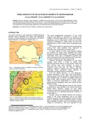
PALEONTOLOGICAL UPDATE OF DEALUL MELCILOR (BRASOV)
PALEONTOLOGICAL UPDATE OF DEALUL MELCILOR (BRASOV)
PALEONTOLOGICAL UPDATE OF DEALUL MELCILOR (BRASOV)

Create successful ePaper yourself
Turn your PDF publications into a flip-book with our unique Google optimized e-Paper software.
D. UNGUREANU<br />
Colospongia catenulata Ott, 1967 in Dragastan<br />
& Gradinaru, 1975<br />
Pl. I, fig. 2-6; Pl. II, fig. 1-3<br />
1936. Colospongia dubia (Münster), Jekelius – pp.<br />
16, 28, 39-40; tab. II, fig. 1-7.<br />
1943. Colospongia dubia (Münster), Simionescu and<br />
Barbu – Pl. I, fig. 17, p. 12; p. 16.<br />
1975. Colospongia catenulata (Ott), Dragastan and<br />
Gradinaru – pp. 248-249.<br />
Small size sphinctozoan sponge, with the<br />
look of a chain with balls, consisting of small<br />
spherical components linked together in chains<br />
up to 13 (Pl. I, fig. 2, 3). The average diameter<br />
of the spherical chambers is up to 3 mm. The<br />
overall size is of up to 3.5 cm long and 4.5 mm<br />
in diameter<br />
The specimens preserved in a very fractured<br />
and brittle altered limestone, together with<br />
echinid spines and coral remains. There could<br />
be noticed no branched specimens, but many in<br />
natural longitudinal or transversal sections.<br />
Internal structure is poorly preserved. The<br />
specimens entirely held in the limestone matrix,<br />
are filled internally with uniform micritic<br />
carbonate crystals and even a sparitic halo<br />
around some specimens can be noticed. On<br />
some specimens near the naturally altered rock<br />
surface, as well as on some naturally sectioned<br />
specimens, the internal separation walls can be<br />
noticed. On one particular specimen, some<br />
intermediate transversal walls remains can be<br />
noticed, also, splitting the chambers in two<br />
halves, probably connected (Pl. I, fig. 4).<br />
The oscula are present either in terminal<br />
position (Pl. I, fig. 5), or laterally, in central<br />
position (Pl. I, fig. 6). There might be 2 or even<br />
3 lateral oscula per segment, in 90° angled<br />
positions. Rarely, the oscula may be located<br />
laterally not centered, and there are no oscula<br />
present on the connection line between the<br />
chambers. The osculum diameter is about 0.1<br />
up to 0.3 mm, with an average of 0.15 mm,<br />
depending on the size of the sponge itself. The<br />
ratio between the osculum diameter and the<br />
chamber external diameter is between 0.07 and<br />
0.2, with the weighted average of 0.1. The<br />
osculum rarely presents a collar externally, but<br />
no particular structure corresponds internally to<br />
it (Pl. II, fig. 1).<br />
The external wall thickness is between 0.12<br />
and 0.57 mm and the ratio between the wall<br />
thickness and the sponge diameter between<br />
0.09 and 0.17. The wall gets thicker in the<br />
chambers connection area. The external walls<br />
are slightly thicker than the internal ones.<br />
The pores are round and evenly distributed<br />
on the sponge surface, without any particular<br />
geometrical pattern. The density of pores is of<br />
25 – 37/mm 2 (Pl. II, fig. 2, 3). They are small<br />
and round. Their diameter is between 20 and<br />
40 µm (30 µm in average) either on the exterior<br />
378<br />
or the interior side of the wall, but they get<br />
much thinner in the wall thickness.<br />
Specimens from Ladinian limestone of<br />
Dealul Melcilor, Brasov.<br />
No. of specimens: 26 and several more<br />
fragments.<br />
Group Chaetetida Sokolov, 1939 in Sokolov,<br />
1971<br />
Genus Chaetetopsis Peterhans 1930 in<br />
Dragastan et al., 1998<br />
Chaetetopsis tithonica sp. n.<br />
Pl. II, fig. 4-6; Pl. III, fig. 1, 2<br />
Type specimen – Holotype: polished colony<br />
+ 2 thin sections, a transversal and an oblique<br />
one – no. 20.644 – National Museum of<br />
Geology, Bucharest<br />
Age: Tithonian<br />
Collecting spot: Temelia quarry, Dealul<br />
Melcilor, Brasov<br />
Species name: Referring to the age of<br />
specimen.<br />
Diagnosis: columnar colony made of thin<br />
fine parallel tubular individuals with polygonal<br />
(mainly hexagonal) section. Macroscopically,<br />
the colony has a radiar development (Pl. II, fig.<br />
4) and a smooth natural surface; on weathered<br />
surfaces, rarely preserved horizontal thin tabula<br />
can be noticed (Pl. II, fig. 5). In thin sections,<br />
the wall between individuals is practically<br />
invisible and the tabula cannot be noticed<br />
anymore.<br />
Dimensions:<br />
Colony height: approx. 6 cm<br />
Colony width: approx. 7 cm<br />
Average individual tube diameter: 0.4 mm<br />
Average wall thickness: 10 – 15 μm<br />
Description: Compact, entirely calcified<br />
colony, with visible brown levels as lateral<br />
stripes. Polygonal uniform individuals, entirely<br />
sparitized, with unusual thin walls, that is a<br />
specific distinctive feature (Pl. II, fig. 6).<br />
Longitudinally, the individuals are long and<br />
horizontal tabula cannot be noticed (Pl. III, fig.<br />
1). The brown stripes in the colony are not<br />
growth levels or tabula levels, but opaque<br />
organic matter accumulation levels, as fluid<br />
secondary inclusions (Pl. III, fig. 2).<br />
Petrologically, the specimen is made of<br />
monocrystalline calcite with adds; on the<br />
surface, the pseudo-morphosis of calcite after<br />
aragonite shows the organic origin of primary<br />
carbonatic skeleton. The specimen has cracks<br />
filled with calcite, but also “growth layers” due to<br />
organic or crystalline matter, and intracrystalline<br />
breaks after crystal forming. It has,<br />
also, signs of levigation.<br />
The species has similarities with<br />
Chaetetopsis crinita Neumayr as figured by