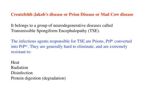
Creutzfeldt-Jakob's disease or Prion Disease or Mad Cow ... - iSites
Creutzfeldt-Jakob's disease or Prion Disease or Mad Cow ... - iSites
Creutzfeldt-Jakob's disease or Prion Disease or Mad Cow ... - iSites

Create successful ePaper yourself
Turn your PDF publications into a flip-book with our unique Google optimized e-Paper software.
<strong>Creutzfeldt</strong>-Jakob’s <strong>disease</strong> <strong>or</strong> <strong>Prion</strong> <strong>Disease</strong> <strong>or</strong> <strong>Mad</strong> <strong>Cow</strong> <strong>disease</strong><br />
It belongs to a group of neurodegenerative <strong>disease</strong>s called<br />
Transmissible Spongif<strong>or</strong>m Encephalopathy (TSE).<br />
The infectious agents responsible f<strong>or</strong> TSE are <strong>Prion</strong>s, PrP c converted<br />
into PrP sc . They are generally hard to eliminate, and are extremely<br />
resistant to: <br />
Heat<br />
Radiation<br />
Disinfection <br />
Protein digestion (degradation)
<strong>Creutzfeldt</strong>-Jakob’s <strong>Disease</strong> <br />
(<strong>Mad</strong> cow <strong>disease</strong> <strong>or</strong> <strong>Prion</strong> <strong>disease</strong>)
Severe Brain Atrophy in CDJ’s patient<br />
www.scienceclarified.com
KURU: laughing death -Papua New Guinea (1957)<br />
http://www.biologie.uni-duesseld<strong>or</strong>f.de<br />
KURU and prion’s <strong>disease</strong>: similarities in the symptoms and in cerebellar ataxia
Models f<strong>or</strong> conf<strong>or</strong>mational conversion of PrP c into PrP sc
!!! vCJD is of <strong>or</strong>al <strong>or</strong>igin,<br />
and PrP sc could take<br />
years bef<strong>or</strong>e converting<br />
PrP c Role<br />
of <strong>or</strong>al mucosas in first<br />
accumulating then<br />
spreading PrP sc <br />
Tissues where PrP sc accumulates
Symptoms in <strong>Creutzfeldt</strong>-Jakob’s <strong>Disease</strong><br />
-It could take years (decades) bef<strong>or</strong>e a carrier of prion <strong>disease</strong> will become fully<br />
symptomatic.<br />
-Symptoms are characterized by cognitive decline, which may be fulminant and<br />
progress to akinetic mutism within few weeks.<br />
-Cerebellar signs are evident (balance and co<strong>or</strong>dination dysfunction -ataxia,<br />
changes in gait, rigid posture, and seizures).<br />
mood swings<br />
depression<br />
anxiety<br />
mem<strong>or</strong>y lapses<br />
social withdrawal<br />
clumsiness <strong>or</strong> lack of co<strong>or</strong>dination<br />
insomnia
As the <strong>disease</strong> rapidly progresses, patients with all f<strong>or</strong>ms of<br />
CJD generally experience:<br />
* visual deteri<strong>or</strong>ation and eventual blindness<br />
* dementia<br />
* involuntary muscle contractions<br />
* muscle paralysis<br />
* slurred speech<br />
* difficulty swallowing<br />
* incontinence<br />
* coma
<strong>Cow</strong> affected by Bovine Spongif<strong>or</strong>m Encephalitis<br />
www.jonbarron.<strong>or</strong>g
Diagnosis:<br />
-Electroencephalography<br />
-MRI<br />
“Probable” CJD is based on the clinical symptoms.<br />
-Post-m<strong>or</strong>tem immunohistochemistry of PrP sc aggregates.<br />
-Biopsy of the tonsils and, in 30% of the cases, of skeletal muscles<br />
can confirm CDJ. Determination of protease-K resistant f<strong>or</strong>m PrP sc .
Absence of Protease-K-digested PrP in CJD used f<strong>or</strong> diagnosis<br />
CJD<br />
Control<br />
<strong>Prion</strong> protein
Spongif<strong>or</strong>m (intracellular vacuolation) change in the c<strong>or</strong>tical<br />
gray matter of the brain, characteristic of TSEs and prions<br />
aggregates<br />
Walker et al.,
*Neuronal death<br />
*Neuronal apoptosis<br />
Features of TSEs and CJD<br />
*Astrogliosis (as a cause <strong>or</strong> a consequence of<br />
inflammation)<br />
*Protein misfolding and aggregation<br />
*Precipitation of aggregates (proteinaceous material) both<br />
at an intracellular and extracellular level (amyloid plaques)
Deposition of fibrillar proteinacious material <br />
in <strong>Creutzfeldt</strong>-Jakob’s <strong>disease</strong> (prion <strong>disease</strong>)<br />
<strong>Prion</strong> <strong>disease</strong>: Alteration in the prion protein lead to both intracellular and extracellular<br />
accumulation of amyloid aggregates, plaques, similar to those characteristic of AD, and<br />
positive to prion protein staining. Probably, replication and accumulation of the protease<br />
insensitive PrP sc results in fibril f<strong>or</strong>mation and plaque deposition. <br />
Alzheimer’s<br />
<strong>Creutzfeldt</strong>-Jakob’s<br />
Aguzzi A, Haass C. Science. 2003 Oct 31;302(5646):814-8. Review.
Epidemiology of <strong>Creutzfeldt</strong>-Jakob’s <strong>disease</strong> (CJD)<br />
CJD is, among the Transmissible Spongif<strong>or</strong>m Encephalopathies, the most diffuse<br />
one. <br />
CJD can be classified as<br />
Sp<strong>or</strong>adic sCJD: etiology not known, caused by both exogenous and endogenous<br />
fact<strong>or</strong>s, represents 85% of all the cases of CJD. In the United States, there are<br />
approximately 200 sp<strong>or</strong>adic CJD cases per year. <br />
Familial fCJD: caused by mutations in the gene f<strong>or</strong> PrP (prion protein). 15% of<br />
CJD cases are inherited.<br />
Iatrogenic iCJD: caused by the spreading of the infectious agent due to<br />
contaminated surgical tools, to the transplantation of tissues, <strong>or</strong> to the<br />
administration of pituitary h<strong>or</strong>mones from deceased patients affected by the<br />
<strong>disease</strong>. 1% of CJD cases.<br />
Variant vCJD: caused by the transmission of Bovine Spongif<strong>or</strong>m<br />
Encephalopathy (BSE) prion to humans (aka <strong>Mad</strong> <strong>Cow</strong> <strong>disease</strong>).
Incidence of BSE rep<strong>or</strong>ted w<strong>or</strong>ldwide
Incidence of vCJD rep<strong>or</strong>ted w<strong>or</strong>ldwide
The prion protein: functional domains and mutations<br />
causing inherited prion’s <strong>disease</strong>s
Functional domains of the prion protein<br />
OR: not required f<strong>or</strong> PrPc function, it might influence the change of<br />
conf<strong>or</strong>mation in PrPsc, as OR KO mice do not propagate the <strong>disease</strong>. Protects<br />
from apoptosis.<br />
CC1: probably involved in protein internalization/trafficking. CC1 KO mice<br />
are viable and could develop the <strong>disease</strong>.<br />
CC2: might w<strong>or</strong>k in concert with HC region, as partial deletion of either <strong>or</strong> the<br />
other domain, <strong>or</strong> ablation of one domain and partial deletion of the other<br />
accelerate the pathology in mice.<br />
C-terminal: gene KO on H2, H3 <strong>or</strong> both domains leads to ataxia and neuron<br />
<strong>disease</strong>, BUT FAIL TO REPLICATE PRIONS. <br />
No transmission of <strong>disease</strong> from H2 KO and H3 KO to other animals. H2 and<br />
H3 might stabilize the conf<strong>or</strong>mation of the protein. <br />
C-terminal deletion prevents GPI anch<strong>or</strong>ing of the protein, no development of<br />
the <strong>disease</strong>.
<strong>Prion</strong> Protein: domains and α-helix structures<br />
www.chemsoc.<strong>or</strong>g<br />
-PrPc contains 208 aminoacid residues and is abundantly expressed in neurons and glial<br />
cells<br />
-Signal peptide sequence<br />
-Octarepeats followed by a sh<strong>or</strong>t Hydrophobic/toxic structure<br />
-The C-terminal p<strong>or</strong>tion of the protein is a globular structure that contains 3 α-helix<br />
domain and 2 β-helical domains. This domain folds rapidly and is extremely stable
Amyloid plaques in TSE<br />
Kuru <strong>disease</strong><br />
GSS <strong>disease</strong> <br />
Gerstmann-Straussler-Sheinker<br />
<strong>disease</strong><br />
Kuru <strong>disease</strong>
Aguzzi et al., Nat Rev Mol Cell Biol. 2007 Jul;8(7):552-61
Physiologic role of PrP c <br />
Caughey and Byron, 2006 Nature 443-19
Antiapoptotic function: <br />
PrP c KO mice are m<strong>or</strong>e susceptible to apoptosis.<br />
Following ischemic injury, PrP c KO mice have increased infarct<br />
volume and increased caspase 3 activation.
Infarcts are larger in PrP c KO mice after ischemic injury
Levels of activated caspase 3 are increased in PrPc KO mice<br />
after ischemic injury
PrP c protects against oxidative stress<br />
PrP c Ko mice are m<strong>or</strong>e susceptible to damage by H 2 O 2 <br />
PrP c KO mice have reduced SOD activity<br />
Brain of PrP c KO mice has increased levels of oxidated proteins,<br />
lipids, DNA.<br />
PrP c also involved in maintaining mitochondrial integrity
Mitochondrial structure is disrupted in <strong>Prion</strong>’s infected hamsters<br />
Control<br />
<strong>Prion</strong>’s infected
PrP c maintains synaptic architecture and function<br />
PrP c localizes mainly at the synaptic terminal<br />
PrP c KO mice have impaired Glutamatergic and GABAergic transmission,<br />
as well as decreased LTP.<br />
Synaptic loss is an early pathologic change in prion’s <strong>disease</strong>.
PrP stains as a flocculate/am<strong>or</strong>phous f<strong>or</strong>m at a synaptic level
PrP c physiological functions
How does the prion f<strong>or</strong>m? <br />
How does it propagate?
Mechanism of replication of PrP Sc from PrP C <br />
Sakaguchi
Nature of the prion<br />
The prion is the minimum required infectious agent able to convert<br />
n<strong>or</strong>mal cellular prion protein Prp C into the scrapie PrP sc .<br />
Is the prion a virus?<br />
NO, the prion is not a virus, as RNA and DNA material are totally absent<br />
in its composition, and the minimal molecular weight necessary f<strong>or</strong><br />
infectivity is ~2x10 5 Da, so small to exclude the size of a virus.<br />
Is the prion proteinaceous material?<br />
YES
The “unf<strong>or</strong>tunate” goal of CDJ <strong>disease</strong> is to convert<br />
n<strong>or</strong>mal cellular prion protein PrP C into the scrapie<br />
f<strong>or</strong>m PrP sc . <br />
This will result in <br />
1-reduction of the f<strong>or</strong>m with α-sheet conf<strong>or</strong>mation<br />
2-accumulation of the f<strong>or</strong>m with β-sheet conf<strong>or</strong>mation,<br />
which will f<strong>or</strong>m aggregates and deposit both at an<br />
intracellular and extracellular level.
Models f<strong>or</strong> conf<strong>or</strong>mational conversion of PrP c into PrP sc
Different biochemical and structural properties <br />
between PrP C and PrP Sc <br />
Sakaguchi
The “protein-only” hypothesis<br />
-In vitro data suggest that prion infectivity is achieved also de novo in the test tube.<br />
-Propagation of conf<strong>or</strong>mationally changed yeast prions has been achieved.<br />
-In vivo, prion protein null mice (PrP -/- <strong>or</strong> PrP KO) are resistant to prion infection,<br />
do not propagate prion infectivity after exposure to PrP Sc , suggesting that PrP C is<br />
necessary to propagate the infectivity caused by PrP Sc .<br />
- <strong>Prion</strong> protein null mice do not propagate prion infection also when infected with<br />
infectious brain tissue. This implies that the protein PrP C ALONE (and not in<br />
combination with other cellular <strong>or</strong> putative viral fact<strong>or</strong>s) is sufficient to propagate<br />
the prion infectivity.<br />
-Brain homogenates spiked with PrP Sc and subjected to sonication and recovery<br />
amplify the infectious species which maintains infectivity when “transferred” to<br />
new tissue.
Fibrils of prion protein: self propagating mechanism?<br />
<strong>Prion</strong> Particles<br />
<strong>Prion</strong> Particles+yeast protein<br />
<strong>Prion</strong> Profile: Far left, infectious prion particles extracted from yeast cells. At<br />
right is an example of what yeast prions can do when mixed with an isolated yeast<br />
protein. These fibrils are essentially long series of prions all linked together after<br />
replicating many times in the presence of the protein.<br />
Soto and Castilla<br />
Nat Med. 2004 Jul;10 Suppl:S63-7. Review.
PMCA Protein Misfolding Cyclic Amplification
PMCA: protein misfolding cyclic amplification:<br />
a technique to amplify prions in vitro
PMCA to generate synthetic strains of prions
Inoculation of synthetic strains of prions to generate<br />
animal models f<strong>or</strong> prion’s <strong>disease</strong>
The “seeding:nucleation” model in <strong>Prion</strong>’s replication
PrP c localizes at the plasma membrane<br />
and undergoes internalization
Upon infection, PrP relocalizes from the plasma<br />
membrane to endocytic compartments
Where does the conversion from PrP c to PrP sc occur?<br />
Transp<strong>or</strong>t of PrP sc ?
Cell biology of PrP in scrapie infected cells
<strong>Prion</strong> protein scrapie accumulation and propagation <br />
from n<strong>or</strong>mal cellular <strong>Prion</strong> protein (PrP C to PrP sc )<br />
Caughey and Byron, 2006 Nature 443-19
Membrane modifications are associated with PrP propagation<br />
Dendrite of natural<br />
sheep scrapie brain<br />
Dendrites of a clinical<br />
<strong>disease</strong>d<br />
intracerebrally<br />
inoculated with a<br />
synthetic PrP source.<br />
Axon terminal from a<br />
22CH scrapieinfected<br />
hamster
Post-translational modifications on <strong>Prion</strong> protein: <br />
Glycosylation and binding to metals <br />
Caughey and Byron, 2006 Nature 443-19
GPI anch<strong>or</strong> as a target f<strong>or</strong> the treatment of CJD?
Inhibition of GPI-anch<strong>or</strong> could reduce the amount of PrP C at the<br />
plasma membrane, reducing propagation of the prion<br />
Caughey and Byron, 2006 Nature 443-19
Can cytosolic aggregates seed f<strong>or</strong> propagation?
PrP aggregates can be found at an intracellular level
Cytosolic PrP C can f<strong>or</strong>m intracellular aggregates in presence<br />
of extracellular PrP fibrils
Induced intracellular aggregates can be transmitted to progeny
F<strong>or</strong>mation of aggregates can also be induced in<br />
neighb<strong>or</strong>ing adjacent cells
Direct transfer of prion cytosolic aggregates from don<strong>or</strong><br />
to recipient cells
PrP sc is toxic both when extracellular and intracellular<br />
MECHANISM?<br />
Loss of physiologic function <strong>or</strong> gain of toxic function?
PrP c KO mice are viable, develop n<strong>or</strong>mally and have no<br />
severe pathologies observed later in life.<br />
PrP c KO do not develop the <strong>disease</strong> as they do not propagate<br />
the f<strong>or</strong>mation of PrP sc <br />
Gain of toxic function rather than loss of physiological function
A role f<strong>or</strong> Autophagy?<br />
Can autophagy target the degradation of PrP sc aggregates<br />
and be used in the therapy of prion’s <strong>disease</strong>?
Accumulation of degenerating lysosomes and other <strong>or</strong>ganelles <br />
at the dark synapse in human vCJD brain
Main steps in macroautophagy
Autophagic vacuoles f<strong>or</strong>m at the synapse of human vCJD brain
Autophagic vacuoles f<strong>or</strong>med in scrapie-infected hamster brain
Autophagy-inducing drugs can regulate autophagy<br />
in scrapie infected cells
Autophagy-inducing drugs can reduce the PK resistant load <br />
of PrP sc aggregates
Autophagy as an approach to reduce prion infection
Small molecules used to regulate autophagy <br />
in neurodegenerative <strong>disease</strong>s
Approaches f<strong>or</strong> the treatment of CJD<br />
-Use of autophagy-inducing molecules<br />
Use of molecules that associate with PrP C and interfere with the capacity of PrP Sc<br />
to bind and to propagate the infectious principle.<br />
-GPI inhibit<strong>or</strong>s<br />
-PrP Sc antibodies: problems in achieving the right level of immunization<br />
(prion protein is a self antigen, and immunesystem is involved in the replication<br />
of the prion agent and its ultimate access to the CNS).<br />
Active immunization with recombinant protein has relatively modest therapeutic<br />
effects in mice.<br />
Passive immunization effective within a month after exposure to the<br />
contaminant. However, too expensive and not effective if perf<strong>or</strong>med in advanced<br />
stages of the <strong>disease</strong>, when the animal is symptomatic.